. 2001 Dec;159(6):2293-301.
doi: 10.1016/S0002-9440(10)63079-9.

Identification of receptor-binding sites of monocyte chemotactic S19 ribosomal protein dimer

Affiliations

Identification of receptor-binding sites of monocyte chemotactic S19 ribosomal protein dimer

Y Shibuya et al. Am J Pathol. 2001 Dec.

Abstract

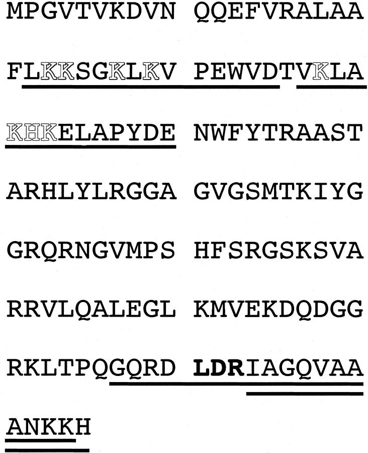
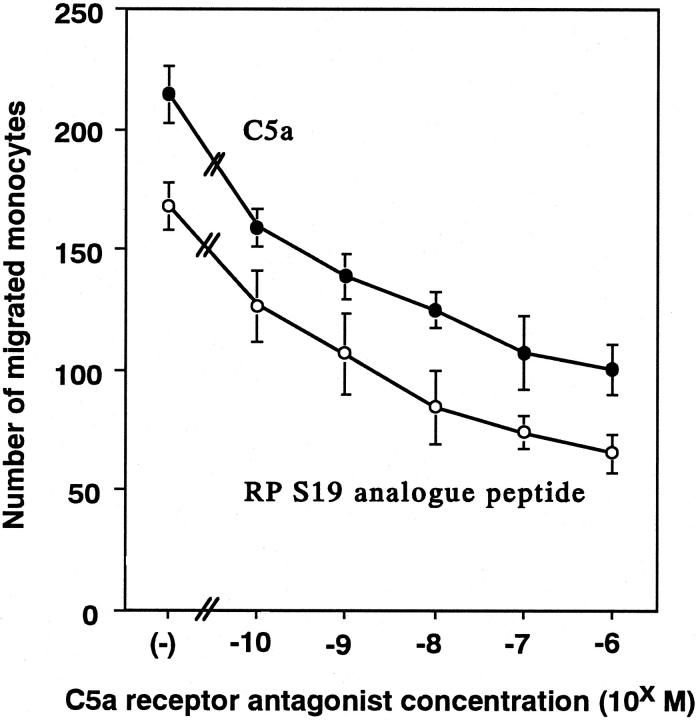
The S19 ribosomal protein (RP S19) cross-linked homo-dimer attracts monocyte migration by binding to C5a receptor on monocytes (H Nishiura, Y Shibuya, T Yamamoto, Laboratory Investigation, 1998, 78:1615-1623). Using site-directed mutants of recombinant RP S19 and synthetic peptides mimicking RP S19 molecular regions, we currently identified the binding sites of the RP S19 dimer to the C5a receptor. The RP S19 dimer activated the receptor by a two-step binding mechanism as in the case of C5a. The first binding site was a basic cluster region containing a -Lys41-His42-Lys43- sequence. The second one was the -Leu131-Asp132-Arg133- moiety, localized 12 residues upstream from the COOH-terminal. The second binding triggered the chemotactic response. The first binding would have a role in achieving a high-binding affinity between the ligand and receptor. The first and second ligand-binding sites of C5a receptor seem to be shared by C5a and the RP S19 dimer, although overall homology between the amino acid sequences of these ligands is only 4%.


Figures

Figure 1.
Figure 1.
S19 ribosomal protein (RP S19) molecular regions as candidates for two C5a receptor-binding sites of the cross-linked RP S19 dimer. Two basic cluster regions as the candidates for the first binding site are indicated with the white letters. The candidate for the second binding site is indicated with the bold letters. The regions mimicked by the synthetic peptides are underlined.
Figure 2.
Figure 2.
Monocyte chemotactic activity of dimers of recombinant RP S19 mutated at one of the basic cluster regions. The cross-linked dimers of wild-type or mutated forms of recombinant RP S19 were subjected to the monocyte chemotaxis assay at the final concentrations of 10−9 mol/L using the multiwell chamber and a Nuclepore filter with a pore size of 5 μm. After a 90-minute incubation, the membrane was stained with Giemsa solution and the total number of monocytes migrated beyond the membrane was counted in five high-power fields of light microscopy. Values are expressed as mean ± SD. Three experiments were performed for each examination. The dotted column, the hatched column with thin slash, and the hatched column with thick slash denote the monocyte chemotactic activity of the dimers of the wild-type RP S19 (Wild), of the Lys23Ala, Lys24Ala, Lys27Ala, and Lys29Ala RP S19 (M 13), and of the Lys38Ala, Lys41Ala, His42Ala, and Lys43Ala RP S19 (M 14), respectively. These dimers were prepared with type II transglutaminase (type II TG). The positive controls (open columns) are 10−9 mol/L, 10−10 mol/L, and 10−11 mol/L of the RP S19 dimerized with factor XIIIa (plasma transglutaminase) in the presence of heparin, and the negative control (open column) is PBS containing 0.1 mg/ml BSA.
Figure 3.
Figure 3.
Inhibition of chemotactic activity of RP S19 dimer by basic cluster analogue peptide, Ac-VKLAKHKELAPYDE. The indicator monocytes in the chemotaxis assay were pretreated with either of the basic cluster analogue peptides or a derivative at a concentration of 10−4 mol/L, or with the vehicle buffer (the medium for the cell suspension, open columns) for 10 minutes at 37°C, and used for the chemotaxis assay attracted by the RP S19 dimer (10−9 mol/L), formyl-Met-Leu-Phe (f-MLF, 10−9 mol/L) or PBS containing 0.1 mg/ml of BSA (PBS). No. 1 peptide (hatched columns with thin slash) denotes Ac-LKKSGKLKVPEWVD mimicking the sequence from Leu22 to Asp35 of RP S19, no. 2 peptide (hatched columns with thick slash) denotes Ac-VKLAKHKELAPYDE mimicking the sequence from Val37 to Asn51, and no. 3 peptide (dotted columns) denotes Ac-VKLAAAAELAPYDE, a derivative of no. 2 peptide substituted the three basic residues in tandem (Lys-His-Lys) by Ala residues. Values are mean ± SD. Three experiments were performed for each examination. The numbers with parentheses on the vertical axis are the values for the f-MLF-induced chemotaxis.
Figure 4.
Figure 4.
Inhibition of chemotactic activity of C5a by first ligand analogue peptide of RP S19 dimer, Ac-VKLAKHKELAPYDE. The indicator monocytes in the chemotaxis assay were pretreated with one of the three peptides described in Figure 3 ▶ , or with the vehicle buffer (open columns) for 10 minutes at 37°C, and used for the chemotaxis assay attracted by zymosan-activated plasma (ZAP, 10%) containing C5a or by PBS containing 0.1 mg/ml of BSA (PBS). The hatched columns with thin slash denote Ac-LKKSGKLKVPEWVD mimicking the sequence from Leu22 to Asp35 of RP S19 (no. 1 peptide), the hatched columns with thick slash denote Ac-VKLAKHKELAPYDE mimicking the sequence from Val37 to Asn51 (no. 2 peptide), and the dotted columns denote Ac-VKLAAAAELAPYDE, a derivative of no. 2 peptide substituted the three basic residues in tandem (Lys-His-Lys) by Ala residues (no. 3 peptide). Values are mean ± SD. Three experiments were performed for each examination.
Figure 5.
Figure 5.
Lack of chemoattracting capacity of first ligand analogue peptide. The basic cluster analogue peptides were subjected to the monocyte chemotaxis assay at the final concentrations of 10−4 mol/L. No. 1 peptide (hatched columns with thin slash) denotes Ac-LKKSGKLKVPEWVD mimicking the sequence from Leu22 to Asp35 of RP S19, and no. 2 peptide (hatched columns with thick slash) denotes Ac-VKLAKHKELAPYDE mimicking the sequence from Val37 to Asn51. The positive control is the RP S19 dimer (10−9 mol/L), and the negative control is PBS containing 0.1 mg/ml of BSA. Values are mean ± SD. Three experiments were performed for each examination.
Figure 6.
Figure 6.
Schematic diagrams of second receptor-binding site of C5a and of candidate for it of RP S19 dimer. In the second binding with the C5a receptor activation, the side chains of Leu72 and Arg74, and the COOH-terminal α-carboxyl group of Arg74 of C5a are essential. The moiety with Leu131-Asp132-Arg133 of RP S19 possesses the same two side chains and β-carboxyl group of Asp132. It is assumed that the β-carboxyl group of Asp132 functions equivalently as the α-carboxyl group of Arg74.
Figure 7.
Figure 7.
Monocyte chemotactic activity of dimers of recombinant RP S19 mutated at Leu131-Asp132-Arg133 moiety. The dimers of the wild-type RP S19 (Wild), of the Leu131Asp RP S19 (L131D), of the Asp132Gly RP S19 (D132G), and of Arg133Ala RP S19 (R133A) were subjected to the monocyte chemotaxis assay at the final concentrations of 10−9 mol/L. The negative control is PBS containing 0.1 mg/ml BSA. Values are mean ± SD. Three experiments were performed for each examination.
Figure 8.
Figure 8.
Monocyte chemotactic activity of peptide analogue of possible second ligand site of RP S19 dimer. An analogue peptide of RP S19 composed of 18 amino acid residues from Gly127 to Lys144 including the Leu-Asp-Arg sequence such as Ac-GQRDLDRIAGQVAAANKK (no. 4 peptide, open circles), and its derivative in which an Asp residue corresponding to Asp132 of RP S19 had been substituted by Gly residue (Ac-GQRDLGRIAGQVAAANKK, no. 5 peptide, filled circles) were subjected for the monocyte chemotaxis assay at various concentrations from 10−10 mol/L to 10−3 mol/L in PBS containing 2 mg/ml BSA. A peptide mimicking the COOH-terminal portion of RP S19 (IAGQVAAANKKH, no. 6 peptide, hatched circles) was also subjected to the chemotaxis assay. The negative control was PBS containing 2 mg/ml BSA. Values are mean ± SD. Three experiments were performed for each examination.
Figure 9.
Figure 9.
Competition between RP S19 dimer and analogue peptides of its second ligand site in monocyte chemotaxis. The indicator monocytes in the chemotaxis assay were pretreated with either the chemotactic analogue peptide (Ac-GQRDLDRIAGQVAAANKK, 10−4 mol/L, hatched columns with thick slash), or the modified one with the Asp-Gly substitution (Ac-GQRDLGRIAGQVAAANKK, 10−4 mol/L, hatched columns with thin slash), or the vehicle buffer (open columns) for 10 minutes at 37°C, and used for the chemotaxis assay attracted by either the RP S19 dimer (10−9 mol/L), f-MLF (10−9 mol/L), or PBS containing 0.1 mg/ml of BSA (PBS). Values are mean ± SD. Three experiments were performed for each examination.
Figure 10.
Figure 10.
Inhibition with synthetic C5a receptor antagonist of monocyte chemotaxis induced by second ligand analogue peptide of RP S19 dimer. Monocytes were pretreated with various concentrations of a synthetic receptor antagonist (NMePhe-Lys-Pro-dCha-dCha-dArg) for 10 minutes at 37°C, then C5a (10−9 mol/L, open circles) or the second ligand analogue peptide of the RP S19 dimer (Ac-GQRDLDRIAGQVAAANKK, 10−7 mol/L, closed circles) was given to induce the chemotaxis. Values are mean ± SD. Three experiments were performed for each examination.

References

    1. Nishiura H, Shibuya Y, Matsubara S, Tanase S, Kambara T, Yamamoto T: Monocyte chemotactic factor in rheumatoid arthritis synovial tissue: probably a cross-linked derivative of S19 ribosomal protein. J Biol Chem 1996, 271:878-882 - PubMed
    1. Nishiura H, Tanase S, Shibuya Y, Nishimura T, Yamamoto T: Determination of the cross-linked residues in homo-dimerization of S19 ribosomal protein concomitant with exhibition of monocyte chemotactic activity. Lab Invest 1999, 79:915-923 - PubMed
    1. Nishimura T, Horino K, Nishiura H, Shibuya Y, Hiraoka T, Tanase S, Yamamoto T: Apoptotic cells of an epithelial cell line, AsPC-1, release monocyte chemotactic S19 ribosomal protein dimer. J Biochem 2001, 129:445-454 - PubMed
    1. Horino K, Nishiura H, Ohsako T, Shibuya Y, Hiraoka T, Kitamura N, Yamamoto T: A monocyte chemotactic factor, S19 ribosomal protein dimer, in phagocytic clearance of apoptotic cells. Lab Invest 1998, 78:603-617 - PubMed
    1. Nishiura H, Shibuya Y, Yamamoto T: S19 ribosomal protein cross-linked dimer causes monocyte-predominant infiltration by means of molecular mimicry to complement C5a. Lab Invest 1998, 78:1615-1623 - PubMed

Publication types

MeSH terms

LinkOut - more resources