. 2001 Dec;108(11):1621-9.
doi: 10.1172/JCI12849.

Podocin, a raft-associated component of the glomerular slit diaphragm, interacts with CD2AP and nephrin

Affiliations

Podocin, a raft-associated component of the glomerular slit diaphragm, interacts with CD2AP and nephrin

K Schwarz et al. J Clin Invest. 2001 Dec.

Abstract

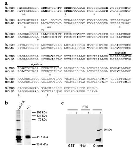
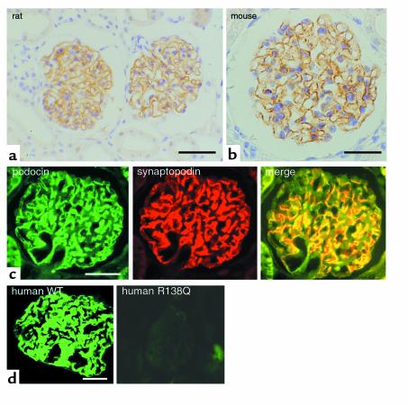
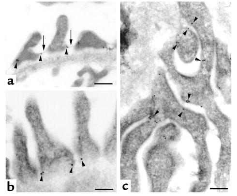
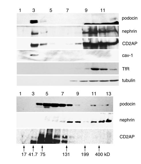
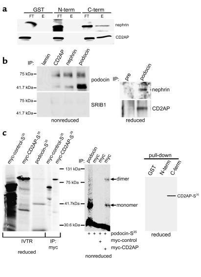
NPHS2 was recently identified as a gene whose mutations cause autosomal recessive steroid-resistant nephrotic syndrome. Its product, podocin, is a new member of the stomatin family, which consists of hairpin-like integral membrane proteins with intracellular NH(2)- and COOH-termini. Podocin is expressed in glomerular podocytes, but its subcellular distribution and interaction with other proteins are unknown. Here we show, by immunoelectron microscopy, that podocin localizes to the podocyte foot process membrane, at the insertion site of the slit diaphragm. Podocin accumulates in an oligomeric form in lipid rafts of the slit diaphragm. Moreover, GST pull-down experiments reveal that podocin associates via its COOH-terminal domain with CD2AP, a cytoplasmic binding partner of nephrin, and with nephrin itself. That podocin interacts with CD2AP and nephrin in vivo is shown by coimmunoprecipitation of these proteins from glomerular extracts. Furthermore, in vitro studies reveal direct interaction of podocin and CD2AP. Hence, as with the erythrocyte lipid raft protein stomatin, podocin is present in high-order oligomers and may serve a scaffolding function. We postulate that podocin serves in the structural organization of the slit diaphragm and the regulation of its filtration function.


Figures

Figure 1
Figure 1
Molecular cloning and Western blot analysis of mouse podocin. (a) Amino acid sequence comparison of human and mouse podocin. Similar residues are shown in boldface, strong similarity in black, and weaker similarity in gray. Different amino acids are marked by asterisks. The transmembrane domain (tm) and the stomatin signature are overlined. The box indicates the peptide used for antibody generation. Most of the amino acid exchanges between human and mouse are restricted to the NH2-terminal part of the protein, whereas the tm domain, the stomatin signature, and the COOH-terminal part are almost identical. The sequence data of mouse podocin are available from GenBank/EMBL/DDBJ under accession no. AJ302048. (b) Western blot analysis of glomerular extracts revealed a 42-kDa band under reducing (red.) conditions (right lane). Under nonreducing (nonred.) conditions (left panel), additional higher molecular bands appeared, indicating podocin dimerization. (c) The specificity of the podocin antibody was confirmed by Western blot analysis of recombinant GST-podocin fragments and GST alone. Only the COOH-terminal fusion protein (C-term) was recognized by the antibody. N-term, NH2-terminal fusion protein.
Figure 2
Figure 2
Expression of podocin in kidney tissues. Immunostaining of rat (a) and mouse (b) adult kidney sections showing the glomerular expression of podocin. (c) Double labeling of podocin (green) with the podocyte foot process marker synaptopodin (red) results in a complete overlap of both signals (yellow in merge). (d) Expression pattern of podocin in normal human kidney (left panel) and in the kidney of a patient with steroid-resistant nephrotic syndrome (right panel). The patient belongs to the group of patients originally described by Boute and coworkers (14). Interestingly, podocin expression can be detected only in the normal kidney, but not in the kidney of the patient with the podocin mutation. WT, wild-type. Bars: a, 30 μm; b, 15 μm; c, 20 μm; d, 25 μm.
Figure 3
Figure 3
Podocin localizes to the SD. Immunogold labeling shows the subcellular distribution of podocin (arrowheads) at the SD (arrows). (a and b) Cross sections of adult rat kidney. (c) A low-power magnification of a flat section. The gold particles are specifically located at the insertion site of the SD in the podocyte cell membrane (arrows in a), whereas no label can be detected in the glomerular basement membrane. b shows a high-power magnification of a. In c, the tangential section shows exclusive labeling of podocyte foot processes (arrowheads). Bars: a and c, 100 nm; b, 200 nm.
Figure 4
Figure 4
Podocin interacts with nephrin and CD2AP in lipid rafts. (a) Flotation analysis of podocin, nephrin, and CD2AP in a sucrose step gradient. The purity of the fractions was confirmed using caveolin-1 (cav-1) as raft marker and transferrin receptor (TfR) showing the absence of contaminating nonraft membranes in the DRM fraction (lane 3). Podocin is preferentially found in the DRM fraction (lane 3), whereas nephrin and CD2AP are found in the DRM fraction and the high sucrose fractions (lanes 9 and 10). The cytosolic protein tubulin is exclusively found in the heavy fractions (lanes 8–12). (b) The ability of podocin, nephrin, and CD2AP to form oligomers was tested by velocity gradient centrifugation. Arrows indicate molecular weight markers. In contrast to CD2AP, both nephrin and podocin were found to form high-order complexes.
Figure 5
Figure 5
The COOH-terminus of podocin interacts with nephrin and CD2AP. (a) The COOH-terminus of podocin associates with nephrin and CD2AP as shown by GST pull-downs. Nephrin and CD2AP are detectable in the final eluate (E) of the GST-podocin-COOH column. GST alone and the podocin NH2-terminus do not interact with these two SD components, as indicated by detection of nephrin and CD2AP in the flow-through (FT) fraction but not in the eluate. (b) The GST pull-down data were confirmed by Co-IPs with cross-linked antibodies against podocin, nephrin, CD2AP, or lamin (negative control) from glomerular extracts. Bound proteins were eluated and analyzed by SDS-PAGE and Western blot under nonreducing conditions (left panel) or in the presence of β-ME (right panel). Blots were then incubated with an irrelevant antibody (SRIB1), or with anti-podocin, anti-CD2AP, or anti-nephrin antibodies. The right panel shows Co-IPs of CD2AP and nephrin with anti-podocin antibody or preimmune serum (pre) under reducing conditions. (c) The ability of podocin to directly interact with CD2AP was tested by coimmunoprecipitation of in vitro translated proteins and by GST pull-downs. The left panel shows the in vitro translated S35-labeled proteins. Co-IP of unlabeled CD2AP and S35-labeled podocin showed direct interaction of both proteins (middle panel). The specificity of the podocin-CD2AP interaction was further confirmed by pull-down experiments, using recombinant podocin-GST fragments and radioactive-labeled CD2AP (right panel). Only with the COOH-terminal part of podocin, CD2AP was coprecipitated. IVTR, in vitro translation reaction.

Comment in

Similar articles

Cited by

References

    1. Mundel P, Kriz W. Structure and function of podocytes: an update. Anat Embryol (Berl) 1995;192:385–397. - PubMed
    1. Daniels BS, Deen WM, Mayer G, Meyer T, Hostetter TH. Glomerular permeability barrier in the rat. Functional assessment by in vitro methods. J Clin Invest. 1993;92:929–936. - PMC - PubMed
    1. Drumond MC, Deen WM. Structural determinants of glomerular hydraulic permeability. Am J Physiol. 1994;266:F1–F12. - PubMed
    1. Smoyer WE, Mundel P. Regulation of podocyte structure during the development of nephrotic syndrome. J Mol Med. 1998;76:172–183. - PubMed
    1. Somlo S, Mundel P. Getting a foothold in nephrotic syndrome. Nat Genet. 2000;24:333–335. - PubMed

Publication types