. 2001 Dec 3;194(11):1561-69.
doi: 10.1084/jem.194.11.1561.

Essential role of the prosurvival bcl-2 homologue A1 in mast cell survival after allergic activation

Affiliations

Essential role of the prosurvival bcl-2 homologue A1 in mast cell survival after allergic activation

Z Xiang et al. J Exp Med. .

Abstract

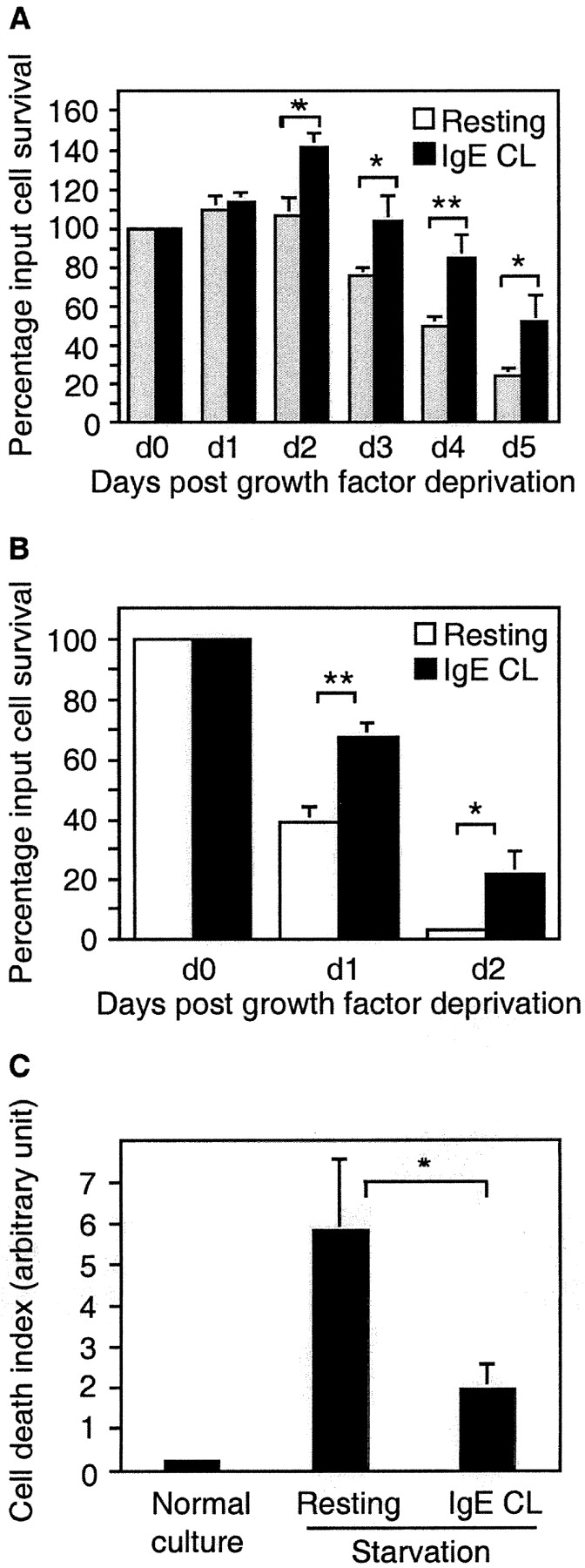
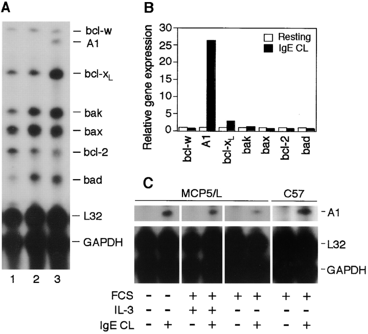
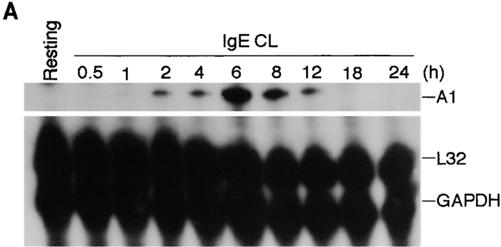
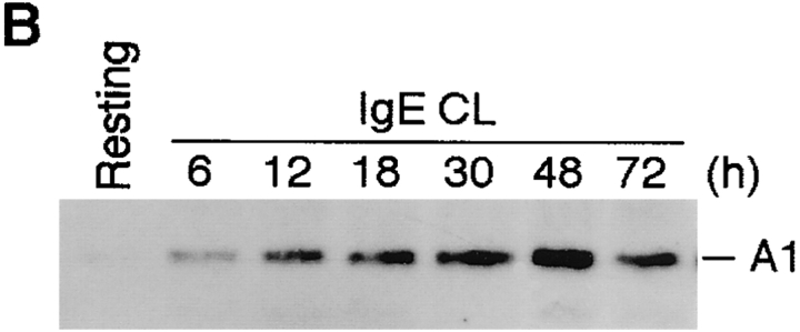
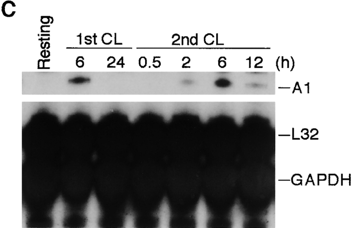
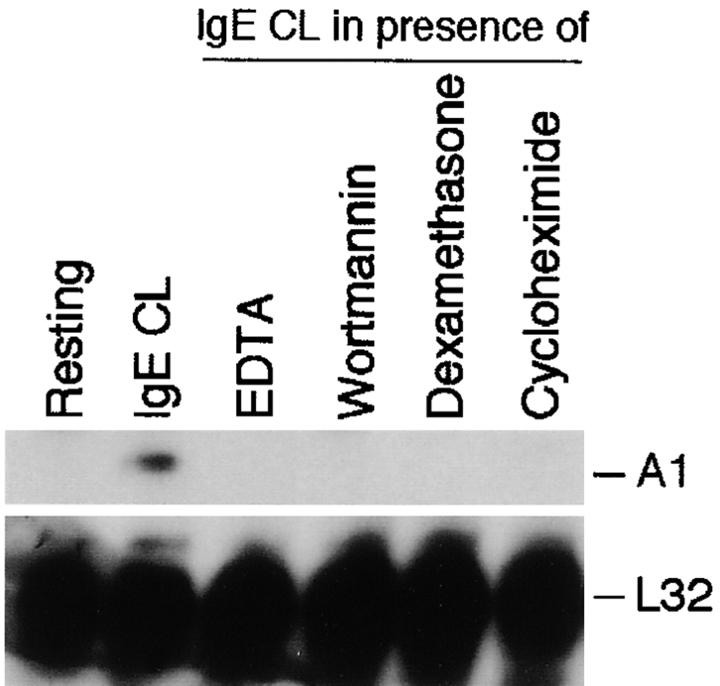
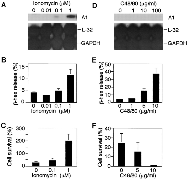
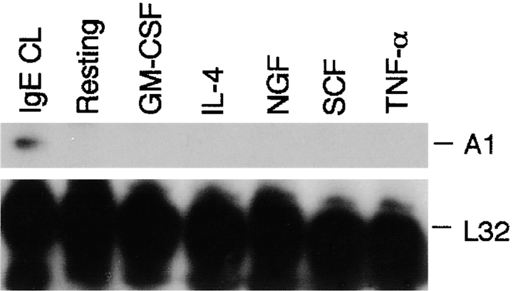
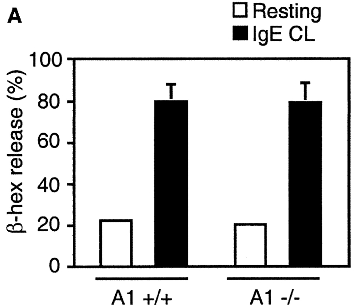
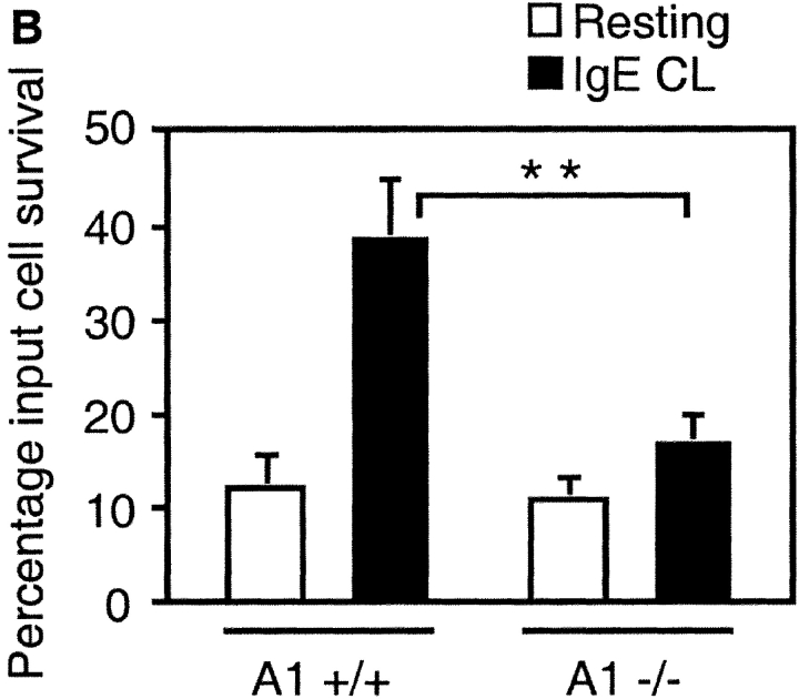
Mast cells reside in tissues, where upon activation through the high-affinity-IgE-receptor (FcepsilonRI) they degranulate and orchestrate the allergic reaction. Mast cells survive this activation and can thus be reactivated. In this study we demonstrate that this process depends on the pro-survival gene A1. Activation of mast cells through FcepsilonRI resulted in degranulation, strong induction of A1 mRNA and protein, and cell survival. In contrast, A1-deficient mast cells released granule mediators similar to the wild-type control, but the cells did not survive an allergic activation. Furthermore, A1(-/-) mice that had been sensitized and provoked with allergen exhibited a lower number of mast cell compared with littermate controls. The induction of A1 was dependent on calcium, as EDTA prevented A1 expression. The calcium ionophore, ionomycin, induced A1 expression and mast cell survival, whereas compound 48/80, a well-known mast cell secretagogue, did not. This study uncovers the importance of A1 for mast cell survival in allergic reactions, and it proposes A1 as a potential target for the treatment of allergic diseases.


Figures

Figure 1.
Figure 1.
Survival promotion after mast cell activation by FcεRI cross-linking. MCP5/L cells (A) or BMCMCs from C57BL/6 (B and C) were either stimulated through cross-linking of FcεRI (IgE CL) or left untreated in RPMI deprived of serum and growth factors. Viability was determined by trypan blue exclusion and presented as the percentage of input cells that are still alive when examined every 24 h (A and B). In C, BMCMC apoptosis was assessed by ELISA measuring the release of nucleosomes into the culture supernatant after 24 h. Data are presented as the mean ± SEM from three to five separate experiments.
Figure 2.
Figure 2.
bcl-2 family gene expression in mast cells after cross-linking of FcεRI. (A) RPA was performed to analyze the expression levels of the indicated genes on total RNA isolated from MCP5/L cells either resting or activated for 6 h through FcεRI cross-linking (IgE CL). Lane 1, resting cells cultured in the presence of WEHI-3–conditioned medium. Lane 2, resting cells that were incubated and washed through the same procedures needed for FcεRI cross-linking (deprived of growth factor). Lane 3, cells that were stimulated through FcεRI cross-linking. A representative experiment of at least six performed independently is shown. (B) Phosphor-Imaging signals presented in A (lanes 2 and 3) are shown as gene expression relative to the average expression of the house keeping genes GAPDH and L32. Data are normalized such that the densitometric level of each gene from the control cells is given a value of 1. (C) RPA was performed as in A to analyze A1 induction in MCP5/L and C57 cells activated for 6 h in the presence or absence of FCS and WEHI as a source of IL-3.
Figure 3.
Figure 3.
Kinetics of A1 mRNA and protein expression in mast cells activated by FcεRI cross-linking. (A) RPA was performed on RNA isolated from MCP5/L cells either at resting state or activated through FcεRI cross-linking (IgE CL) for various time points as indicated to detect A1 transcript levels. (B) Accumulation of A1 protein in resting and activated BMCMCs was analyzed by Western blot analysis. (C) MCP5/L cells were activated through FcεRI cross-linking (1st CL) up to 24 h, and then were washed and incubated under normal culture condition. 48 h after the first activation, cellular IgE receptors were cross-linked again (2nd CL). A1 induction at time points both in the first and the second activation periods were analyzed by RPA. Data shown are representative of three separate experiments.
Figure 3.
Figure 3.
Kinetics of A1 mRNA and protein expression in mast cells activated by FcεRI cross-linking. (A) RPA was performed on RNA isolated from MCP5/L cells either at resting state or activated through FcεRI cross-linking (IgE CL) for various time points as indicated to detect A1 transcript levels. (B) Accumulation of A1 protein in resting and activated BMCMCs was analyzed by Western blot analysis. (C) MCP5/L cells were activated through FcεRI cross-linking (1st CL) up to 24 h, and then were washed and incubated under normal culture condition. 48 h after the first activation, cellular IgE receptors were cross-linked again (2nd CL). A1 induction at time points both in the first and the second activation periods were analyzed by RPA. Data shown are representative of three separate experiments.
Figure 4.
Figure 4.
Effects of a number of inhibitors on A1 induction in mast cells after activation by FcεRI cross-linking. RPA was performed on RNA from MCP5/L cells which were activated through FcεRI cross-linking (IgE CL) in the absence or presence of various inhibitors as indicated. Data shown represent comparable results from at least three separate experiments.
Figure 5.
Figure 5.
The effects of ionomycin and compound 48/80 on mast cell A1 expression, mediator release, and survival. RPA was performed on RNA derived from MCP5/L cells incubated for 6 h in the presence of ionomycin or compound 48/80 at concentrations indicated (A and D). Release of β-hexosaminidase (β-hex) after 30 min was measured using an enzymatic colorimetric method and was indicative of activation and degranulation of mast cells (B and E). Survival rate of mast cells after 4 days in medium deprived of serum and growth factors was measured (C and F) as explained in Fig. 1. Data shown in A and D are a representative of three separate experiments and data shown in the rest are expressed as the mean ± SEM from three experiments.
Figure 6.
Figure 6.
Absence of A1 induction in cells treated with cytokines. MCP5/L cells were incubated in the presence of various cytokines as indicated for 6 h and A1 expression was measured by RPA. Cells that were activated by FcεRI cross-linking (IgE CL) were presented as positive control.
Figure 7.
Figure 7.
Absence of survival promotion of mast cells from A1−/− mice after FcεRI cross-linking (IgE CL). (A) β-hexosaminidase (β-hex) release from A1−/− mast cells was assessed as explained in Fig. 5 to demonstrate that the cells were properly activated. (B) BMCMCs from A1 knockouts and wild-type littermates were stimulated and viability was determined as explained in Fig. 1. (C) The mast cell number was quantified in the footpad skin sections of A1-a−/− and wild-type control mice before and after OVA treatment as detailed in Materials and Methods. Values presented are the means ± SEM from three to six separate experiments.
Figure 7.
Figure 7.
Absence of survival promotion of mast cells from A1−/− mice after FcεRI cross-linking (IgE CL). (A) β-hexosaminidase (β-hex) release from A1−/− mast cells was assessed as explained in Fig. 5 to demonstrate that the cells were properly activated. (B) BMCMCs from A1 knockouts and wild-type littermates were stimulated and viability was determined as explained in Fig. 1. (C) The mast cell number was quantified in the footpad skin sections of A1-a−/− and wild-type control mice before and after OVA treatment as detailed in Materials and Methods. Values presented are the means ± SEM from three to six separate experiments.

Similar articles

Cited by

References

    1. Metcalfe, D.D., D. Baram, and Y.A. Mekori. 1997. Mast cells. Physiol. Rev. 77:1033–1079. - PubMed
    1. Nilsson, G., J.J. Costa, and D.D. Metcalfe. 1999. Mast cells and basophils. Inflammation: Basic Principles and Clinical Correlates. J.I. Gallin and R. Snyderman, editors. Lippincott-Raven publications, Philadelphia, PA. 97–117.
    1. Galli, S.J. 2000. Allergy. Curr. Biol. 10:R93–R95. - PubMed
    1. Otsuka, H., J. Denburg, J. Dolovich, D. Hitch, P. Lapp, R.S. Rajan, J. Bienenstock, and D. Befus. 1985. Heterogeneity of metachromatic cells in human nose: Significance of mucosal mast cells. J. Allergy Clin. Immunol. 76:695–702. - PubMed
    1. Wardlaw, A.J., S. Dunnette, G.J. Gleich, J.V. Collins, and A.B. Kay. 1988. Eosinophils and mast cells in bronchoalveolar lavage in subjects with mild asthma. Relationship to bronchial hyperreactivity. Am. Rev. Respir. Dis. 137:62–69. - PubMed

MeSH terms