Isotype-specific selection of high affinity memory B cells in nasal-associated lymphoid tissue
- PMID: 11733574
- PMCID: PMC2193529
- DOI: 10.1084/jem.194.11.1597
Isotype-specific selection of high affinity memory B cells in nasal-associated lymphoid tissue
Abstract
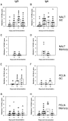
Mucosal immunoglobulin (Ig)A dominance has been proposed to be associated with preferential class switch recombination (CSR) to the IgA heavy chain constant region, Calpha. Here, we report that B cell activation in nasal-associated lymphoid tissue (NALT) upon stimulation with the hapten (4-hydroxy-3-nitrophenyl)acetyl (NP) coupled to chicken gamma globulin caused an anti-NP memory response dominated by high affinity IgA antibodies. In the response, however, NP-specific IgG(+) B cells expanded and sustained their number as a major population in germinal centers (GCs), supporting the view that CSR to IgG heavy chain constant region, Cgamma, operated efficiently in NALT. Both IgG(+) and IgA(+) GC B cells accumulated somatic mutations, indicative of affinity maturation to a similar extent, suggesting that both types of cell were equally selected by antigen. Despite the selection in GCs, high affinity NP-specific B cells were barely detected in the IgG memory compartment, whereas such cells dominated the IgA memory compartment. Taken together with the analysis of the V(H) gene clonotype in GC and memory B cells, we propose that NALT is equipped with a unique machinery providing IgA-specific enrichment of high affinity cells into the memory compartment, facilitating immunity with high affinity and noninflammatory secretory antibodies.
Figures


Similar articles
-
Altered affinity maturation in primary response to (4-hydroxy-3-nitrophenyl) acetyl (NP) after autologous reconstitution of irradiated C57BL/6 mice.Dev Immunol. 2002 Sep;9(3):119-25. doi: 10.1080/1044667031000137593. Dev Immunol. 2002. PMID: 12885152 Free PMC article.
-
Affinity maturation without germinal centres in lymphotoxin-alpha-deficient mice.Nature. 1996 Aug 1;382(6590):462-6. doi: 10.1038/382462a0. Nature. 1996. PMID: 8684487
-
B cell-specific deficiencies in mTOR limit humoral immune responses.J Immunol. 2013 Aug 15;191(4):1692-703. doi: 10.4049/jimmunol.1201767. Epub 2013 Jul 15. J Immunol. 2013. PMID: 23858034 Free PMC article.
-
Affinity enhancement of antibodies: how low-affinity antibodies produced early in immune responses are followed by high-affinity antibodies later and in memory B-cell responses.Cancer Immunol Res. 2014 May;2(5):381-92. doi: 10.1158/2326-6066.CIR-14-0029. Cancer Immunol Res. 2014. PMID: 24795350 Review.
-
Potential of nasopharynx-associated lymphoid tissue for vaccine responses in the airways.Am J Respir Crit Care Med. 2011 Jun 15;183(12):1595-604. doi: 10.1164/rccm.201011-1783OC. Epub 2011 Mar 18. Am J Respir Crit Care Med. 2011. PMID: 21471092 Review.
Cited by
-
Non-Reflex Defense Mechanisms of Upper Airway Mucosa: Possible Clinical Application.Physiol Res. 2020 Mar 27;69(Suppl 1):S55-S67. doi: 10.33549/physiolres.934404. Physiol Res. 2020. PMID: 32228012 Free PMC article. Review.
-
Turbinate-homing IgA-secreting cells originate in the nasal lymphoid tissues.Nature. 2024 Aug;632(8025):637-646. doi: 10.1038/s41586-024-07729-x. Epub 2024 Jul 31. Nature. 2024. PMID: 39085603
-
Local and systemic antibody responses in mice immunized intranasally with native and detergent-extracted outer membrane vesicles from Neisseria meningitidis.Infect Immun. 2004 May;72(5):2528-37. doi: 10.1128/IAI.72.5.2528-2537.2004. Infect Immun. 2004. PMID: 15102760 Free PMC article.
-
In vivo activation of naive CD4+ T cells in nasal mucosa-associated lymphoid tissue following intranasal immunization with recombinant Streptococcus gordonii.Infect Immun. 2006 May;74(5):2760-6. doi: 10.1128/IAI.74.5.2760-2766.2006. Infect Immun. 2006. PMID: 16622213 Free PMC article.
-
Nasal immunity to staphylococcal toxic shock is controlled by the nasopharynx-associated lymphoid tissue.Clin Vaccine Immunol. 2011 Apr;18(4):667-75. doi: 10.1128/CVI.00477-10. Epub 2011 Feb 16. Clin Vaccine Immunol. 2011. PMID: 21325486 Free PMC article.
References
-
- Neutra, M.R., E. Pringault, and J.P. Kraehenbuhl. 1996. Antigen sampling across epithelial barriers and induction of mucosal immune responses. Annu. Rev. Immunol. 14:275–300. - PubMed
-
- Neutra, M.R. 1999. M cells in antigen sampling in mucosal tissues. Curr. Top. Microbiol. Immunol. 236:17–32. - PubMed
-
- Brandtzaeg, P., E.S. Baekkevold, I.N. Farstad, F.L. Jahnsen, F.E. Johansen, E.M. Nilsen, and T. Yamanaka. 1999. Regional specialization in the mucosal immune system: what happens in the microcompartments? Immunol. Today. 20:141–151. - PubMed
-
- Corthesy, B., and J.P. Kraehenbuhl. 1999. Antibody-mediated protection of mucosal surfaces. Curr. Top. Microbiol. Immunol. 236:93–111. - PubMed
-
- Weinstein, P.D., and J.J. Cebra. 1991. The preference for switching to IgA expression by Peyer's patch germinal center B cells is likely due to the intrinsic influence of their microenvironment. J. Immunol. 147:4126–4135. - PubMed
Publication types
MeSH terms
Substances
LinkOut - more resources
Full Text Sources
Miscellaneous

