

Effect of three different media on serum free culture of donor corneas and isolated human corneal endothelial cells
- PMID: 11734511
- PMCID: PMC1723804
- DOI: 10.1136/bjo.85.12.1416
Effect of three different media on serum free culture of donor corneas and isolated human corneal endothelial cells
Abstract
Background: Removal of bovine serum from organ culture medium is necessary because of the variability in serum composition and the potential risk of infection. Two specific endothelial cell media (F99 and Endothelial-SFM) were compared with the routinely used medium MEM for their use in serum free cultivation of human corneal endothelial cells (HCEC) and donor corneas.
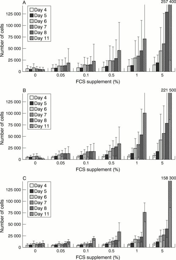
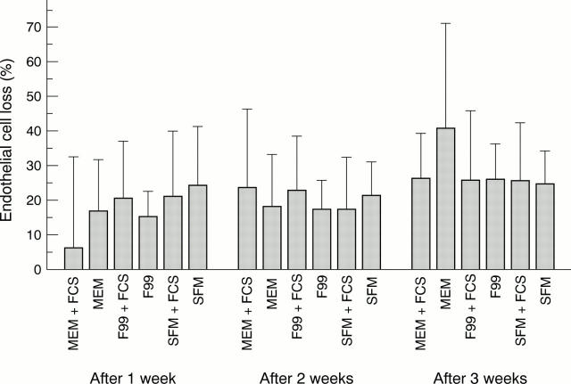
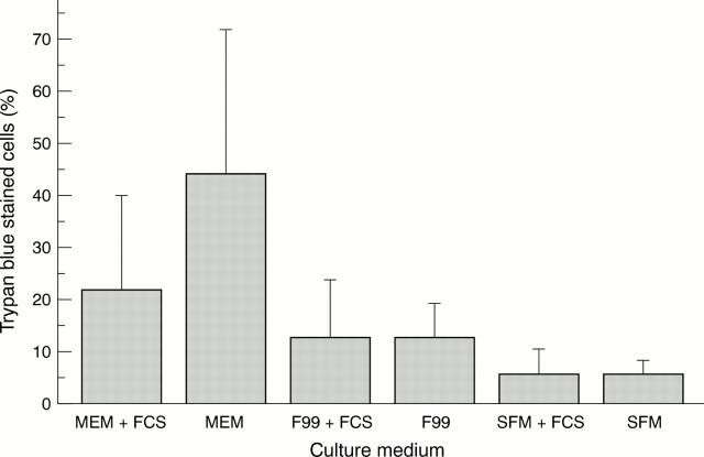
Methods: HCEC were incubated in three test media with or without increasing serum content and a growth assay was performed. Seven pairs of donor corneas were cultured in each of three media for 3 weeks, one cornea with serum supplementation and one without. Endothelial cell density was determined once each week. Trypan blue staining of the endothelium and vital staining of keratocytes was performed after 3 weeks.
Results: All three media promoted proliferation of cultured HCEC when supplemented with serum. Endothelial cell density of donor corneas was comparable after 3 weeks of cultivation in the different media. Only corneas cultured in medium MEM without serum exhibited a higher endothelial cell loss. Trypan blue staining of the endothelium after cultivation revealed the lowest number of damaged cells on corneas cultured in the medium Endothelial-SFM. The highest densities of keratocytes were found in corneas cultured in Endothelial-SFM and the lowest densities occurred after culture in MEM.
Conclusion: After incubation in Endothelial-SFM even under serum free conditions corneas were found to be of higher quality with respect to endothelial cell survival, cell membrane integrity, and keratocyte density. This medium may replace MEM, which is routinely used in European eye banks but requires supplementation with serum.
Figures
References
Publication types
MeSH terms
Substances
LinkOut - more resources
Full Text Sources
Other Literature Sources
Medical