. 2001 Dec 18;98(26):15191-6.
doi: 10.1073/pnas.261452898. Epub 2001 Dec 4.

The coxsackievirus and adenovirus receptor is a transmembrane component of the tight junction

Affiliations

The coxsackievirus and adenovirus receptor is a transmembrane component of the tight junction

C J Cohen et al. Proc Natl Acad Sci U S A. .

Abstract

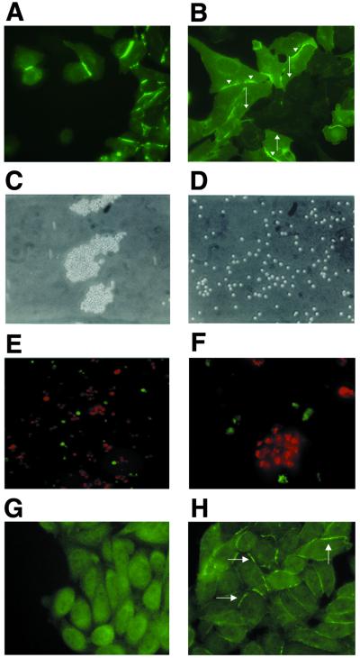
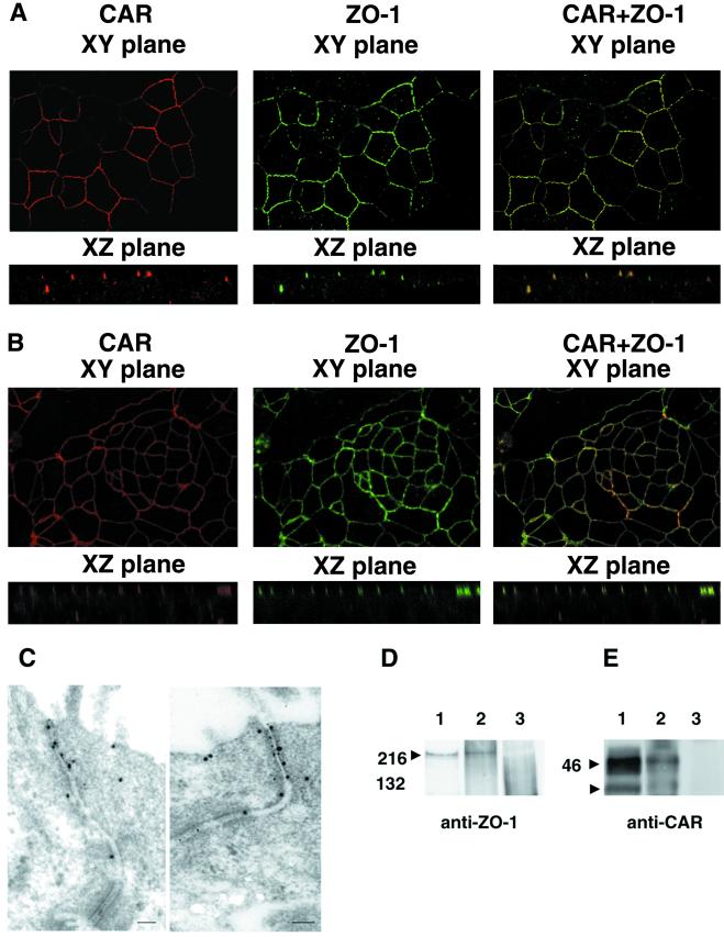
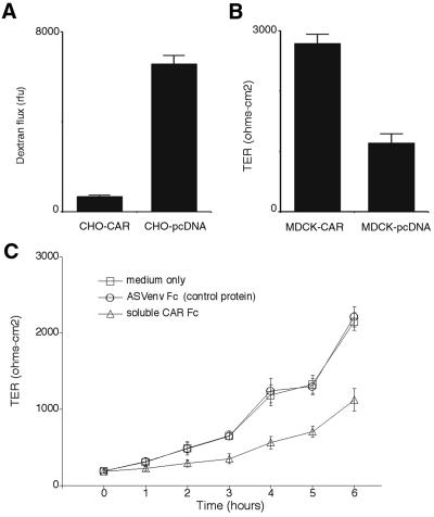
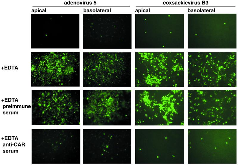
The coxsackievirus and adenovirus receptor (CAR) mediates viral attachment and infection, but its physiologic functions have not been described. In nonpolarized cells, CAR localized to homotypic intercellular contacts, mediated homotypic cell aggregation, and recruited the tight junction protein ZO-1 to sites of cell-cell contact. In polarized epithelial cells, CAR and ZO-1 colocalized to tight junctions and could be coprecipitated from cell lysates. CAR expression led to reduced passage of macromolecules and ions across cell monolayers, and soluble CAR inhibited the formation of functional tight junctions. Virus entry into polarized epithelium required disruption of tight junctions. These results indicate that CAR is a component of the tight junction and of the functional barrier to paracellular solute movement. Sequestration of CAR in tight junctions may limit virus infection across epithelial surfaces.


Figures

Figure 1
Figure 1
CAR in transfected cells. (A and B) CAR is localized at homotypic cell junctions in CAR-expressing CHO cells. (A) CHO cells stably transfected with CAR were stained with anti-CAR antibody and examined by immunofluorescence microscopy. CAR staining is enhanced at sites of intercellular contact. (B) Mock-transfected (CAR-negative) and CAR-transfected CHO cells were plated together on coverslips and then stained for CAR 48 h later. CAR is concentrated at contacts between CAR-positive cells (arrowheads), but not at contacts between CAR-positive and CAR-negative cells (filled arrows). (C and D) CAR mediates aggregation of CAR-transfected CHO cells. Clustering of CAR-transfected (C) and mock-transfected (D) CHO cells viewed by light microscopy. (E and F) CAR-mediated aggregation is caused by homophilic interactions. CHO-CAR cells labeled with DiI (red) and mock-transfected CHO cells labeled with DiO (green) were mixed in equal numbers then incubated together for 5 h. (E, ×200; F, ×600.) (G and H) CAR-expressing and mock-transfected CHO cells were stained for ZO-1. (G) Mock-transfected CHO cells show diffuse cytoplasmic staining of ZO-1. (H) ZO-1 localized at intercellular contacts between CHO-CAR cells.
Figure 2
Figure 2
Association between CAR and ZO-1 in epithelial cells. (A and B) CAR and ZO-1 colocalize when examined by confocal microscopy. (A) Human well-differentiated tracheobronchial epithelial cells were stained with anti-CAR polyclonal rabbit serum and Cy3-conjugated secondary antibody (red) or with monoclonal anti-ZO-1 antibody and FITC-conjugated secondary antibody (green). (B) Polarized T-84 colonic epithelial cells were examined as in A. (C) Colocalization of CAR and ZO-1 by immunoelectron microscopy. T-84 cell monolayers were fixed and stained with rabbit polyclonal anti-CAR and mouse monoclonal anti-ZO-1 antibody, then with 8-nm gold-labeled anti-mouse Ig to detect ZO-1 and 18-nm gold-labeled anti-rabbit Ig antibodies to detect CAR. Cells were examined by electron microscopy as described in Materials and Methods. (D and E) CAR and ZO-1 coprecipitate. T-84 cell lysates were immunoprecipitated with anti-ZO-1 antibody (D lane 1 and E lane 2), anti-CAR antibody (D lane 2 and E lane 1), or with a control antibody (D and E lane 3). Immunoprecipitates were electrophoresed in SDS/polyacrylamide gels, transferred to a poly(vinylidene difluoride) membrane, and probed with a monoclonal anti-ZO-1 antibody (D) or anti-CAR antiserum (E) as described in Materials and Methods. Arrowheads mark the location of ZO-1 (D) and CAR (E), which immunoprecipitates as a doublet (22).
Figure 3
Figure 3
CAR is a barrier to paracellular solute and ion movement. (A) FITC dextran flux was measured across CHO cell monolayers. Confluent monolayers of CHO-CAR and mock-transfected CHO cells were grown on transwell membranes. FITC-labeled dextran was added to the upper chamber and after 2 h, aliquots of fluid from the lower chamber were collected and assayed by fluorimetry. Graph shows mean and standard deviation for triplicate monolayers. Results are representative of three experiments. (B) TER of MDCK cell monolayers. Confluent monolayers of mock-transfected (MDCK-pcDNA) or CAR-expressing MDCK cells were grown on Transwell membranes at confluency for 5 days. TER was measured with an epithelial voltohmmeter. Graph shows mean and standard deviation for quadruplicate monolayers. Results are representative of three experiments. (C) Soluble CAR inhibits the formation of tight junctions. Monolayers of T-84 cells were exposed to EDTA to disrupt tight junctions. EDTA was replaced with medium alone, medium containing 5 μg of soluble CAR, or medium containing 5 μg of the avian sarcoma virus envelope protein [ASVenv Fc (control protein)]. The TER was measured as an indication of tight junction reassembly. Graph shows mean and standard deviation for triplicate monolayers. Results are representative of three experiments.
Figure 4
Figure 4
CAR-mediated coxsackievirus and adenovirus infection of polarized T-84 cells requires disruption of tight junctions. Confluent monolayers of T-84 cells were exposed to coxsackievirus B3 or to adenovirus 5 encoding GFP from either the apical or basolateral surface. Cells were examined for coxsackievirus protein expression 16 h later or for GFP expression 48 h later. In some experiments, tight junctions were disrupted with EDTA before monolayers were exposed to virus. In some experiments, monolayers were incubated with rabbit anti-CAR antiserum or with preimmune rabbit serum after EDTA treatment.

References

    1. Bergelson J M, Cunningham J A, Droguett G, Kurt-Jones E A, Krithivas A, Hong J S, Horwit M S, Crowell R L, Finberg R W. Science. 1997;275:1320–1323. - PubMed
    1. Tomko R P, Xu R, Philipson L. Proc Natl Acad Sci USA. 1997;94:3352–3356. - PMC - PubMed
    1. Roelvink P W, Lizonova A, Lee J G, Li Y, Bergelson J M, Finberg R W, Brough D E, Kovesdi I, Wickham T J. J Virol. 1998;72:7909–7915. - PMC - PubMed
    1. Bewley M, Springer K, Zhang Y-B, Freimuth P, Flanagan J. Science. 1999;286:1579–1583. - PubMed
    1. He Y, Chipman P R, Howitt J, Bator C M, Whitt M A, Baker T S, Kuhn R J, Anderson C W, Freimuth P, Rossmann M G. Nat Struct Biol. 2001;8:874–878. - PMC - PubMed

Publication types

Substances