. 2001 Dec 18;98(26):14991-6.
doi: 10.1073/pnas.261567198. Epub 2001 Dec 4.

A genetic screen identifies a cellular regulator of adeno-associated virus

Affiliations

A genetic screen identifies a cellular regulator of adeno-associated virus

T Cathomen et al. Proc Natl Acad Sci U S A. .

Abstract

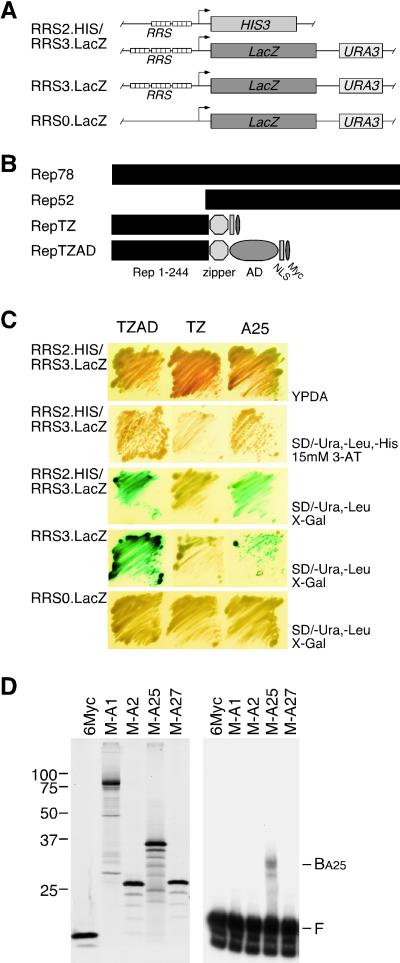
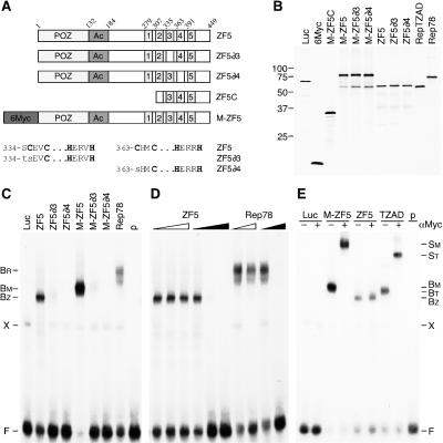
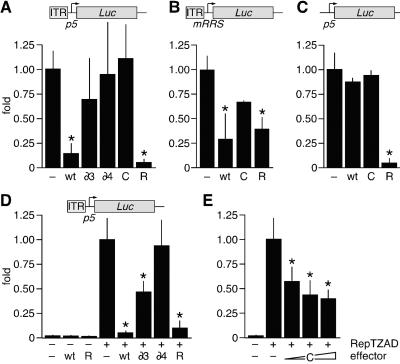
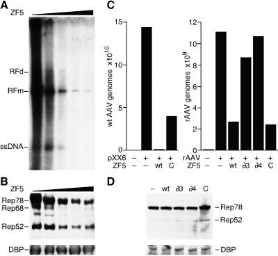
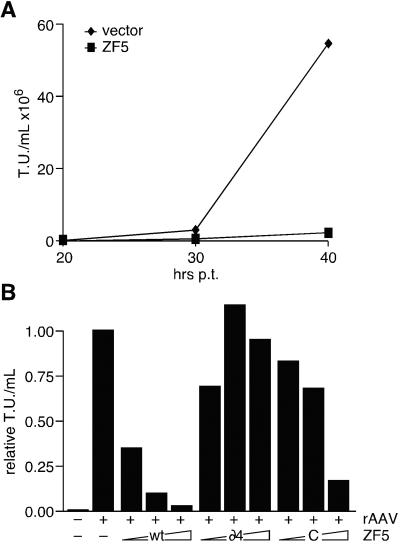
Adeno-associated virus type 2 (AAV2) is a human parvovirus that has attracted attention as a vector for gene transfer. Replication and site-specific integration of the wild-type virus requires binding of the AAV2 Rep proteins to a cis-regulatory element named the Rep recognition sequence (RRS). RRS motifs are found within the cellular AAVS1 integration locus, the viral p5 promoter, and the inverted terminal repeats (ITRs). Here we report the design of a genetic screen based on the yeast one-hybrid assay to identify cellular RRS-binding proteins. We show that the human zinc finger 5 protein (ZF5) binds specifically to RRS motifs in vitro and in vivo. ZF5 is a highly conserved and ubiquitously expressed transcription factor that contains five C-terminal zinc fingers and an N-terminal POZ domain. Ectopic expression of ZF5 leads to an ITR-dependent repression of the autologous p5 promoter and reduces both AAV2 replication and the production of recombinant AAV2. By using deletion and substitution mutants we show that two different domains of ZF5 contribute to AAV2 repression. Negative regulation of the p5 promoter requires the POZ domain, whereas viral replication is inhibited by the zinc finger domain, likely by competing with Rep for binding to the ITR. Identification and characterization of proteins that bind the ITR, the only viral genetic element retained in AAV2 vectors, will lead to new insights into the unique life cycle of AAV2 and will suggest improvements important for its application as a gene therapy vector.


Figures

Figure 1
Figure 1
A genetic screen identifies cellular proteins that bind the RRS motif. (A) Schematic of yeast strains. Reporter strain YM.RRS2.HIS/RRS3.LacZ contains integrated HIS3 and LacZ reporter cassettes driven from minimal yeast promoters with two or three upstream tandem copies of the RRS. Control strains YM.RRS3.LacZ and YM.RRS0.LacZ contain a LacZ cassette with three upstream tandem copies or no RRS. (B) Schematic of wild-type and chimeric Rep proteins. Protein RepTZ comprises residues 1–244 of Rep, a modified leucine zipper (RepTZ), a nuclear localization signal (NLS), and a Myc epitope tag. RepTZAD additionally contains the transcriptional activation domain (AD) of VP16. (C) Yeast in vivo plate assays demonstrate RRS binding. Strains expressing RepTZAD or RepTZ served as positive and negative controls, and clone A25 was isolated in the one-hybrid screen. Transformed yeast cells were grown on nonselective medium (YPDA) and on selection medium [SD/−Ura,−Leu,−His, 15 mM 3-amino-1,2,4-triazole (3-AT)]. Interaction was confirmed on plates supplemented with X-gal to detect β-galactosidase activity. Specificity of the DNA-binding activity was confirmed in strains YM.RRS3.LacZ and YM.RRS0.LacZ. (D) EMSA identifies ITR-binding proteins. Positive clones were translated in vitro in the presence of [35S]methionine and separated on a 12% SDS-polyacrylamide gel (Left). Size markers are indicated on the left. In vitro synthesized proteins were analyzed for binding to a 32P-labeled ITR probe by EMSA (Right). The positions of free (F) and bound (BA25) DNA substrate are indicated.
Figure 2
Figure 2
ZF5 binding to the RRS in vitro is sequence-specific. (A) Schematic of wild-type and mutant ZF5 proteins. ZF5 consists of a POZ domain, a stretch of acidic residues (Ac), and five C2H2-type zinc fingers. Mutations in ZF5∂3 and ZF5∂4 are shown below. The C-terminal fragment of ZF5 isolated in the screen (ZF5C) contains residues 308–449. Proteins were tagged with an N-terminal Myc epitope (6Myc). (B) In vitro translation of ZF5 proteins. Proteins were synthesized in the presence of [35S]methionine and separated on a 12% SDS polyacrylamide gel. Size markers are indicated on the left. (C) Mutations in zinc fingers 3 and 4 disrupt binding of ZF5 to the RRS. In vitro translated proteins were incubated with a 32P-labeled RRS oligonucleotide probe, and DNA binding was analyzed by EMSA. (D) ZF5 binds specifically to the RRS motif. Increasing molar ratios (1, 5, 25× for ZF5, and 1, 25× for Rep78) of unlabeled DNA fragments containing the RRS (black triangles) or a mutant RRS (open triangles) were added as competitors. (E) The tagged ZF5-DNA complex can be supershifted by a Myc-specific antibody. The presence (+) or absence (−) of antibody is indicated above. Rep78, RepTZAD, and luciferase (Luc) were included as controls. The positions of free (F), bound (B), and supershifted (S) DNA substrate are indicated. Shifted complexes are shown for ZF5 (BZ), M-ZF5 (BM and SM), Rep78 (BR), and RepTZAD (BT and ST). X, indicates a nonspecific band; p, lanes that contain probe alone.
Figure 3
Figure 3
ZF5 is a repressor of the AAV2 p5 promoter. (A–C) ZF5 represses the AAV2 p5 promoter through binding to the ITR. 293T cells were transfected with reporter plasmids pGL3.ITR/p5.Luc (A), pGL3.ITR/M1.Luc (B), or pGL2.p5.Luc (C) as shown schematically above. Cells were cotransfected with expression plasmids for ZF5, ZF5∂3, ZF5∂4, ZF5C, or Rep78. (D and E) ZF5 competes with Rep for binding to the ITR. HeLa cells were transfected with reporter plasmid pGL3.ITR/p5.Luc, plasmid pcDNA.RepTZAD where indicated (+) and either pRK5.ZF5, pRK5.ZF5∂3, pRK5.ZF5∂4, and pcDNA.Rep78 (D) or increasing amounts (0.5, 1, and 2 μg) of pRK5.ZF5C (E). Luciferase activity was normalized for transfection efficiency and is shown relative to transfection with empty vector (−). The graphs reflect average value and standard deviation of at least two experiments performed in duplicate. *, statistical significance (P < 0.01); R, Rep78; wt, ZF5; ∂3, ZF5∂3; ∂4, ZF5∂4; C, ZF5C.
Figure 4
Figure 4
ZF5 inhibits AAV2 replication. (A and B) ZF5 inhibits replication of wild-type AAV2. 293T cells in 60-mm wells were transfected with increasing amounts of pRK5.ZF5 (0, 1, 2, 4, and 6 μg) and 0.5 μg of an infectious AAV2 clone (pNTC244) and subsequently infected with Ad5. Cell pellets were processed to analyze either AAV2 replication (A) or protein expression (B). Southern blot analysis of LMW DNA was performed with a 32P-labeled AAV2-specific probe. The positions of single-stranded AAV2 genome (ssDNA) and monomeric (RFm) and dimeric (RFd) replicative forms are indicated. Protein expression was assessed by immunoblotting for Rep or Ad5-DNA-binding protein (DBP). (C and D) ZF5 inhibits AAV2 replication. 293T cells were transfected with pXX6,ZF5 expression vectors as indicated and either superinfected with AAV2 (C, Left) or cotransfected with pAAV.GFP and pcDNA.Rep78 (C, Right). After 40 h the cells were harvested, and the viral replication was quantified by real-time PCR with Rep or GFP-specific primers. The columns reflect the average value of a representative experiment performed in duplicate. Protein expression was analyzed by immunoblotting for Rep or Ad5-DBP (D). wt, ZF5; ∂3, ZF5∂3; ∂4, ZF5∂4; C, ZF5C.
Figure 5
Figure 5
ZF5 inhibits AAV2 vector production. (A) 293T cells in 35-mm wells were transfected with pAAV.GFP, pXX2, pXX6, and empty vector or pRK5.ZF5. Virus was harvested at 20, 30, and 40 h posttransfection, and vector titers were determined. (B) Cells were transfected as described for A with increasing amounts of pRK5.ZF5, pRK5.ZF5∂4, or pRK5.ZF5C (0.1, 0.3, and 1.0 μg). Viral vectors were harvested after 40 h and quantified. The titer is shown relative to transfection with empty vector. The graphs reflect the average value of a representative experiment performed in duplicate. −, empty vector; wt, ZF5; ∂4, ZF5∂4; C, ZF5C; T.U., transducing units.

References

    1. Berns K I. In: Virology. Fields B N, Knipe D M, Howley P M, editors. Vol. 2. Philadelphia: Lippincott-Raven; 1996. pp. 2173–2197.
    1. Laughlin C A, Cardellichio C B, Coon H C. J Virol. 1986;60:515–524. - PMC - PubMed
    1. Cheung A K, Hoggan M D, Hauswirth W W, Berns K I. J Virol. 1980;33:739–748. - PMC - PubMed
    1. Kotin R M, Siniscalco M, Samulski R J, Zhu X D, Hunter L, Laughlin C A, McLaughlin S, Muzyczka N, Rocchi M, Berns K I. Proc Natl Acad Sci USA. 1990;87:2211–2215. - PMC - PubMed
    1. Samulski R J, Zhu X, Xiao X, Brook J D, Housman D E, Epstein N, Hunter L A. EMBO J. 1991;10:3941–3950. - PMC - PubMed

Publication types

LinkOut - more resources