

MC(1) receptors are constitutively expressed on leucocyte subpopulations with antigen presenting and cytotoxic functions
- PMID: 11737060
- PMCID: PMC1906236
- DOI: 10.1046/j.1365-2249.2001.01604.x
MC(1) receptors are constitutively expressed on leucocyte subpopulations with antigen presenting and cytotoxic functions
Abstract
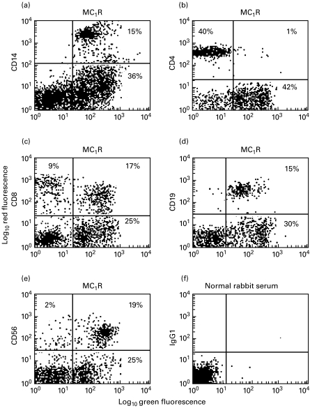
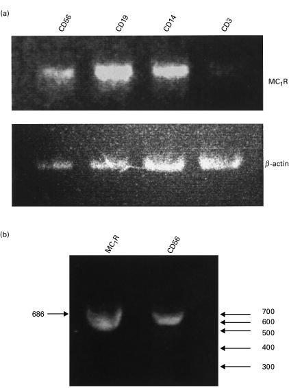
The expression of melanocortin MC(1) receptors on human peripheral lymphocyte subsets was analysed by flow cytometry using rabbit antibodies selective for the human MC(1) receptor and a panel of monoclonal antibodies against lymphocyte differentiation markers. The MC(1) receptor was found to be constitutively expressed on monocytes/macrophages, B-lymphocytes, natural killer (NK) cells and a subset of cytotoxic T-cells. Interestingly T-helper cells appeared to be essentially devoid of MC(1) receptors. The results were confirmed by RT-PCR which indicated strong expression of MC(1) receptor mRNA in CD14(+), CD19(+) and CD56(+) cells. However, only a faint RT-PCR signal was seen in CD3(+) cells, in line with the immuno-staining results that indicated that only part of the CD3(+) cells (i.e. some of the CD8(+) cells) expressed the MC(1) receptor. The MC(1) receptors' constitutive expression on immune cells with antigen-presenting and cytotoxic functions implies important roles for the melanocortic system in the modulation of immune responses.
Figures
Similar articles
-
Quantitative measurement of the levels of melanocortin receptor subtype 1, 2, 3 and 5 and pro-opio-melanocortin peptide gene expression in subsets of human peripheral blood leucocytes.Scand J Immunol. 2005 Mar;61(3):279-84. doi: 10.1111/j.1365-3083.2005.01565.x. Scand J Immunol. 2005. PMID: 15787746
-
Perforin expression can define CD8 positive lymphocyte subsets in pigs allowing phenotypic and functional analysis of natural killer, cytotoxic T, natural killer T and MHC un-restricted cytotoxic T-cells.Vet Immunol Immunopathol. 2006 Apr 15;110(3-4):279-92. doi: 10.1016/j.vetimm.2005.10.005. Epub 2005 Dec 1. Vet Immunol Immunopathol. 2006. PMID: 16325923
-
Reference ranges of lymphocyte subsets in healthy adults and adolescents with special mention of T cell maturation subsets in adults of South Florida.Immunobiology. 2014 Jul;219(7):487-96. doi: 10.1016/j.imbio.2014.02.010. Epub 2014 Mar 2. Immunobiology. 2014. PMID: 24661720
-
Microenvironment abnormalities and lymphomagenesis: Immunological aspects.Semin Cancer Biol. 2015 Oct;34:36-45. doi: 10.1016/j.semcancer.2015.07.004. Epub 2015 Jul 29. Semin Cancer Biol. 2015. PMID: 26232774 Free PMC article. Review.
-
[Deep lung--cellular reaction to HIV].Rev Port Pneumol. 2007 Mar-Apr;13(2):175-212. Rev Port Pneumol. 2007. PMID: 17492233 Review. Portuguese.
Cited by
-
Nanoparticles for Topical Application in the Treatment of Skin Dysfunctions-An Overview of Dermo-Cosmetic and Dermatological Products.Int J Mol Sci. 2022 Dec 15;23(24):15980. doi: 10.3390/ijms232415980. Int J Mol Sci. 2022. PMID: 36555619 Free PMC article. Review.
-
Diminishment of alpha-MSH anti-inflammatory activity in MC1r siRNA-transfected RAW264.7 macrophages.J Leukoc Biol. 2008 Jul;84(1):191-8. doi: 10.1189/jlb.0707463. Epub 2008 Apr 3. J Leukoc Biol. 2008. PMID: 18388300 Free PMC article.
-
Repository corticotrophin injection exerts direct acute effects on human B cell gene expression distinct from the actions of glucocorticoids.Clin Exp Immunol. 2018 Apr;192(1):68-81. doi: 10.1111/cei.13089. Epub 2018 Jan 12. Clin Exp Immunol. 2018. PMID: 29205315 Free PMC article.
-
Differential haemoparasite intensity between black sparrowhawk (Accipiter melanoleucus) morphs suggests an adaptive function for polymorphism.PLoS One. 2013 Dec 31;8(12):e81607. doi: 10.1371/journal.pone.0081607. eCollection 2013. PLoS One. 2013. PMID: 24391707 Free PMC article.
-
Negative regulators that mediate ocular immune privilege.J Leukoc Biol. 2018 Feb 12:10.1002/JLB.3MIR0817-337R. doi: 10.1002/JLB.3MIR0817-337R. Online ahead of print. J Leukoc Biol. 2018. PMID: 29431864 Free PMC article. Review.
References
-
- Wikberg JES, Muceniece R, Mandrika I, Prusis J, Post C, Skottner A. New aspects on the melanocortins and their receptors. Pharmacol Res. 2000;42:393–420. 10.1006/phrs.2000.0725. - DOI - PubMed
-
- Chhajlani V, Wikberg JES. Molecular cloning and expression of the human melanocyte stimulating hormone receptor cDNA. FEBS Lett. 1992;309:417–20. - PubMed
-
- Loir B, Perez Sanches C, Ghanem G, Lozano JA, Garcia-Borron JC, Jimenez-Cervantes C. Expression of the MC1 receptor gene in normal and malignant human melanocytes. A semiquantitative RT-PCR study. Cell Mol Biol. 1999;45:1083–92. - PubMed
-
- Bhardwaj R, Becher E, Mahnke K, Hartmeyer M, Schwarz T, Scholzen T, Luger TA. Evidence for the differential expression of the functional alpha-melanocyte-stimulating hormone receptor MC-1 on human monocytes. J Immunol. 1997;158:3378–84. - PubMed
-
- Taherzadeh S, Sharma S, Chhajlani V, et al. α-MSH and its receptors in regulation of tumor necrosis factor-α production by human monocyte/macrophages. Am J Physiol. 1999;276:R1289–94. - PubMed
Publication types
MeSH terms
Substances
LinkOut - more resources
Full Text Sources
Other Literature Sources
Research Materials
