

IFN-gamma synergizes with TNF-alpha but not with viable H. pylori in up-regulating CXC chemokine secretion in gastric epithelial cells
- PMID: 11737065
- PMCID: PMC1906237
- DOI: 10.1046/j.1365-2249.2001.01634.x
IFN-gamma synergizes with TNF-alpha but not with viable H. pylori in up-regulating CXC chemokine secretion in gastric epithelial cells
Abstract
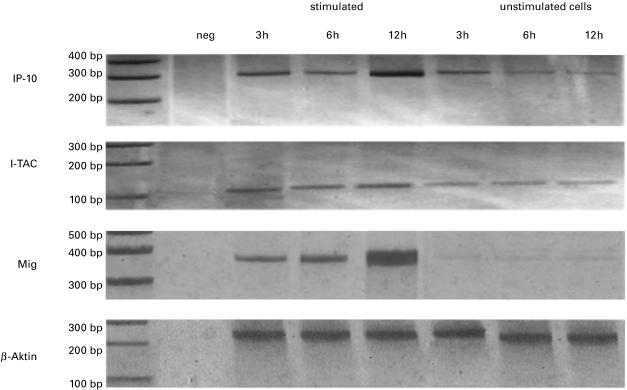
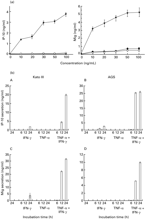
Helicobacter pylori colonizes the gastric epithelial surface and induces epithelial cells to increase production of the neutrophil attractant IL-8. Little is known about the role of the gastric epithelium in regulating mucosal T cell trafficking. We therefore characterized constitutive and regulated epithelial expression of the CXC chemokines IP-10, I-TAC and Mig, which specifically attract CXCR3 expressing CD4(+) T cells. Human gastric epithelial cell lines (AGS, Kato III, NCI) were used to characterize the constitutive and regulated expression of three CXC chemokines in response to IFN-gamma, TNF-alpha and different H. pylori preparations. Chemokine mRNA and protein production were measured by RT-PCR and ELISA. Gastric epithelial cells constitutively expressed mRNA for IP-10, Mig and I-TAC. IFN-gamma in combination with TNF-alpha strongly induced secretion of those chemokines. Soluble or membranous fractions of H. pylori significantly inhibited IFN-gamma/TNF-alpha induced epithelial cell IP-10 and Mig production. Gastric epithelial cells may contribute to mucosal T cell trafficking. The capacity of H. pylori products to inhibit IP-10 and Mig secretion may explain, at least in part, the failure to induce protective immunity against this bacterium and the ability of H. pylori to affect the presentation of the local inflammation.
Figures
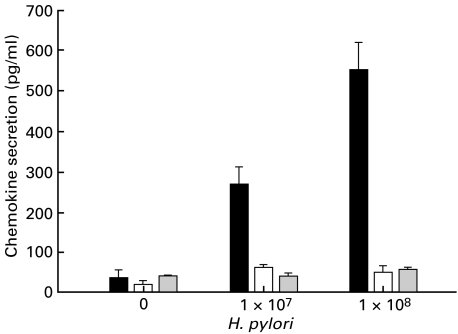
 Mig secretion). Confluent monolayers of AGS cell line (2 × 10⁶ cells) were coincubated with different concentrations of living H. pylori for 20 h. Supernatants were harvested, centrifuged and analysed for chemokine-secretion by ELISA. Values are expressed as mean ± CI. Representative results from one of four experiments are shown.
References
-
- Genta RM, Lew GM, Graham DY. Changes in the gastric mucosa following eradication of Helicobacter pylori. Mod Pathol. 1993;6:281–9. - PubMed
-
- Genta RM. The immunobiology of Helicobacter pylori gastritis. Semin Gastrointest Dis. 1997;8:2–11. - PubMed
-
- Crabtree JE. Role of cytokines in pathogenesis of Helicobacter pylori-induced mucosal damage. Dig Dis Sci. 1998;43:46–55. - PubMed
-
- Ando T, Kusugami K, Ohsuga M, et al. Interleukin-8 activity correlates with histological severity in Helicobacter pylori-associated antral gastritis. Am J Gastroenterol. 1996;91:1150–6. - PubMed
Publication types
MeSH terms
Substances
LinkOut - more resources
Full Text Sources
Research Materials
