

doi: 10.1046/j.1365-2249.2001.01702.x.
Differential roles of osteopontin/Eta-1 in early and late lpr disease
Affiliations
- PMID: 11737079
- PMCID: PMC1906233
- DOI: 10.1046/j.1365-2249.2001.01702.x
Item in Clipboard
Differential roles of osteopontin/Eta-1 in early and late lpr disease
Clin Exp Immunol.
2001 Dec.
Abstract
The cytokine osteopontin (Eta-1) leads to macrophage-dependent polyclonal B-cell activation and is induced early in autoimmune prone mice with the lpr mutation, suggesting a significant pathogenic role for this molecule. Indeed, C57BL/6-Fas(lpr/lpr) mice crossed with osteopontin(-/-) mice display delayed onset of polyclonal B-cell activation, as judged by serum immunoglobulin levels. In contrast, they are subject to normal onset, but late exacerbation of lymphoproliferation and evidence of kidney disease. These observations define two stages of Fas(lpr/lpr) disease with respect to osteopontin-dependent pathogenesis that should be taken into account in the design of therapeutic approaches to the clinical disease.
Figures
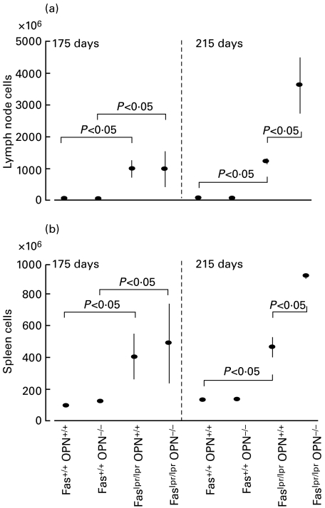
Disease progression in lymph nodes and spleen. (a,b) Proliferation was assessed in early (175 days) and late (215 days) disease by determination of cell numbers of combined inguinal, brachial, submandibular and mesenteric lymph nodes (a) or of whole spleens (b). Each data point represents mean ± standard error from three to six mice. (c) The composition of subpopulations of cells in lymph nodes was determined by flow cytometry. Various hatches represent the percentages of total for the indicated cell types (CD4 stands for CD3+CD4+CD8−, CD8 is short for CD3+CD4−CD8+).
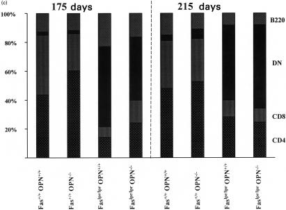
Disease progression in lymph nodes and spleen. (a,b) Proliferation was assessed in early (175 days) and late (215 days) disease by determination of cell numbers of combined inguinal, brachial, submandibular and mesenteric lymph nodes (a) or of whole spleens (b). Each data point represents mean ± standard error from three to six mice. (c) The composition of subpopulations of cells in lymph nodes was determined by flow cytometry. Various hatches represent the percentages of total for the indicated cell types (CD4 stands for CD3+CD4+CD8−, CD8 is short for CD3+CD4−CD8+).
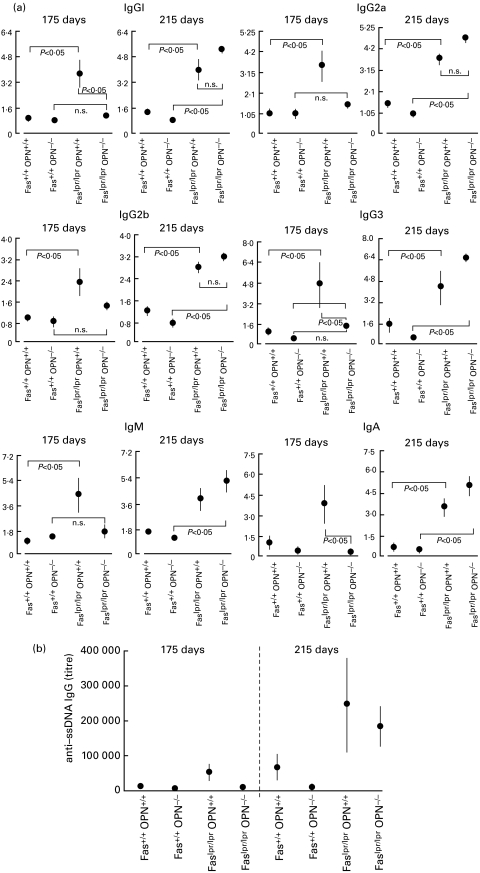
Immunoglobulin levels in serum. (a) Immunoglobulin isotypes in serum were measured with a commercially available kit (PharMingen) and results are plotted as -fold induction with Fas+/+OPN+/+ equal to 1. (b) Antibody of the IgG class to single-stranded DNA and double-stranded DNA (not shown) were determined by ELISA. Each data point represents mean value ± standard error. Statistical significance was analysed with the non-parametric U-test.
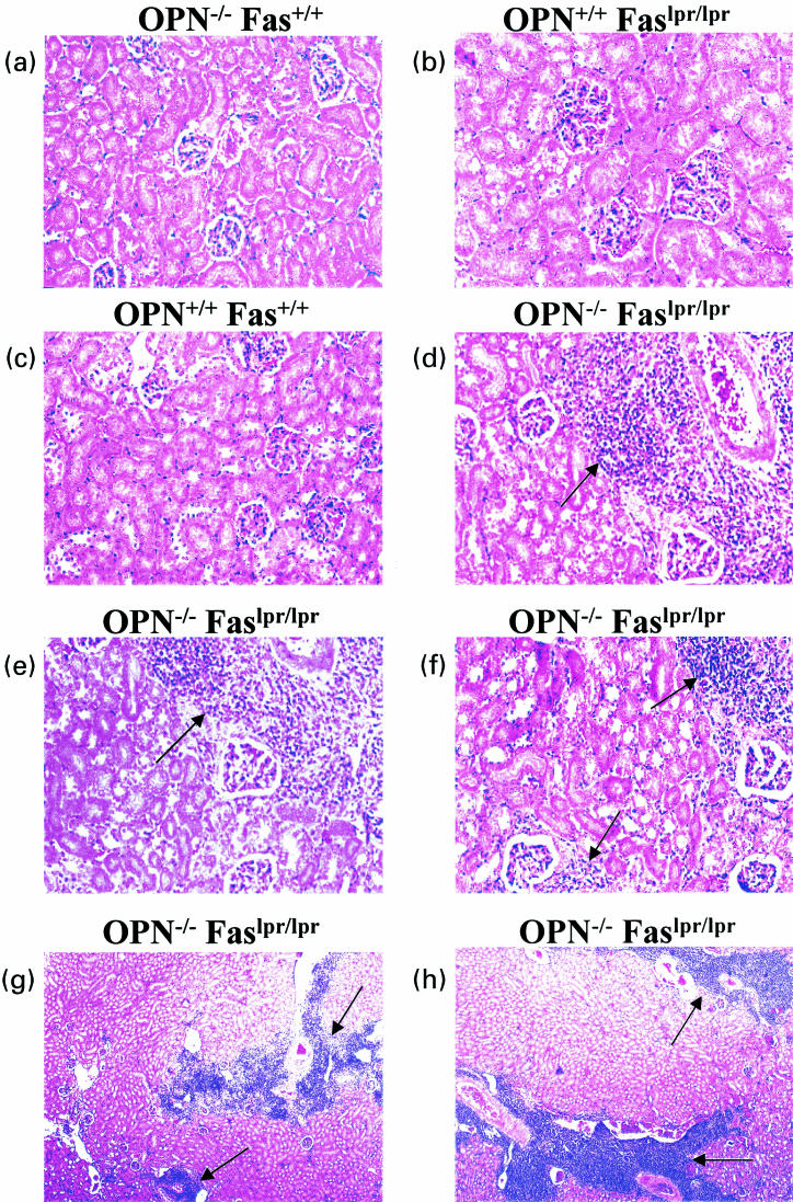
Autoimmune nephritis. At 215 days, kidneys were extracted from mice with the indicated genotypes and fixed in 10% buffered formalin. Histology slides were prepared and stained with haematoxilin/eosin. (a–f) One microscopic field, representative for 3–5 mice, is shown for each genotype under study, with the exception of Faslpr/lprOPN−/–, for which three representative fields (from distinct mice) are shown to illustrate the lymphocytic infiltrates. (g,h) A lower magnification of the kidneys from two distinct Faslpr/lprOPN−/– mice indicates the wide distribution of the lymphocytic infiltrates.
References
-
- Lampe MA, Patarca R, Iregui MV, Cantor H. Polyclonal B cell activation by the Eta-1 cytokine and the development of systemic autoimmune disease. J Immunol. 1991;147:2902–6. - PubMed
-
- Iizuka J, Katagiri Y, Tada N, et al. Introduction of an osteopontin gene confers the increase in B1 cell population and the production of anti-DNA autoantibodies. Lab Invest. 1998;78:1523–33. - PubMed
-
- Weber GF, Ashkar S, Glimcher MJ, Cantor H. Receptor–ligand interaction between CD44 and osteopontin (Eta-1) Science. 1996;271:509–12. - PubMed
Publication types
MeSH terms
Substances
Grants and funding
LinkOut - more resources
Full Text Sources
Medical
Molecular Biology Databases
Research Materials
Miscellaneous
