. 2001 Dec 10;155(6):1043-53.
doi: 10.1083/jcb.200108026. Epub 2001 Dec 10.

Thin filaments elongate from their pointed ends during myofibril assembly in Drosophila indirect flight muscle

Affiliations

Thin filaments elongate from their pointed ends during myofibril assembly in Drosophila indirect flight muscle

M Mardahl-Dumesnil et al. J Cell Biol. .

Abstract

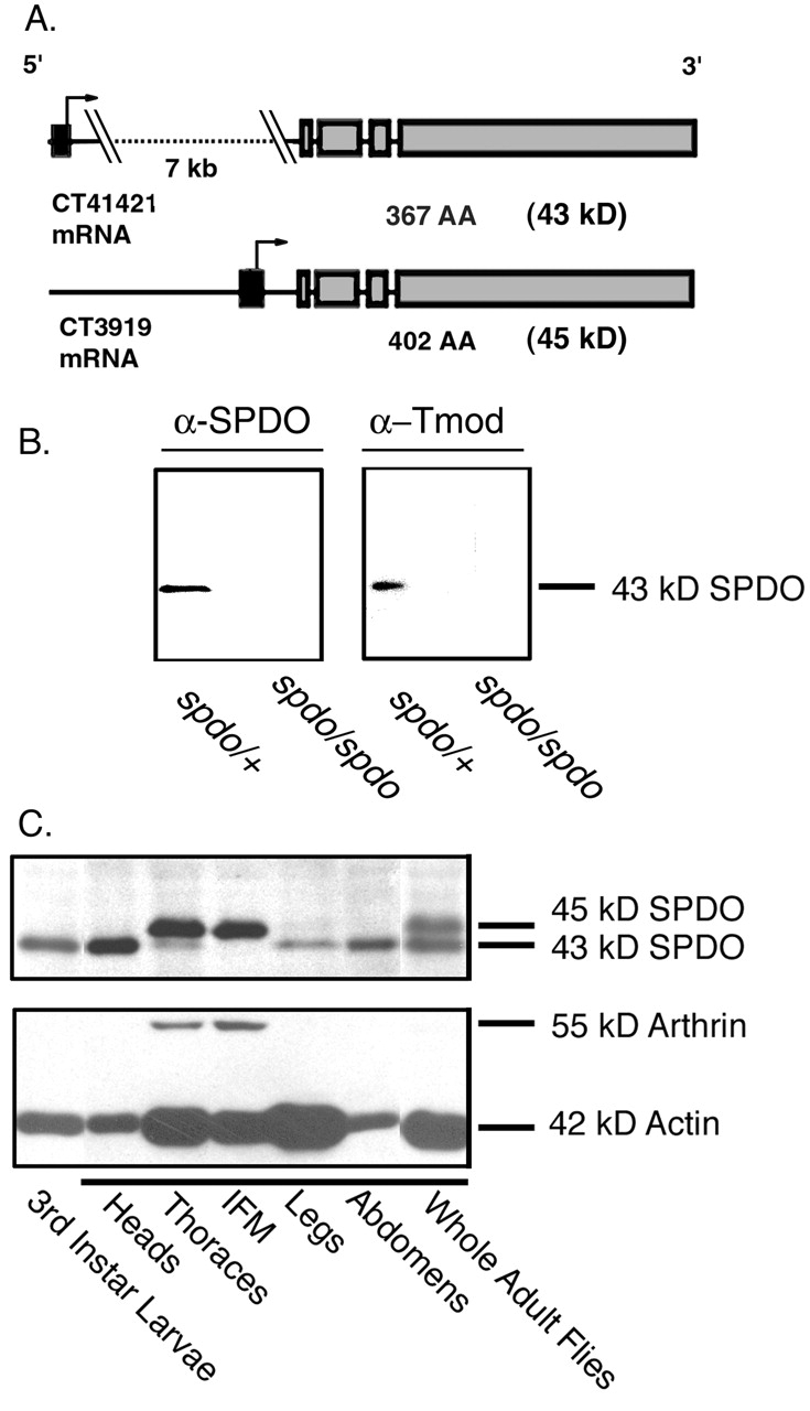
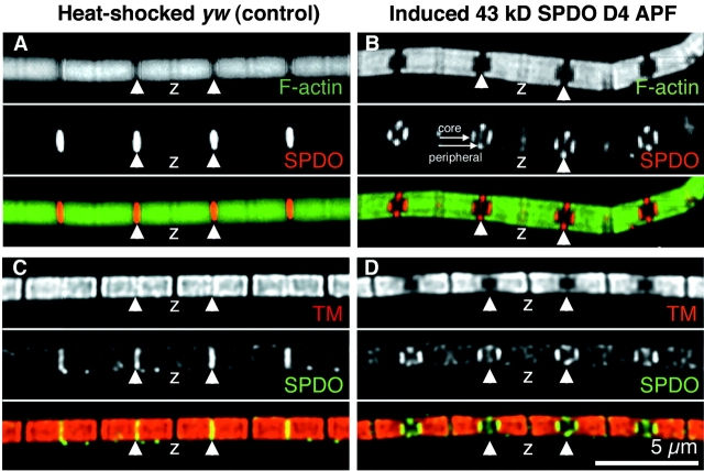
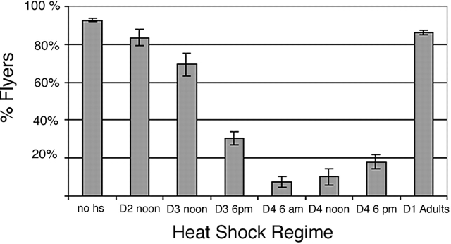
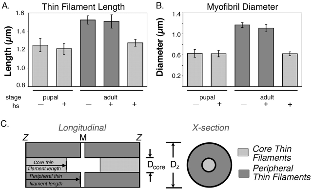
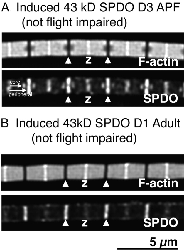
Tropomodulin (Tmod) is an actin pointed-end capping protein that regulates actin dynamics at thin filament pointed ends in striated muscle. Although pointed-end capping by Tmod controls thin filament lengths in assembled myofibrils, its role in length specification during de novo myofibril assembly is not established. We used the Drosophila Tmod homologue, sanpodo (spdo), to investigate Tmod's function during muscle development in the indirect flight muscle. SPDO was associated with the pointed ends of elongating thin filaments throughout myofibril assembly. Transient overexpression of SPDO during myofibril assembly irreversibly arrested elongation of preexisting thin filaments. However, the lengths of thin filaments assembled after SPDO levels had declined were normal. Flies with a preponderance of abnormally short thin filaments were unable to fly. We conclude that: (a) thin filaments elongate from their pointed ends during myofibril assembly; (b) pointed ends are dynamically capped at endogenous levels of SPDO so as to allow elongation; (c) a transient increase in SPDO levels during myofibril assembly converts SPDO from a dynamic to a permanent cap; and (d) developmental regulation of pointed-end capping during myofibril assembly is crucial for specification of final thin filament lengths, myofibril structure, and muscle function.


Figures

Figure 1.
Figure 1.
SPDO isoforms are expressed differentially during development and in adult tissues. (A) A data analysis program (GadFly) (http://flybase.bio.indiana.edu:82/) which searches expressed sequence tags (ESTs) from the Drosophila genome project predicts two alternative promoters (black exons) used to generate SPDO isoforms. CT41421 and CT3919 are mRNA transcripts predicted to generate 366 AA (43 kD) and 402 AA (45 kD) protein products, respectively. Arrow represents translational start sites that contribute 2 or 36 AA for CT4121 and CT 3919, respectively. (B) Western Blot prepared from heterozygous (spdo/+) or homozygous (spdo/spdo) spdo embryos probed with mouse anti-SPDO antibody (α-SPDO) (left) or anti–human erythrocyte Tmod antibody (α-Tmod) (right). (C) Western blot prepared from larvae or dissected adult flies probed with anti-SPDO antibodies.
Figure 2.
Figure 2.
A developmental SPDO isoform switch occurs in the IFM during early adulthood. (A) Schematic of the Drosophila life cycle at 25°C (for review see Ashburner, 1989). After ∼1 d, fertilized embryos hatch to larvae. The larval period lasts ∼6 d before formation of an immobile pupa. Over the next 4 d APF, larval tissues remodel to give rise to a winged adult that emerges from the pupa at eclosion (hatching) (∼10-d cycle). White prepupae formation marks the first day of pupation (D1 APF). Adult flies on the first day after eclosion are indicated as D1 Adults. (B) Whole thoraces were dissected from pupae (12 PM D4 APF), or D1 adult flies aged 2–3 or 8–10 h, or from D2 adults aged 25–30 h after eclosion. Triton X-100 extracted myofibrils from thoraces were isolated and prepared for Western blots. Blots were probed with anti-SPDO (top), and then stripped and reprobed with the C4 anti-actin monoclonal antibody to control for loading (bottom).
Figure 9.
Figure 9.
Model of IFM thin filament assembly in wild-type flies and in the presence of excess 43-kD SPDO. Based on the ultrastructural analysis of Reedy and Beall (1993), by the D2 APF, small sarcomeres of ∼1.7 μm in length have already formed with clear interdigitation of thick and thin filaments (a). In wild-type flies (left panel), the sarcomeres continue to develop throughout pupation by elongation of preexisting “core” filaments from their pointed ends (b–c) and by addition of new filaments to the periphery of the myofibril (c–d). The 43-kD SPDO isoform is present throughout thin filament elongation during pupation in noninduced hs = 43 spdo or wild-type flies (a–d), and is replaced by the 45-kD SPDO isoform in early adulthood, after the majority of assembly is completed (e). In contrast (right panel) transient overexpression of the 43-kD SPDO isoform by heat-shock (HS) induction during pupation (c) prevents uncapping and thin filament elongation of the preexisting “core” thin filaments (c–d). Normal replacement of the 43-kD SPDO isoform by the 45-kD isoform at adulthood is also prevented (d–e). However, sarcomere elongation is unaffected as are addition and elongation of peripheral thin filaments, and these peripheral thin filaments appear to be capped by the 45-kD isoform at their pointed ends (c–e) (Fig. 7). Black lines, thin filaments; filled black arrowheads, 43-kD SPDO isoform; hollow arrowheads, 45-kD SPDO isoform; small arrows indicate dynamic exchange of SPDO; Z, Z line.
Figure 7.
Figure 7.
Transiently overexpressed 43 kD-SPDO bound to thin filaments irreversibly. (A) Relative amounts of 43-kD or 45-kD SPDO isoforms associated with Triton X-100 extracted pupal myofibrils from whole thoraces (D4 APF), adult myofibrils from thoraces of D3 adult hs-43 kD spdo flies heat shocked (HS) on D4 APF (+), or from untreated D3 adult hs-43 kD spdo flies (−). Samples from soluble (S) and myofibril pellet fractions (P) were electrophoresed on SDS-PAGE followed by immunoblotting with anti-SPDO (top), and then stripped and reprobed with the C4 anti-actin monoclonal antibody to control for loading (bottom). Pupae were dissected immediately after 1 h of heat shock at 12 PM D4 APF, or allowed to develop to adulthood at 25°C. (B) Staged hs-43 kD spdo flies were heat shocked (HS) for 1 h on D4 APF (+) or untreated (−) and allowed to develop to adulthood. Adults were collected and aged up to 10 d. The IFMs were dissected and prepared for SDS-PAGE and immunoblotting. The blots were stripped as above. Both the 43-kD actin and the 55-kD IFM-specific ubiquinated actin (Arthrin) (Ball et al., 1987) were detected at similar levels with or without heat-shock treatment.
Figure 4.
Figure 4.
Transient induction of 43-kD SPDO expression during myofibril assembly produces abnormally short core thin filaments. Isolated IFM myofibrils from heat-treated (12 PM D4 APF) yw adult flies (A and C) or heat-treated hs-43 kD spdo adult flies (B and D) were fixed and stained with Bodipy-phallacidin (green) (top panels in A, B) or anti-TM (top panel in C and D) followed by staining with antibodies to SPDO (middle panels). Images were selected from optically sectioned (100 nm increments) myofibrils obtained from deconvolution microscopy and correspond to the center section of the myofibril. White arrowheads, pointed ends; Z, Z-discs. Bar, 5 μm.
Figure 3.
Figure 3.
Overexpression of the 43-kD SPDO isoform during mid- to late-myofibril assembly during pupation affects adult flight ability. Staged hs-43 kD spdo lines were treated to one 37°C heat-shock treatment for 1 h during pupation (ranging from D2 APF to adult; Fig. 2 A), and then allowed to develop at 25°C. Adults were collected for flight testing. The percentage of 2–3-d-old adult flies that were capable of upward or horizontal flight (% Fliers) was graphed in relation to the timing of heat-shock induction. In the control groups, >70% of adult yw flies that were heat shocked any time between D2 and D4 APF could fly (unpublished data).
Figure 8.
Figure 8.
Overexpression of SPDO during assembly affects myofibril morphology. Ultrastructure of IFM from D2 adult flies that had been heat shocked on D4 APF (hs-43 kD spdo (A–C and F) from control yw flies heat shocked on D4 APF (D and E). Longitudinal sections are shown in A and D, cross-sections shown in B, C, E, and F. The ultrastructure is normal for heat-shocked control flies (D and E). Black arrows (A and F), missing thick and thin filaments. g, glycogen granules; M, M-line; MT, mitochondria; myf, myofibril; SR, sarcoplasmic reticulum; Z, Z line. Bars: (A and D) 3.0 μm; (B and E) 1.0 μm; (C, F, and E inset) 0.5 μm.
Figure 5.
Figure 5.
Transient induction of 43-kD SPDO during myofibril assembly irreversibly prevents thin filament elongation. Thin filament lengths (A) and myofibril diameters (B) were measured as indicated in the diagram (C) using Deltavision software, from deconvolved images of Bodipy- phallacidin stained IFM myofibrils isolated from either pupal flies (1 PM D4 APF), D1 adult hs-43 kD spdo flies heat-shocked at 12 PM D4 APF, or untreated D2 adult flies. Error bars, SEM. (C) Schematic diagram of longitudinal and cross sections of an IFM myofibil from an adult hs-43 kD spdo fly that was heat shocked during pupation. Light gray shading indicates Bodipy-phallacidin stained “core” of the myofibril with shorter thin filaments and dark gray shading indicates Bodipy-phallacidin stained “peripheral” thin filaments.
Figure 6.
Figure 6.
Overexpression of 43-kD SPDO during early- or late-myofibril assembly affects myofibril structure without affecting muscle function. Myofibrils from D2 adult hs-43 kD spdo flies (A) heat shocked at 12 PM D3 APF or (B) 12 PM D1 after eclosion. (Top) Bodipy-phallacidin staining for F-actin; (Bottom) anti-SPDO staining. Short white arrow, short “core” thin filaments; long white arrow, longer “peripheral” thin filaments; white arrowheads, pointed ends; Z, Z-disc. Bar, 5 μm.

Similar articles

Cited by

References

    1. Almenar-Queralt, A., C.C. Gregorio, and V.M. Fowler. 1999. a. Tropomodulin assembles early in myofibrillogenesis in chick skeletal muscle: Evidence that thin filaments rearrange to form striated myofibrils. J. Cell Sci. 112:1112–1123. - PubMed
    1. Almenar-Queralt, A., A. Lee, C.A. Conley, L. Ribas de Pouplana, and V.M. Fowler. 1999. b. Identification of a novel tropomodulin isoform, skeletal tropomodulin, that caps actin filament pointed ends in fast skeletal muscle. J. Biol Chem. 274:28466–28475 (erratum published 275[17]:13164). - PubMed
    1. Ashburner, M. 1989. Drosophila, a Laboratory Manual. Cold Spring Harbor Laboratory Press, Cold Spring Harbor, NY.
    1. Ball, E., C.C. Karlik, C.J. Beall, D.L. Saville, J.C. Sparrow, B. Bullard, and E.A. Fyrberg. 1987. Arthrin, a myofibrillar protein of insect flight muscle, is an actin-ubiquitin conjugate. Cell. 51:221–228. - PubMed
    1. Beall, C.J., M.A. Sepanski, and E.A. Fyrberg. 1989. Genetic dissection of Drosophila myofibril formation: effects of actin and myosin heavy chain null alleles. Genes Dev. 3:131–140. - PubMed

Publication types

MeSH terms