. 2001 Dec 15;21(24):9519-28.
doi: 10.1523/JNEUROSCI.21-24-09519.2001.

Caspase-9 activation results in downstream caspase-8 activation and bid cleavage in 1-methyl-4-phenyl-1,2,3,6-tetrahydropyridine-induced Parkinson's disease

Affiliations

Caspase-9 activation results in downstream caspase-8 activation and bid cleavage in 1-methyl-4-phenyl-1,2,3,6-tetrahydropyridine-induced Parkinson's disease

V Viswanath et al. J Neurosci. .

Abstract

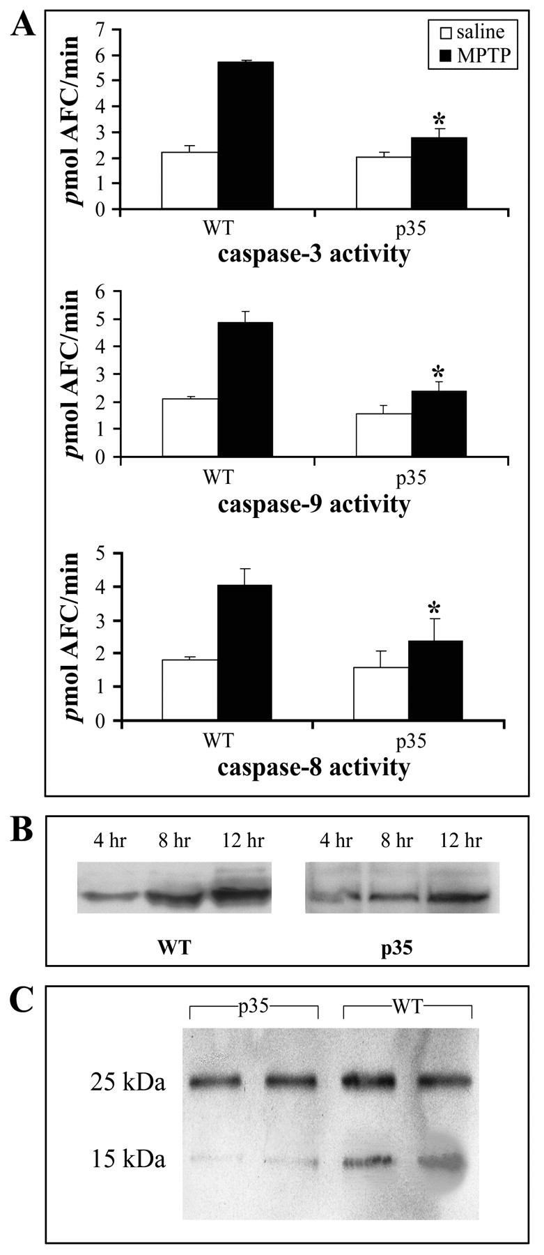
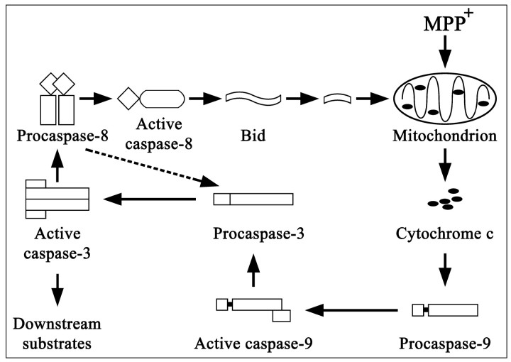
Parkinson's disease (PD) and 1-methyl-4-phenyl-1,2,3,6-tetrahydropyridine (MPTP) toxicity are both associated with dopaminergic neuron death in the substantia nigra (SN). Apoptosis has been implicated in this cell loss; however, whether or not it is a major component of disease pathology remains controversial. Caspases are a major class of proteases involved in the apoptotic process. To evaluate the role of caspases in PD, we analyzed caspase activation in MPTP-treated mice, in cultured dopaminergic cells, and in postmortem PD brain tissue. MPTP was found to elicit not only the activation of the effector caspase-3 but also the initiators caspase-8 and caspase-9, mitochondrial cytochrome c release, and Bid cleavage in the SN of wild-type mice. These changes were attenuated in transgenic mice neuronally expressing the general caspase inhibitor protein baculoviral p35. These mice also displayed increased resistance to the cytotoxic effects of the drug. MPTP-associated toxicity in culture was found temporally to involve cytochrome c release, activation of caspase-9, caspase-3, and caspase-8, and Bid cleavage. Caspase-9 inhibition prevented the activation of both caspase-3 and caspase-8 and also inhibited Bid cleavage, but not cytochrome c release. Activated caspase-8 and caspase-9 were immunologically detectable within MPP(+)-treated mesencephalic dopaminergic neurons, dopaminergic nigral neurons from MPTP-treated mice, and autopsied Parkinsonian tissue from late-onset sporadic cases of the disease. These data demonstrate that MPTP-mediated activation of caspase-9 via cytochrome c release results in the activation of caspase-8 and Bid cleavage, which we speculate may be involved in the amplification of caspase-mediated dopaminergic cell death. These data suggest that caspase inhibitors constitute a plausible therapeutic for PD.


Figures

Fig. 1.
Fig. 1.
Toxicity induced by MPTP is attenuated in transgenic mice neuronally expressing the general caspase inhibitor protein baculoviral p35. A, Stereological counts of TH+ neurons from wild-type (WT) versus p35 transgenic mice after MPTP administration. Both wild-type and p35 transgenics were killed 7 d after MPTP injection, and SN tissue was immunostained with TH antibody. The cell number was assessed stereologically in every alternate section. Data are means ± SD of five to seven animals per group; *p < 0.01. B, Effects of MPTP administration on dopamine and HVA levels in WT versus p35. For all animals the striatum was dissected for measurement of dopamine and HVA 7 d after MPTP administration. Data are means ± SD from three to four mice per group; *p < 0.01.
Fig. 2.
Fig. 2.
Caspase-3, caspase-8, and caspase-9 activation, cytochrome c release, and Bid cleavage after MPTP administration. A, Caspase-3, caspase-9, and caspase-8 activities. Substantia nigra (SN) dissected from MPTP versus saline-treated wild-type and p35 transgenic animals was used to measure caspase activities with specific fluorogenic tetrapeptide substrates (n = 5–7 animals per group for all assays). The caspase activities were measured 24 hr after MPTP injection. Values represent the means ± SD from three individual experiments; *p < 0.01. B, MPTP-induced cytochrome c release from the mitochondria. Cytosolic extracts were prepared at the indicated times, and cytochromec was evaluated by Western blot analysis with the use of a monoclonal antibody. C, In vivo Bid cleavage 24 hr after MPTP administration in WT and p35 mice. SN was dissected, and total cell lysates were subjected to immunoblotting. Molecular weights are shown on the left. The 25 kDa band represents full-length Bid; the 15 kDa fragment represents the cleaved form.
Fig. 3.
Fig. 3.
MPP+ induces time-dependent activation of various caspases, cytochrome c release, and Bid cleavage in PC12 cells. Values represent the means ± SD from three experiments; p < 0.01.A, PC12 cells were incubated with 150 μmMPP+ for the indicated times. Then the cell lysates were analyzed for caspase activities with the use of specific fluorogenic substrates. B, MPP+-induced cytochrome c release from the mitochondria. Mitochondrial and cytosolic extracts were prepared as described, and cytochrome c release was evaluated by using a monoclonal antibody at 2, 4, and 8 hr after MPP+ treatment. C, Cleavage of Bid in PC12 cells at various times after treatment with MPP+; the 15 kDa fragment was not observed at either 0 or 2 hr after MPP+ application (data not shown).
Fig. 4.
Fig. 4.
Effects of treatment of PC 12 cells with specific cell-permeable inhibitors of caspase-9 and caspase-8 before treatment with MPP+ on the activation of caspase-9, caspase-3, and caspase-8, cytochrome c release, and Bid cleavage. Values for all assays represent the means ± SD from three experiments; p < 0.01. A, Effects of pretreatment with a caspase-9 specific inhibitor on the activation of caspase-8, caspase-9, and caspase-3. PC12 cells were incubated with 25 μm LEHD-CHO 1 hr before treatment with MPP+. B, Effects of pretreatment with the specific peptide inhibitor to caspase-8 on the activation of caspase-8, caspase-3, and caspase-9. PC12 cells were incubated with 25 μm IETD-CHO at 1 hr before treatment with MPP+. C, Bid cleavage, but not cytochrome c release, is attenuated in PC12 cells after preincubation with LEHD-CHO. Data represent three individual experiments.
Fig. 5.
Fig. 5.
TH+ cell counts in MPP+-treated mesencephalic cultures from p35 transgenics (Tg) versus wild-type (WT) animals. A, Percentage of TH+ neurons in p35 transgenic versus wild-type mice mesencephalic cultures after 6, 12, and 24 hr of MPP+ (5 μm) treatment compared with untreated WT; *p < 0.01. B, Representative morphology of TH+ neurons in MPP+-treated wild-type cultures after 0, 6, 12, and 24 hr. Magnification, 40×. Data represent three independent experiments.
Fig. 6.
Fig. 6.
Temporal MPP+-induced activation of caspase-9, caspase-3, and caspase-8 in TH+ neurons in mesencephalic cultures. Triple labeling shows immunostaining for the active forms of caspase-9, caspase-3, and caspase-8 in apoptotic (DAPI-stained) TH+ neurons at the time of first induction, i.e., 2, 6, and 12 hr, respectively. Magnification, 60×. Data represent three independent experiments.
Fig. 7.
Fig. 7.
Presence of activated caspase-8 and caspase-9 in TH+ neurons of the substantia nigra of MPTP-treated mice and Parkinsonian brain. A, Wild-type mice were treated with MPTP, and 40 μm sections were double immunolabeled for antibody against TH and the activated form of either caspase-9 or caspase-8. B, Postmortem human brain samples from Parkinson's patients were double immunostained for TH and activated caspase-9 or activated caspase-8. Magnification, 40×.
Fig. 8.
Fig. 8.
Possible pathway of caspase activation in MPTP-induced dopaminergic cell death. MPTP administration results in the release of mitochondrial cytochrome c and the activation of procaspase-9, leading to the subsequent activation of procaspase-3. Active caspase-3 can cleave downstream substrates, resulting in apoptosis. Active caspase-3 also can activate procaspase-8. Active caspase-8 in turn can cleave Bid, leading to cytochrome c release and setting up a self-amplification loop. Caspase-8 also has been reported to lead directly to the cleavage of caspase-3 (Kumar, 1999). A similar pattern may be at work in the SN of PD patients; alternatively, protein aggregates may lead to caspase-8 oligomerization and activation.

References

    1. Andersen J. Does neuronal loss in Parkinson's disease involve programmed cell death? BioEssays. 2001;23:640–646. - PubMed
    1. Anglade P, Vyas S, Javoy-Agid F, Herrero MT, Michel PP, Marquez J, Mouatt-Prigent A, Ruberg M, Hirsch EC, Agid Y. Apoptosis and autophagy in nigral neurons of patients with Parkinson's disease. Histol Histopathol. 1997;12:25–31. - PubMed
    1. Beal MF, Kowall NW, Swartz KJ, Ferrante R. Homocysteic acid striatal lesions in rats spare somatostatin-neuropeptide Y neurons. Neurosci Lett. 1990;108:36–42. - PubMed
    1. Beal MF, Hyman BT, Koroshetz W. Do defects in mitochondrial energy metabolism underlie the pathology of neurodegenerative diseases? Trends Neurosci. 1993;16:125–131. - PubMed
    1. Bump NJ, Hackett M, Hugunin M, Seshagiri S, Brady K, Chen P, Ferenz C, Franklin S, Ghayur T, Li P, Licari P, Mankovitch J, Shi L, Greenberg AH, Miller LK, Wong W. Inhibition of ICE family proteases by baculovirus anti-apoptotic protein p35. Science. 1995;269:1885–1888. - PubMed

Publication types

MeSH terms

Substances