. 2001 Dec 15;21(24):9792-800.
doi: 10.1523/JNEUROSCI.21-24-09792.2001.

Retardation of cochlear maturation and impaired hair cell function caused by deletion of all known thyroid hormone receptors

Affiliations

Retardation of cochlear maturation and impaired hair cell function caused by deletion of all known thyroid hormone receptors

A Rusch et al. J Neurosci. .

Abstract

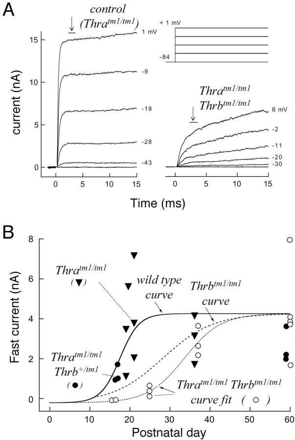
The deafness caused by early onset hypothyroidism indicates that thyroid hormone is essential for the development of hearing. We investigated the underlying roles of the TRalpha1 and TRbeta thyroid hormone receptors in the auditory system using receptor-deficient mice. TRalpha1 and TRbeta, which act as hormone-activated transcription factors, are encoded by the Thra and Thrb genes, respectively, and both are expressed in the developing cochlea. TRbeta is required for hearing because TRbeta-deficient (Thrb(tm1/tm1)) mice have a defective auditory-evoked brainstem response and retarded expression of a potassium current (I(K,f)) in the cochlear inner hair cells. Here, we show that although TRalpha1 is individually dispensable, TRalpha1 and TRbeta synergistically control an extended array of functions in postnatal cochlear development. Compared with Thrb(tm1/tm1) mice, the deletion of all TRs in Thra(tm1/tm1)Thrb(tm1/tm1) mice produces exacerbated and novel phenotypes, including delayed differentiation of the sensory epithelium, malformation of the tectorial membrane, impairment of electromechanical transduction in outer hair cells, and a low endocochlear potential. The induction of I(K,f) in inner hair cells was not markedly more retarded than in Thrb(tm1/tm1) mice, suggesting that this feature of hair cell maturation is primarily TRbeta-dependent. These results indicate that distinct pathways mediated by TRbeta alone or by TRbeta and TRalpha1 together facilitate control over an extended range of functions during the maturation of the cochlea.


Figures

Fig. 1.
Fig. 1.
ABR thresholds in mice with single or combined deletions of TRα1 and TRβ. A, Mean ABR thresholds ± SEM (in decibels of SPL) for wild-type (wt),Thratm1/tm1 (atm1/tm1),Thrbtm1/tm1(btm1/tm1),Thratm1/tm1Thrbtm1/tm1(atm1/tm1btm1/tm1) mice, or other combined mutant strains. All genotypes were on a uniform, congenic C57BL/6J (N > 10) background. Responses to click, 8, 16, and 32 kHz stimuli are shown. Groups shown contained n = 7–8 mice at 5–13 weeks of age. Thresholds were undetectable or were significantly elevated (p < 0.01) inThratm1/tm1Thrbtm1/tm1mice compared with wt or Thrbtm1/tm1mice for all stimulus frequencies tested. B,C, Representative ABR waveforms for wt andThratm1/tm1Thrbtm1/tm1 mice in response to click (B) and 16 kHz (C) stimuli. Waveforms are shown on a 4 μV fixed scale for comparison (actual thresholds were determined on a normalized scale for sensitivity; see Materials and Methods). Thresholds are underlined. No waveform was detectable forThratm1/tm1Thrbtm1/tm1mice for the click stimulus. Only weak, atypical waveforms were detected for the 16 kHz stimulus shown.
Fig. 2.
Fig. 2.
Cochlear malformations inThratm1/tm1Thrbtm1/tm1(A–H) andThrbtm1/tm1(I–L) mouse strains. A–H,Thratm1/tm1Thrbtm1/tm1 and wt control mice at postnatal day 8 (A–D) and as adults (7- to 8-week-old) (E–H).A, C, Low (A) and higher (C) magnification view of a mid-modiolar, midbasal turn of the cochlea from a wt pup at P8 showing the tectorial membrane (TM) extending over the inner sulcus (IS) to the hair cells (the TM is slightly retracted from the OHCs because of shrinkage during fixation). The tunnel of Corti (TC) has opened between the inner and outer pillar cells. The filled arrowhead indicates an IHC, and thethree arrows indicate OHCs. Abbreviations and symbols are the same in other panels; see M, for full description. B, D, Low (B) and higher (D) magnification view of a midbasal turn from aThratm1/tm1Thrbtm1/tm1pup. The TM is enlarged, the IS has not formed, the tunnel of Corti has not opened, and the pillar cells protrude (open arrowhead) above the epithelium between the IHC and OHCs. The greater epithelial ridge below the TM is markedly thicker than in the wt pup. Scale bars: A (same for B),C (same for D), 100 μm. E, Apical turn of the cochlea from an adult wild-type mouse.F, G, H, Apical (F), mid (G), and basal (H) turns of the cochlea of aThratm1/tm1Thrbtm1/tm1adult. The tunnel of Corti and IS have opened. An occasional but minimal loss of hair cells was evident in mid- and basal turns. The TC is present but appears somewhat misshapen. The TM is enlarged and deformed, and in basal turns is often shriveled and retracted into the IS. Scale bar: E (same in F–H), 50 μm. I–L, Cochlear basal turn fromThrbtm1/tm1 and wt control mice at P9 (I, J) and at P20 (K, L). J, InThrbtm1/tm1 pups at P9, the formation of the IS is retarded, the TM is slightly enlarged, the underlying epithelial cell layer is thicker than in wt controls, and the tunnel of Corti (TC) is unopened. K, In wt mice at P20, the organ of Corti has matured (the TM in this example is slightly lifted above the OHCs because of shrinkage during fixation).L, In Thrbtm1/tm1 mice at P20, the IS has opened, but the TM is slightly enlarged; the epithelium lining the IS is slightly thicker than in wt controls. TheThrbtm1/tm1 phenotype is milder than inThratm1/tm1Thrbtm1/tm1mice. I–L represent 1 μm sections stained with toluidine blue (and thus differ from the 3 μm, thionin-stained sections in A–H). Cochleas fromn ≥ 3 mice per genotype per age were examined.M, Schematic diagram of major structures of the cochlea. The hair cells reside on the basilar membrane and lie below the tectorial membrane. IHC, Inner hair cell;OHC, outer hair cell; PC, pillar cell;TC, tunnel of Corti; GER, greater epithelial ridge; LER, lesser epithelial ridge of the organ of Corti.
Fig. 3.
Fig. 3.
Tectorial membrane malformation inThratm1/tm1Thrbtm1/tm1and Thrbtm1/tm1 mice.A, B, Transmission electron micrographs showing the ultrastructure of the matrix in the region of the TM that overlies the sensory hair cells in the organ of Corti in adult wt (A) andThratm1/tm1Thrbtm1/tm1 (B) mice. In wt mice, the major 20 nm diameter collagen fibrils (two arrowheads) are seen embedded in a striated sheet matrix formed from alternating light and dark staining, fine diameter filaments (five small arrows). In theThratm1/tm1Thrbtm1/tm1mouse, collagen fibrils are present, but the striated sheet matrix is disorganized throughout the TM. C, D, Partial disarray of TM ultrastructure inThrbtm1/tm1 mice. The TM displays a similar disarray asThratm1/tm1 Thrbtm1/tm1mice in the upper region (C) (i.e., those regions that are located furthest from the apical surface of the organ of Corti). The lower region(D) appears normal. Micrographs shown are from a 7-month-old wt mouse (A), a 6-month-oldThratm1/tm1Thrbtm1/tm1mouse (B), and a 4-month-oldThrbtm1/tm1 mouse (C, D). Scale bar: C (same in A, B, D), 200 nm. E, F, Representative immunostaining for α-tectorin (E) and β-tectorin (F) in cochlear sections of adult wt andThratm1/tm1 Thrbtm1/tm1mice. InThratm1/tm1Thrbtm1/tm1mice, the malformation of the TM is evident, but it is still immunoreactive for the tectorins. Similar results were found for otogelin (data not shown).
Fig. 4.
Fig. 4.
Delayed expression of theIK,f fast current in inner hair cells ofThratm1/tm1Thrbtm1/tm1mice. A, Fast-activating currents (IK,f) in IHCs of aThratm1/tm1 mouse at P21 and aThratm1/tm1Thrbtm1/tm1mouse at P25. The fast component IK,f was largely missing in theThratm1/tm1Thrbtm1/tm1IHC. Arrows and bars indicate the time window during which the amplitude of IK,fwas measured at −25 mV. The voltage step protocol used is indicated above the traces of theThratm1/tm1Thrbtm1/tm1cell. B, Developmental profile ofIK,f at −25 mV in IHCs ofThratm1/tm1Thrbtm1/tm1mice. Currents were measured as indicated by the arrowand bar in A, and they were plotted versus the depolarizing membrane potential to obtainI–V plots. IK,f was then measured at −25 mV from these plots by interpolating data points negative and positive to −25 mV and plotted versus the postnatal age of the mouse. The solid line is the previously determined curve for IK,f in IHCs of wt mice and the broken (dashed) line the curve forThrbtm1/tm1 mice as reported (Rüsch et al., 1998). The dotted line is the logistic growth function fitted to the data points plotted for IHCs ofThratm1/tm1Thrbtm1/tm1mice (circular points), where the current maximum and minimum were fixed to the values of the wt fit [4.28 nA and −208 pA (−0.208 pA)]; t1/2 = 33.6 d and slope = 0.22/d. Data points for IK,ffrom IHCs of normal-hearingThratm1/tm1Thrb+/tm1(filled circles), andThratm1/tm1 littermates (inverted triangles) followed the normal wt developmental profile.
Fig. 5.
Fig. 5.
Reduced nonlinear capacitance in outer hair cells inThratm1/tm1Thrbtm1/tm1mice. A, Measurements of nonlinear capacitance in aThratm1/tm1Thrbtm1/tm1 mouse and a control (Thratm1/tm1Thrb+/tm1) mouse. B, Plot of values of the nonlinear capacitance in five OHCs of threeThratm1/tm1Thrb+/tm1control mice and in 10 OHCs of threeThratm1/tm1 Thrbtm1/tm1mice at P8. The capacitance was significantly smaller in OHCs fromThratm1/tm1Thrbtm1/tm1mice (p < 0.001).

Similar articles

Cited by

References

    1. Bradley DJ, Towle HC, Young WS., III α and β thyroid hormone receptor (TR) gene expression during auditory neurogenesis: evidence for TR isoform-specific transcriptional regulation in vivo. Proc Natl Acad Sci USA. 1994;91:439–443. - PMC - PubMed
    1. Brucker-Davis F, Skarulis MC, Pikus A, Ishizawar D, Mastroianni M-A, Koby M, Weintraub BD. Prevalence and mechanisms of hearing loss in patients with resistance to thyroid hormone (RTH). J Clin Endocrinol Metab. 1996;81:2768–2772. - PubMed
    1. Campos-Barros A, Amma LL, Faris JS, Shailam R, Kelley MW, Forrest D. Type 2 iodothyronine deiodinase expression in the cochlea before the onset of hearing. Proc Natl Acad Sci USA. 2000;97:1287–1292. - PMC - PubMed
    1. Corey DP, Breakefield XO. Transcription factors in inner ear development. Proc Natl Acad Sci USA. 1994;91:433–436. - PMC - PubMed
    1. Dallos P, Popper A, Fay R. The cochlea. Springer; New York: 1996.

Publication types

MeSH terms

Substances

LinkOut - more resources