. 2001 Dec 15;21(24):9888-95.
doi: 10.1523/JNEUROSCI.21-24-09888.2001.

Functional regeneration in a rat Parkinson's model after intrastriatal grafts of glial cell line-derived neurotrophic factor and transforming growth factor beta1-expressing extra-adrenal chromaffin cells of the Zuckerkandl's organ

Affiliations

Functional regeneration in a rat Parkinson's model after intrastriatal grafts of glial cell line-derived neurotrophic factor and transforming growth factor beta1-expressing extra-adrenal chromaffin cells of the Zuckerkandl's organ

E F Espejo et al. J Neurosci. .

Abstract

Intrabrain transplantation of chromaffin cell aggregates of the Zuckerkandl's organ, an extra-adrenal paraganglion that has never been tested for antiparkinsonian treatment, induced gradual improvement of functional deficits in parkinsonian rats. These beneficial effects were related to long survival of grafted cells, striatal reinnervation, and enhancement of dopamine levels in grafted striatum. Grafted cells were not dopaminergics, but they expressed glial cell line-derived neurotrophic factor (GDNF) and transforming growth factor-beta(1). These factors were detected in the host striatal tissue, indicating that chromaffin cells secreted them after grafting. Because glial cell line-derived neurotrophic factor possesses neurorestorative properties over dopaminergic neurons, and transforming growth factor-beta(1) is a cofactor that potentiates the neurotrophic actions of GDNF, functional regeneration was likely caused by the chronic trophic action of neurotrophic factors delivered by long-surviving grafted cells. This work should stimulate research on the clinical applicability of transplants of the Zuckerkandl's organ in Parkinson's disease.


Figures

Fig. 1.
Fig. 1.
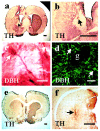
Morphological features of the Zuckerkandl's organ (a–c) and adrenal medulla (d).a, Coronal section of the Zuckerkandl's organ used for transplantation, without enzymatic treatment, after double labeling for TH (red) and DBH (green). Confocal fluorescent micrograph shows dispersed cell groups and cell nests (Cn) of noradrenergic chromaffin cells (TH+/DBH+;yellow–green) surrounded by mesenchyma (Ms). b, Higher magnification of a cell nest labeled for DBH showing many DBH+ rounded chromaffin cells of 20–30 μm in diameter (green).c, Higher magnification of chromaffin cells with green and yellow patches in the cytoplasm indicative of the presence of DBH and TH–DBH, respectively, and hence norepinephrine. d,Piece of an adrenal medulla used for transplantation, without enzymatic treatment, after double labeling for TH and DBH immunofluorescence. Confocal fluorescent micrograph shows many rounded chromaffin cells (15–25 μm in diameter) with green and yellow patches in the cytoplasm, indicative of the presence of DBH and TH–DBH. Scale bars:a, 200 μm; b, d, 100 μm; c, 50 μm.
Fig. 2.
Fig. 2.
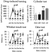
Time course of drug-induced turning, right forepaw use (cylinder test), and sensorimotor orientation (whisker-touch and odor tests), after grafts of the Zuckerkandl's organ and adrenal tissue. Groups: ●, control (nonparkinsonian rats); ▪, 6-OHDA-induced lesion and Zuckerkandl's organ graft (Zuckerkandl group); ▴, 6-OHDA-induced lesion and sham graft (sham-grafted group); and ▾, 6-OHDA-induced lesion and adrenal transplant (adrenal group). Two-way ANOVA for repeated measures indicated significant group (3, 41 df) and interaction effects (15, 210 df) for drug-induced turning (group, F = 12.4; interaction,F = 7.3; p < 0.001), whisker-touch latency (group, F = 133.2; interaction, F = 36.7; p < 0.001), and odor latency (group, F = 80.8; interaction, F = 26.6; p < 0.001). Maximum latency in sensorimotor tests, 25 sec. Mean ± SEM. *p < 0.05; **p < 0.01 versus sham-grafted rats (Newman–Keuls test, or Student'st test in the cylinder test). pre, Before lesion; les, 2 weeks after lesion; 1m, 2m, 3m, 5m, 1, 2, 3 and 5 months after extra-adrenal, adrenal, or sham grafting, respectively; ctrl, control group;adrl, adrenal group; Zuck, Zuckerkandl group; sham, sham-grafted group. Grafts were implanted 2 weeks after nigra lesion (arrow). The cylinder test corresponds to values 5 months after grafting.
Fig. 3.
Fig. 3.
Morphological features of 5-month-old transplants of the Zuckerkandl's organ (a–d), as well as striatal appearance 5 months after sham-grafting (e) or adrenal cell transplants (f). a, b, Coronal sections of the striatum grafted with extra-adrenal cells of the Zuckerkandl's organ where a TH-positive transplant (arrow) and a broad TH+ reinnervated area in the host left striatum (brown) can be clearly observed.c, Higher magnification of the transplant labeled for DBH where DBH+ chromaffin cells (arrows) with long neuritic processes (arrowheads) are observed.d, Confocal fluorescent micrographs of the graft revealing the presence of DBH+ cells (green, arrows) and many neuritic processes running out of the graft.e, Coronal section of a sham-grafted striatum showing the nearly absent TH positivity in the left denervated striatum.f, Coronal section through the brain striatum with an adrenal medullary transplant, where a necrotic graft can be observed (arrow), as well as lack of striatal TH positivity except for a halo of TH positivity surrounding the graft, and a small TH+ area at the border of the left ventricle. g, Graft. Scale bars: a, b, e,f, 1 mm; d, 100 μm; c, 50 μm.
Fig. 4.
Fig. 4.
Immunohistochemistry for GDNF and TGF-β1 in transplants of the Zuckerkandl's organ at 5 months after grafting, showing the presence of GDNF-positive (a) and TGF-β1-positive chromaffin cells (b). g, Graft. Scale bars, 100 μm.

Similar articles

Cited by

References

    1. Aebischer P, Ridet J-L. Recombinant proteins for neurodegenerative diseases: the delivery issue. Trends Neurosci. 2001;24:533–540. - PubMed
    1. Ahonen M, Soinila S, Joh TH. Pre- and postnatal development of rat retroperitoneal paraganglia. J Auton Nerv Syst. 1987;18:11–120. - PubMed
    1. Bankiewicz KS, Palmatie M, Plunkett RJ, Cummins A, Oldfield EH. Reversal of hemiparkinsonian syndrome in nonhuman primates by amnion implantation into caudate nucleus. J Neurosurg. 1994;81:869–876. - PubMed
    1. Batchelor PE, Liberatore GT, Wong JYF, Porritt MJ, Frerichs F, Donnan GA, Howells DW. Activated macrophages and microglia induce dopaminergic sprouting in the injured striatum and express brain-derived neurotrophic factor and glial cell line-derived neurotrophic factor. J Neurosci. 1999;19:1708–1716. - PMC - PubMed
    1. Beck KD, Valverde J, Alexi T, Poulsen K, Moffat B, Vandlen RA, Rosenthal A, Hefti F. Mesencephalic dopaminergic neurons protected by GDNF from axotomy-induced degeneration in the adult brain. Nature. 1995;373:339–341. - PubMed

Publication types

MeSH terms

LinkOut - more resources