Comparative Study
. 2001 Dec 15;21(24):9930-43.
doi: 10.1523/JNEUROSCI.21-24-09930.2001.

cAMP response element-binding protein is required for dopamine-dependent gene expression in the intact but not the dopamine-denervated striatum

Affiliations
Comparative Study

cAMP response element-binding protein is required for dopamine-dependent gene expression in the intact but not the dopamine-denervated striatum

M Andersson et al. J Neurosci. .

Abstract

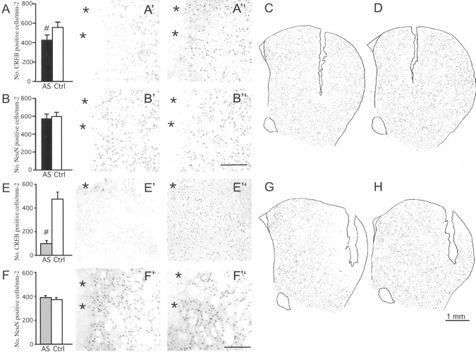
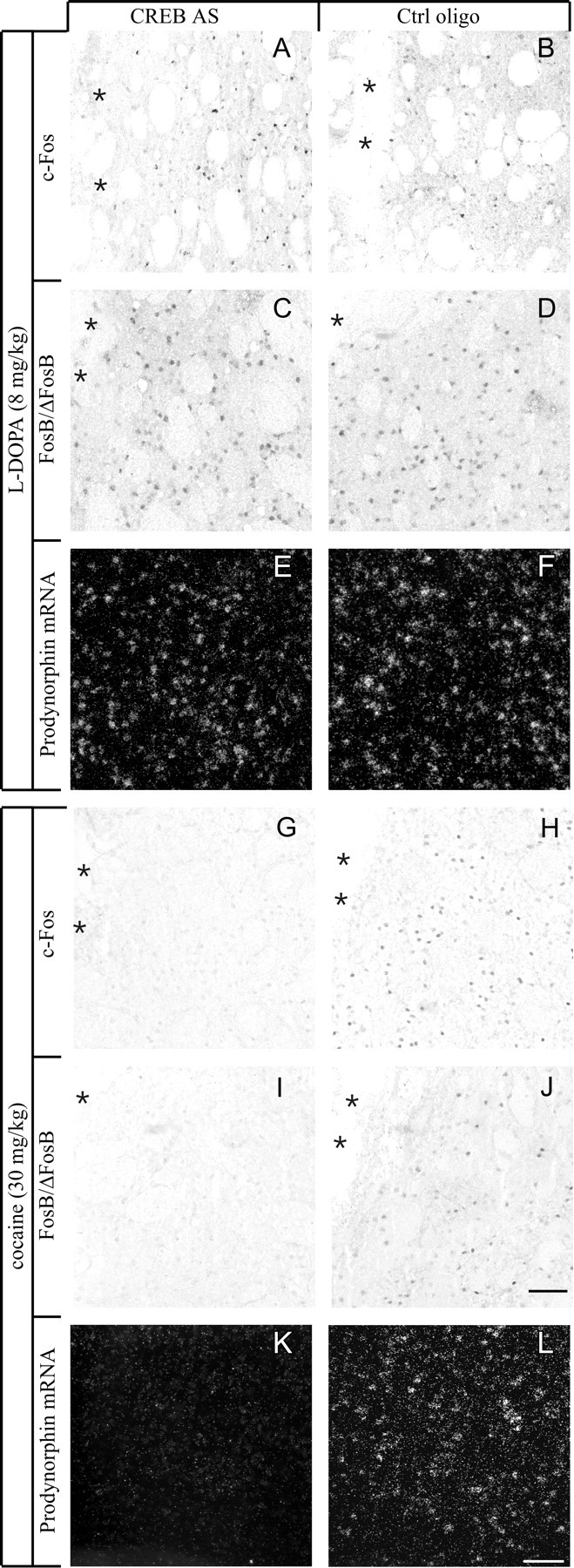
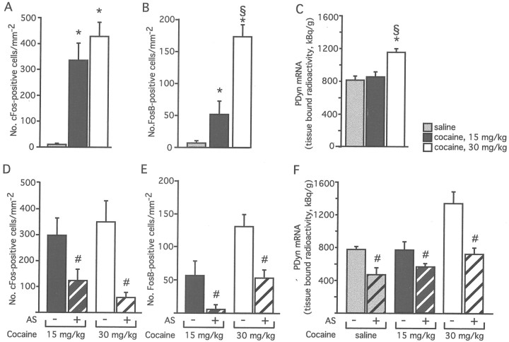
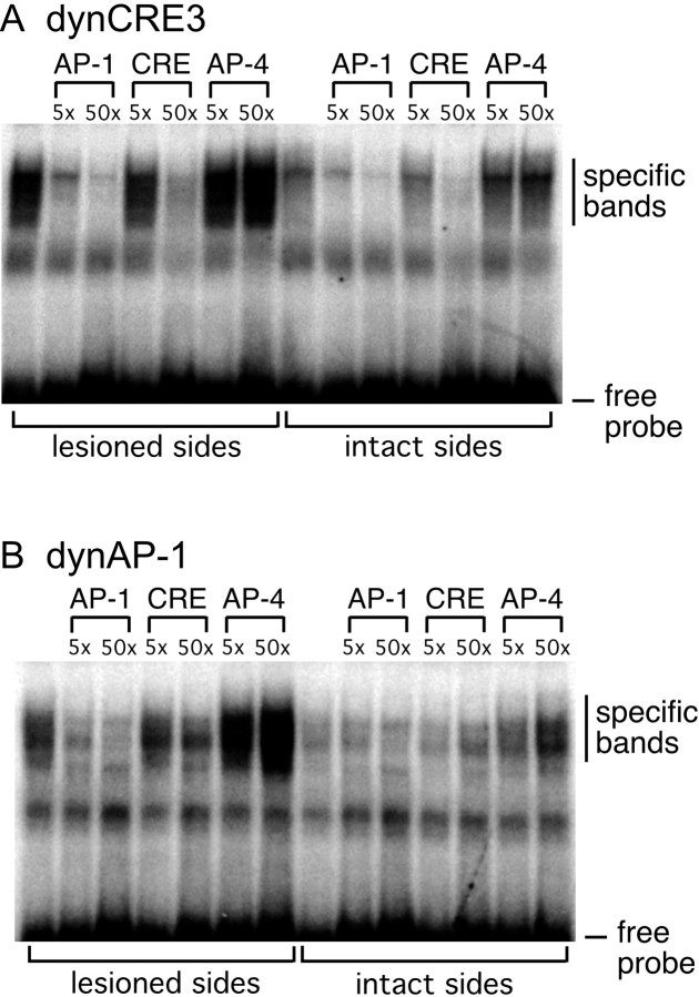
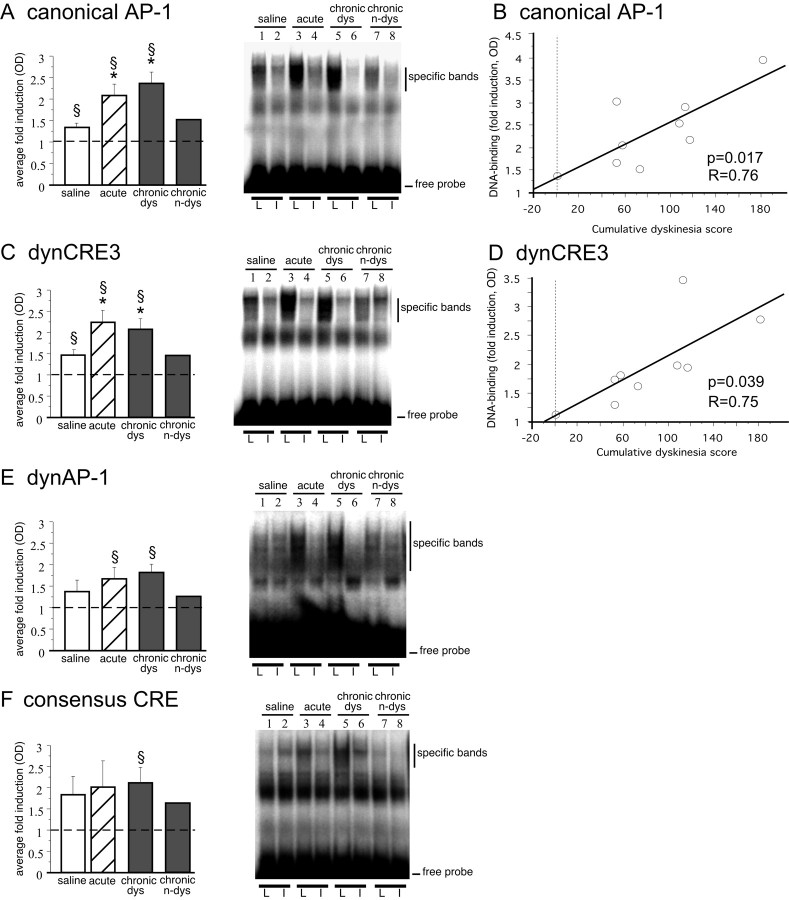
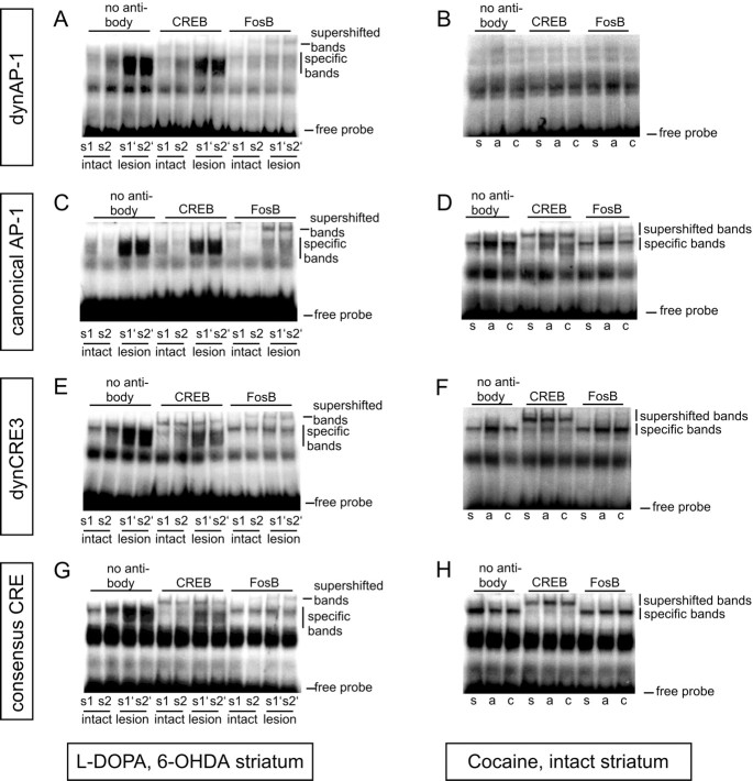
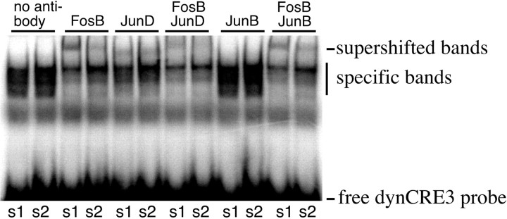
The cAMP response element-binding protein (CREB) is believed to play a pivotal role in dopamine (DA) receptor-mediated nuclear signaling and neuroplasticity. Here we demonstrate that the significance of CREB for gene expression depends on the experimental paradigm. We compared the role of CREB in two different but related models: l-DOPA administration to unilaterally 6-hydroxydopamine lesioned rats, and cocaine administration to neurologically intact animals. Antisense technology was used to produce a local knockdown of CREB in the lateral caudate-putamen, a region that mediates the dyskinetic or stereotypic manifestations associated with l-DOPA or cocaine treatment, respectively. In intact rats, CREB antisense reduced both basal and cocaine-induced expression of c-Fos, FosB/DeltaFosB, and prodynorphin mRNA. In the DA-denervated striatum, CREB was not required for l-DOPA to induce these gene products, nor did CREB contribute considerably to DNA binding activity at cAMP responsive elements (CREs) and CRE-like enhancers. DeltaFosB-related proteins and JunD were the main contributors to both CRE and AP-1 DNA-protein complexes in l-DOPA-treated animals. In behavioral studies, intrastriatal CREB knockdown caused enhanced activity scores in intact control animals and exacerbated the dyskinetic effects of acute l-DOPA treatment in 6-OHDA-lesioned animals. These data demonstrate that CREB is not required for the development of l-DOPA-induced dyskinesia in hemiparkinsonian rats. Moreover, our results reveal an unexpected alteration of nuclear signaling mechanisms in the parkinsonian striatum treated with l-DOPA, where AP-1 transcription factors appear to supersede CREB in the activation of CRE-containing genes.


Figures

Fig. 1.
Fig. 1.
Different methods of CREB antisense delivery produce different extents of CREB knockdown in the striatum. The effects of single oligonucleotide injections are shown inAD, whereas those produced by a 14 d oligonucleotide infusion are shown inEH. The number of CREB-immunoreactive cells in the region flanking the injection cannula is shown inA (single injection) or E (continuous infusion), and the corresponding photomicrographs are shown inA′ or E′ (antisense group), andA" or E" (control oligonucleotide). NeuN-immunoreactive neurons within the same regions are shown inBB" andFF". Camera lucida drawings illustrate the regional distribution of CREB knockdown after single oligonucleotide injection (C, antisense;D, control) or continuous oligonucleotide infusion (G, antisense; H, control).#p < 0.01; n ≥ 7 in all groups; values give means + SEM. Asterisks inA′, A", B′,B", E′, E", F′, andF" mark the cannula track. AS, CREB antisense;Ctrl, control oligo. Scale bars in B" andF", 200 μm.
Fig. 2.
Fig. 2.
l-DOPA regulates c-Fos, FosB/ΔFosB, and prodynorphin independently of CREB in 6-OHDA-lesioned rats. The effects of acute doses of l-DOPA on the number of c-Fos (A) or FosB/ΔFosB (B) immunoreactive cells and on the levels of PDyn mRNA (C) were measured on the cross-sectional area of the caudate–putamen at mid-rostrocaudal levels. l-DOPA induced levels of Fos family proteins (A,B) and PDyn (C) mRNA dose dependently. A single injection of CREB antisense oligonucleotides did not prevent the induction of c-Fos (D), FosB/ΔFosB (E), or PDyn mRNA (F) by l-DOPA. CREB antisense-treated animals are marked as AS + and represented usingbars with diagonal stripes, whereas animals receiving control oligonucleotides are marked as AS − and represented using bars with no texture. Antisense effects were measured in 0.54-mm2-large areas next to the injection track. *p < 0.01 versus lesion-only; #p < 0.05 versus 5 mg/kg; and §p < 0.05 versus 3 mg/kgl-DOPA; n = 3–5 per group inAC, and ≥5 per group inDF; values give means + SEM.
Fig. 3.
Fig. 3.
CREB knockdown differentially affects the induction of c-Fos (A, B,G, H), FosB/ΔFosB (C, D, I,J), or PDyn mRNA (E,F, K, L) byl-DOPA or cocaine. In the absence of CREB,l-DOPA was able to induce all gene expression markers in the 6-OHDA-lesioned striatum (compare antisense group inA, C, and E with control group in B, D, andF), whereas cocaine was unable to induce the same gene products in the intact striatum (G, I, K, antisense group; H, J, L, control group). Photomicrographs were taken under bright-field (AD,GJ) or dark-field illumination (E, F, K,L) from animals that sustained intrastriatal oligonucleotide infusion and acute drug injections.Asterisks mark the cannula track. Scale bar, 100 μm.
Fig. 4.
Fig. 4.
Loss of CREB increases abnormal involuntary movements (orolingual, limb, and axial AIMs) in 6-OHDA-lesioned rats treated with l-DOPA. In the acutelyl-DOPA-treated rats (A), CREB antisense produced levels of AIM scores that were similar to chronicl-DOPA-treated rats (compare AS group inA with Ctrl group in B). A trend toward more severe dyskinesias in the CREB AS group was also seen during chronic l-DOPA treatment (B). AIMs scores were collected during a single testing session inA and during four testing sessions in B(the AIM score in the last of these sessions is shown in the diagram). Each dot in the scattergram represents one single animal; the group median is shown as a horizontal bar.#p < 0.01.
Fig. 5.
Fig. 5.
Cocaine-induced expression of c-Fos, FosB/ΔFosB, and PDyn mRNA is dependent on CREB. Acute treatment with cocaine produced a dose-dependent increase in the number of c-Fos (A) and FosB/ΔFosB immunoreactive cells (B), and in the levels of PDyn mRNA (C), as measured on the cross-sectional area of the caudate–putamen at mid-rostrocaudal levels. Knockdown of CREB prevented the cocaine-stimulated increase in c-Fos (D), FosB/ΔFosB (E), and PDyn mRNA (F). CREB antisense-treated animals are marked as AS + and represented using bars withdiagonal stripes, whereas animals receiving control oligonucleotides are marked as AS − and represented usingbars with no texture. The data presented here are derived from two sets of experiments. Animals injected with 15 mg/kg cocaine received a single intrastriatal injection of CREB AS, whereas animals injected with 30 mg/kg cocaine or saline received a 14 d intrastriatal oligonucleotide infusion. *p < 0.05 versus saline; §p < 0.01 versus 15 mg/kg cocaine; #p < 0.05 versus control oligo; n ≥ 5 in each group; values give means + SEM.
Fig. 6.
Fig. 6.
Loss of CREB increases activity/stereotypy scores in intact rats challenged with an injection of saline (A) but not cocaine (B). CREB AS was infused continuously for 14 d before the acute challenge. The rats were neurologically intact. Each dotin the scattergram represents one single animal; the group median is shown as a horizontal bar.#p < 0.01.
Fig. 7.
Fig. 7.
Gel mobility shift assay and competition experiment determine the specificity of DNA–protein complexes formed with dynCRE3 (A) and dynAP-1 (B). Unlabeled consensus AP-1, CRE, and the unrelated AP-4 sequences were used to compete for proteins bound to radiolabeled dynCRE3 (A) and dynAP1 (B) promoter elements. Striatal samples were pooled from several l-DOPA-treated rats. The experiment was repeated three times.
Fig. 8.
Fig. 8.
Acute and chronic treatment withl-DOPA induces striatal DNA binding activity to the canonical AP-1 (A), the dynCRE3 (C), the dynAP-1 (E), and the consensus CRE (F). Optical densities of specific bands are expressed as the fold induction on the lesioned over the intact side. *p < 0.05 versus saline group; §p < 0.05 versus intact side; values give means + SEM; n ≥ 5 in all groups but chronic n-dys (i.e., chronicallyl-DOPA-treated but nondyskinetic case). As shown inB and D, only one animal remained nondyskinetic (0 cumulative dyskinesia scores) during chronicl-DOPA treatment. In B and D, levels of binding to the canonical AP-1 (B) and dynCRE3 (D) in the chronic l-DOPA cases are plotted on the cumulative axial, limb, and orolingual AIM scores recorded from the same animals. The probability value (p) and correlation coefficient (R) of the corresponding simple regression equation are given in the bottom right corner. L, 6-OHDA-lesioned side; I, contralateral intact side.
Fig. 9.
Fig. 9.
FosB/ΔFosB-related proteins are the predominant transcription factors bound to CRE and AP-1 enhancers inl-DOPA-treated rats. To study the contribution of CREB- or FosB/ΔFosB-related proteins to the DNA binding activity induced byl-DOPA (A, C,E, G) or cocaine (B,D, F, H), supershift assays were performed using antisera against CREB or FosB/ΔFosB. Antibodies were preincubated with striatal protein extracts before addition of the radioactively labeled promoter elements dynAP-1 (A, B), canonical AP-1 (C, D), dynCRE3 (E,F), and consensus CRE (G,H). The experiments were repeated at least three times with samples from different rats. In the left-hand pictures, s1 and s2 are protein extracts from intact striata of two chronicallyl-DOPA-treated rats, whereas s1′ ands2′ are extracts from the corresponding 6-OHDA-lesioned side. Rats treated with acute l-DOPA showed comparable results. In the right-hand pictures, s,a, and c are protein extracts from animals treated with saline, acute cocaine, or chronic cocaine, respectively.
Fig. 10.
Fig. 10.
Like FosB/ΔFosB-related proteins, JunD can bind at AP-1 and CRE enhancers in 6-OHDA-lesioned,l-DOPA-treated rats. To characterize possible binding partners to FosB/ΔFosB proteins in the l-DOPA-induced dynCRE3 complex, supershift assays were performed with JunD or JunB antisera. s1 and s2 are samples from the lesioned striata of two l-DOPA-treated rats. This experiment was repeated with samples from additional animals, and with the dynAP-1 probe, it obtained similar results.
Fig. 11.
Fig. 11.
l-DOPA induces FosB/ΔFosB proteins to a higher level than cocaine does. Immunoblots with an antibody to FosB/ΔFosB are shown in A, and the corresponding densitometric analysis is shown in B. All the immunoblots used for analysis were processed simultaneously. A representative immunoblot stained with a PanFos antibody is shown in C. Arrows indicate the expected molecular weights of c-Fos (55 kDa), ΔFosB (33 kDa), and the chronic FRAs (35–37 kDa). *p < 0.001 versus saline, acute cocaine, and chronic cocaine group.§p < 0.05 versus saline controls in the cocaine paradigm. #p < 0.01 versus saline controls in the l-DOPA paradigm;n ≥ 5; values give means + SEM.

References

    1. Andersson M, Hilbertson A, Cenci MA. Striatal fosB expression is causally linked with l-DOPA-induced abnormal involuntary movements and the associated upregulation of striatal prodynorphin mRNA in a rat model of Parkinson's disease. Neurobiol Dis. 1999;6:461–474. - PubMed
    1. Calon F, Tahar AH, Blanchet PJ, Morissette M, Grondin R, Goulet M, Doucet JP, Robertson GS, Nestler E, Di Paolo T, Bedard PJ. Dopamine-receptor stimulation: biobehavioral and biochemical consequences. Trends Neurosci. 2000;23:S92–100. - PubMed
    1. Carlezon WA, Jr, Thome J, Olson VG, Lane-Ladd SB, Brodkin ES, Hiroi N, Duman RS, Neve RL, Nestler EJ. Regulation of cocaine reward by CREB. Science. 1998;282:2272–2275. - PubMed
    1. Cenci MA, Lee CS, Björklund A. l-DOPA-induced dyskinesia in the rat is associated with striatal overexpression of prodynorphin- and glutamic acid decarboxylase mRNA. Eur J Neurosci. 1998;10:2694–2706. - PubMed
    1. Cenci MA, Tranberg A, Andersson M, Hilbertson A. Changes in the regional and compartmental distribution of FosB- and JunB-like immunoreactivity induced in the dopamine-denervated rat striatum by acute or chronic l-DOPA treatment. Neuroscience. 1999;94:515–527. - PubMed

Publication types

MeSH terms

LinkOut - more resources