

Review
doi: 10.1128/jvi.76.1.1-8.2002.
Directed egress of animal viruses promotes cell-to-cell spread
Affiliations
- PMID: 11739666
- PMCID: PMC135733
- DOI: 10.1128/jvi.76.1.1-8.2002
Item in Clipboard
Review
Directed egress of animal viruses promotes cell-to-cell spread
J Virol.
2002 Jan.
No abstract available
Figures
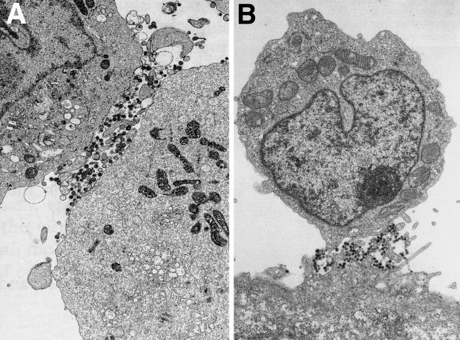
Polarized egress of HIV particles on T lymphocytes. (A) HIV-infected C8166 cells, a CD4+ T-cell line, were analyzed by electron microscopy. Virions were found preferentially at sites of cell contact. (Reprinted from AIDS [26] with permission of the publisher.) (B) HIV-infected CD4+ T cells (upper cell) adhering to a BeWo epithelial cell in the lower part of the panel. Virions were observed at sites of cell-cell contact, between microvilli extending from both cells. (Reprinted from AIDS [65] with permission of the publisher.)
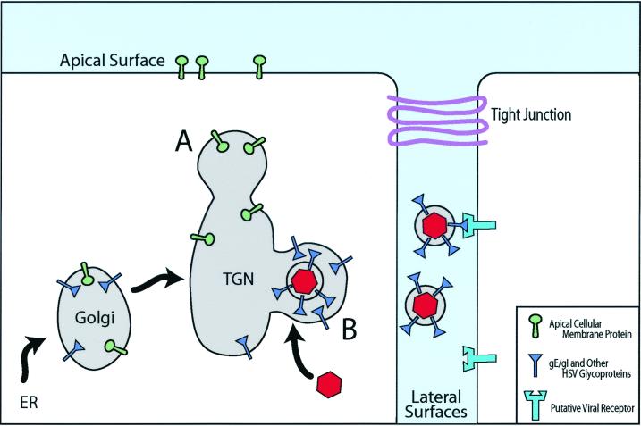
Directed egress of alphaherpesviruses by sorting in the TGN. Alphaherpesvirus glycoproteins accumulate in the TGN, and this process involves the cytoplasmic domains of gE/gI (blue) as well as other viral membrane proteins, such as gM. There is assembly of tegument components around these sites of glycoprotein accumulation and binding of nucleocapsids (red). In epithelial cells, assembly of viral particles occurs selectively in sorting compartments of the TGN that will ultimately be delivered to cell basolateral or lateral surfaces (B), e.g., through gE/gI coupling to μ1B-substituted AP1 clathrin adapters (46). Other viral or cellular membrane proteins are sorted in different domains of the TGN to apical surfaces (A). Virions reach the lateral surfaces of cells in transport vesicles, which fuse with the plasma membrane and deliver virus particles into the space between cells. There are specific interactions of HSV glycoproteins with components of cell junctions—gD with nectins and apparently gE/gI with other molecules—which facilitate entry into adjacent cells. When both cells are infected, there can be accumulation of HSV particles between cells as receptors are blocked. ER, endoplasmic reticulum.

Egress of VV particles from cells on actin tails. Poxvirus nucleocapsids are formed and acquire envelopes in virus factories, producing IMV, which subsequently acquire two additional membranes by budding into the TGN, forming IEV. Microtubules are involved in the transport of IMV and IEV to the plasma membrane (PM). There is fusion of the outer envelope with the plasma membrane, and CEV (bound to the cell surface) rest on platforms, composed in part of A33R, A34R, and A36R, that sit at one pole of the virus particle. A36R promotes the nucleation of actin filaments through interactions with Nck and recruitment of WIP, N-WASP, and Arp2/3 (inset), leading to actin polymerization. Actin tails produce microvilli that project CEV toward adjacent cells, promoting virus entry.
References
-
- Arvin, A. M. 1996. Varicella-zoster virus, p. 2547–2585. In B. N. Fields, D. M. Knipe, and P. M. Howley (ed.), Fields virology, 3rd ed., vol. 2. Lippincott-Raven Publishers, Philadelphia, Pa.
Publication types
MeSH terms
Grants and funding
LinkOut - more resources
Full Text Sources
