Comparative Study
. 2002 Jan;76(1):105-17.
doi: 10.1128/jvi.76.1.105-117.2002.

The late domain of human immunodeficiency virus type 1 p6 promotes virus release in a cell type-dependent manner

Affiliations
Comparative Study

The late domain of human immunodeficiency virus type 1 p6 promotes virus release in a cell type-dependent manner

Dimiter G Demirov et al. J Virol. 2002 Jan.

Abstract

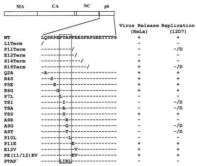
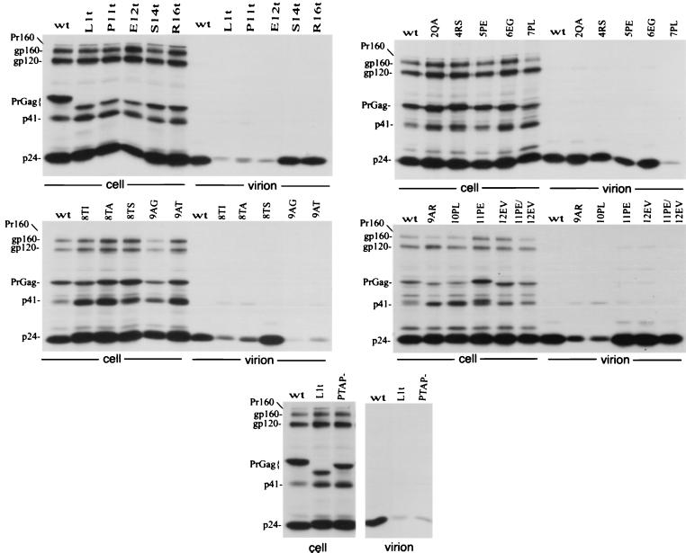
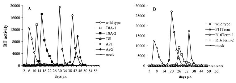
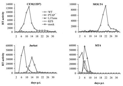
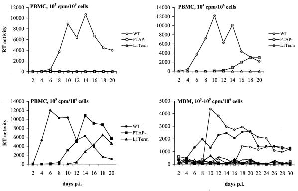
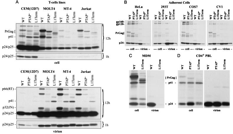
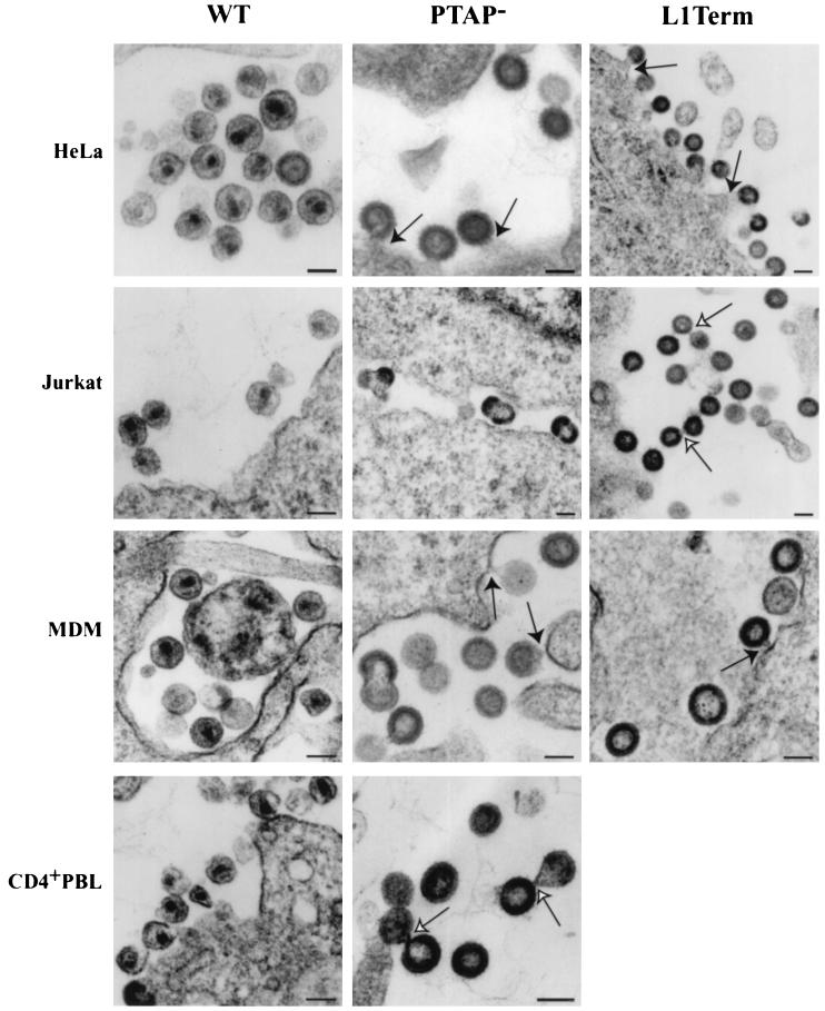
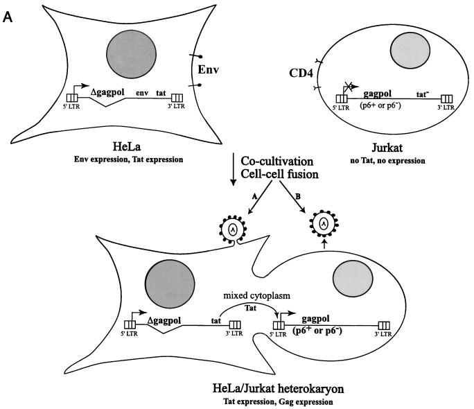
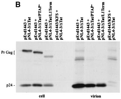
The p6 domain of human immunodeficiency virus type 1 (HIV-1) is located at the C terminus of the Gag precursor protein Pr55(Gag). Previous studies indicated that p6 plays a critical role in HIV-1 particle budding from virus-expressing HeLa cells. In this study, we performed a detailed mutational analysis of the N terminus of p6 to map the sequences required for efficient virus release. We observed that the highly conserved P-T/S-A-P motif located near the N terminus of p6 is remarkably sensitive to change; even conservative mutations in this sequence imposed profound virus release defects in HeLa cells. In contrast, single and double amino acid substitutions outside the P-T/S-A-P motif had no significant effect on particle release. The introduction of stop codons one or two residues beyond the P-T/S-A-P motif markedly impaired virion release, whereas truncation four residues beyond P-T/S-A-P had no effect on particle production in HeLa cells. By examining the effects of p6 mutation in biological and biochemical analyses and by electron microscopy, we defined the role of p6 in particle release and virus replication in a panel of T-cell and adherent cell lines and in primary lymphocytes and monocyte-derived macrophages. We demonstrated that the effects of p6 mutation on virus replication are markedly cell type dependent. Intriguingly, even in T-cell lines and primary lymphocytes in which p6 mutations block virus replication, these changes had little or no effect on particle release. However, p6-mutant particles produced in T-cell lines and primary lymphocytes exhibited a defect in virion-virion detachment, resulting in the production of tethered chains of virions. Virus release in monocyte-derived macrophages was markedly inhibited by p6 mutation. To examine further the cell type-specific virus release defect in HeLa versus T cells, transient heterokaryons were produced between HeLa cells and the Jurkat T-cell line. These heterokaryons display a T-cell-like phenotype with respect to the requirement for p6 in particle release. The results described here define the role of p6 in virus replication in a wide range of cell types and reveal a strong cell type-dependent requirement for p6 in virus particle budding.


Figures

FIG. 1.
FIG. 1.
Schematic representation of p6 mutants analyzed in this study. The arrangement of the HIV-1 Gag domains is depicted at the top. The amino acid sequence of the N-terminal region of p6 encompassing the L domain is shown below. The P-T/S-A-P motif is boxed. Backslashes represent stop codons; dashes denote amino acid sequence identity with the WT. The columns at the right summarize the phenotypes of the mutants in HeLa and CEM(12D-7) cells: +, virus release or replication kinetics similar to WT; −, severe defect in virus release or a block in virus replication; D, delayed virus replication. The L1Term, Q2A, R4S, E6G, A9R, and PTAP mutants were constructed previously (24) and were analyzed in more detail in this study. P7L, T8I, and P10L (24) were reconstructed here to increase the number of nt substitutions. (Table 1).
FIG. 2.
FIG. 2.
Immunoprecipitation of cell- and virion-associated proteins produced in HeLa cells. Transfected cells were metabolically labeled overnight with [35S]Met/Cys; cell and virion lysates were immunoprecipitated with anti-HIV-Ig. For the mutants containing termination codons, Term is abbreviated t. At the left side of each panel are indicated the positions of the Gag-Pol precursor Pr160GagPol (Pr160); the Env glycoproteins gp160 and gp120; the Gag precursor (PrGag); the Gag processing intermediate p41; and the CA protein (p24).
FIG. 3.
FIG. 3.
Replication kinetics of p6 mutants in the CEM(12D-7) T-cell line. CEM(12D-7) cells were transfected in parallel with the indicated molecular clones. Cells were split every 2 days, and RT activity was determined at each time point. Only those mutants that replicated with a delay relative to the WT (i.e., that reverted in culture) are shown. (A) Point mutants within P-T/S-A-P; (B) truncation mutants. p.t., posttransfection.
FIG. 4.
FIG. 4.
Replication kinetics of the L1Term and PTAP mutants in T-cell lines. The indicated T-cell lines were transfected in parallel with WT or mutant molecular clones. Cells were split every 2 days, and RT activity was determined at each time point. Untransfected cells (mock) and cells transfected with the env-negative molecular clone pNL4-3KFS (KFS) served as negative controls. p.t., posttransfection.
FIG. 5.
FIG. 5.
Replication kinetics of the L1Term and PTAP mutants in primary cell types. In each case, 106 cells (PBMC or MDM) were infected at the indicated RT input with virus stocks prepared by triple transfection of 293T cells with WT or mutant molecular clones, pHCMV-G, and pCMVNLGagPolRRE (Materials and Methods). pNL4-3 and pNL(AD8) were used as the parental molecular clones in PBMC and MDM infections, respectively. Supernatants from infected cells were collected every 2 days postinfection (p.i.) for measurement of RT activity. Virus inputs (in RT cpm) per 106 cells are indicated as follows: white symbols, 103; gray symbols, 104; and black symbols, 105. WT, PTAP, and L1Term are indicated by circles, squares, and triangles, respectively.
FIG. 6.
FIG. 6.
Immunoprecipitation of cell- and virion-associated proteins produced in diverse cell types. In each case, 106 cells were infected with 107 cpm of virus stocks prepared by triple transfection of 293T cells with WT or mutant pNL4-3 molecular clones, pHCMV-G, and pCMVNLGagPolRRE (Materials and Methods). At 24 h postinfection, cells were labeled overnight with [35S]Met-Cys; cell and virion lysates were immunoprecipitated with anti-HIV-Ig. (A) T-cell lines; (B) adherent cell lines; (C) MDM; (D) CD4+ PBLs. In panel A, 12-h and 1-h exposures are shown. The positions of the following viral proteins are indicated: the Env glycoproteins gp160 and gp120; the Gag precursor (PrGag); the Gag processing intermediates p41 and p25; the CA protein (p24); and (in virions) p32 (IN) and p66 (RT).
FIG. 7.
FIG. 7.
Visualization of virus assembly and budding in diverse cell types by EM. The WT and mutant molecular clones were introduced into Jurkat cells, MDM, and PBLs as indicated in Fig. 6; clones were introduced into HeLa cells by transfection. Solid arrows indicate particles tethered to the plasma membrane or (in MDM) intracellular membranes. Open arrows indicate particles tethered to each other, occasionally forming long chains (e.g., L1Term in Jurkat). Bar, 100 nm.
FIG. 8.
FIG. 8.
Immunoprecipitation of cell- and virion-associated proteins produced in transient HeLa/Jurkat heterokaryons. (A) Strategy for generating and expressing WT and p6-mutant Gag in heterokaryons. Details are provided in the text (Materials and Methods and Results). (B) Immunoprecipitation analysis. Cocultivated cells were metabolically labeled for 16 h with [35S]Met/Cys. Cell- and virion-associated proteins were immunoprecipitated with rabbit anti-p24. The positions of the Gag precursor (PrGag) and CA (p24) are shown.
FIG. 8.
FIG. 8.
Immunoprecipitation of cell- and virion-associated proteins produced in transient HeLa/Jurkat heterokaryons. (A) Strategy for generating and expressing WT and p6-mutant Gag in heterokaryons. Details are provided in the text (Materials and Methods and Results). (B) Immunoprecipitation analysis. Cocultivated cells were metabolically labeled for 16 h with [35S]Met/Cys. Cell- and virion-associated proteins were immunoprecipitated with rabbit anti-p24. The positions of the Gag precursor (PrGag) and CA (p24) are shown.

References

    1. Accola, M. A., B. Strack, and H. G. Gottlinger. 2000. Efficient particle production by minimal Gag constructs which retain the carboxy-terminal domain of human immunodeficiency virus type 1 capsid-p2 and a late assembly domain. J. Virol. 74:5395–5402. - PMC - PubMed
    1. Adachi, A., H. E. Gendelman, S. Koenig, T. Folks, R. Willey, A. Rabson, and M. A. Martin. 1986. Production of acquired immunodeficiency syndrome-associated retrovirus in human and nonhuman cells transfected with an infectious molecular clone. J. Virol. 59:284–291. - PMC - PubMed
    1. Bork, P., and M. Sudol. 1994. The WW domain: a signalling site in dystrophin? Trends Biochem Sci. 19:531–533. - PubMed
    1. Craven, R. C., R. N. Harty, J. Paragas, P. Palese, and J. W. Wills. 1999. Late domain function identified in the vesicular stomatitis virus M protein by use of rhabdovirus-retrovirus chimeras. J. Virol. 73:3359–3365. - PMC - PubMed
    1. Dalgarno, D. C., M. C. Botfield, and R. J. Rickles. 1997. SH3 domains and drug design: ligands, structure, and biological function. Biopolymers 43:383–400. - PubMed

Publication types

Substances