Comparative Study
. 2002 Jan;76(1):165-77.
doi: 10.1128/jvi.76.1.165-177.2002.

Multiple eIF4GI-specific protease activities present in uninfected and poliovirus-infected cells

Affiliations
Comparative Study

Multiple eIF4GI-specific protease activities present in uninfected and poliovirus-infected cells

Miguel Zamora et al. J Virol. 2002 Jan.

Abstract

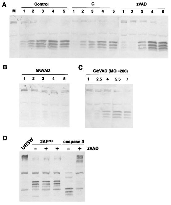
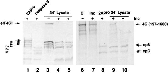
Cleavage of eukaryotic translation initiation factor 4GI (eIF4GI) is required for shutoff of host cell translation during poliovirus (PV) infection of HeLa cells. Reports published by several groups have led to confusion whether this cleavage is mediated by viral 2A protease (2A(pro)) or a putative cellular enzyme (termed eIF4Gase) which is activated by 2A(pro) or other aspects of viral infection. Here we have further investigated eIF4Gase activities in PV-infected cells. Column purification of eIF4GI cleavage activity separated two activities which generated N-terminal cleavage products of different lengths. Both activities were detected using either native eIF4G or radiolabeled recombinant eIF4G as the substrate. Analysis of cleavage products formed by each activity on native and mutant substrates suggests that one activity cleaves eIF4G1 at or very near the 2A(pro) cleavage site and the other activity cleaves approximately 40 residues upstream of the 2A(pro) cleavage site. When PV infections in HeLa cells were supplemented with 2 mM guanidine, which indirectly limits expression of 2A(pro), two distinct C-terminal cleavage fragments of eIF4GI were detected. These C-terminal cleavage fragments of eIF4GI were purified from infected cells, and a new eIF4GI cleavage site was mapped to a unique site 43 amino acids upstream of the known 2A(pro) cleavage site. Further, eIF4GI cleavage in vivo could be blocked by addition of zVAD to PV-guanidine infections. zVAD is a broad-spectrum caspase inhibitor which had no effect on 2A(pro) cleavage activity or PV polyprotein processing. Lastly, similar types of eIF4Gase cleavage activities were also detected in uninfected cells under various conditions, including early apoptosis or during cell cycle transit. The data suggest that the same types of eIF4GI cleavage activities which are generated in PV-infected cells can also be generated in the absence of virus. Taken together, the data support a model in which multiple cellular activities process eIF4GI in PV-infected cells, in addition to 2A(pro).


Figures

FIG. 1.
FIG. 1.
Schematic of eIF4GI complete amino acid sequence and isoforms derived from internal translation initiation at in-frame AUG codons. All isoforms of eIF4G are depicted cleaved at the known 2Apro cleavage site. The black box represents additional amino acids found in the new extended cDNA sequence. Hatched or gray boxes represent binding domains for the proteins PABP, eIF4E, eIF4A, eIF3, and mnk-1 as indicated. The mapped location of epitopes recognized by the polyclonal anti-eIF4GI antisera used in this study are also indicated. Numbers to the left of each isoform identify which amino acid in the open reading frame begins the isoforms, and the numbers to the right indicate total number of amino acids in the isoform, along with apparent mobility in SDS-polyacrylamide gels.
FIG. 2.
FIG. 2.
D gel analysis of eIF4GI cleavage fragments. (A) RSW from PV-infected HeLa cells (harvested at 4 h postinfection) was subjected to isoelectric focusing in the first dimension and SDS-PAGE in the second dimension, followed by immunoblot analysis with a mixture of antibodies specific for N-terminal or C-terminal domains of eIF4GI. Positions of isoelectric points (indicated above the panel) were determined by migration of known isoelectric focusing protein standards. The right panel shows migration of eIF4G cleavage products in the one-dimensional SDS-PAGE lane of the same gels. (B) Migration of CPCs from guanidine-PV infection. The right panel shows migration of eIF4G cleavage products in the one-dimensional SDS-PAGE lane of the same gels. (C) Appearance of eIF4GI-CPC doublet in guanidine infections. Shown is an enlarged view of the boxed region in panel B.
FIG. 3.
FIG. 3.
Chromatographic separation of different eIF4G cleavage activities. (A) Separation on m7GTP-Sepharose. Pooled partly purified material was applied to m7GTP-Sepharose, and fractions were eluted and assayed for eIF4G cleavage activity as described in Materials and Methods using antisera specific for N-terminal domain of eIF4GI. Immunoblot controls are URSW and PvRSW incubated alone for the period of the assay. The flowthrough fraction is in lane F, and eluted fractions are in lanes labeled with numbers. Arrows indicate CPN produced by eIF4Gase-α or 2Apro activity, and arrowheads indicate CPN cleavage products produced by eIF4Gase-β. (B) Pooled material from S300 Sepharose was applied to a nickel-affinity column, and then fractions were eluted with either pH 6.0 buffer (fraction 1) or 25 mM EDTA (fraction 2). Fractions were tested for eIF4G cleavage activity as described in Materials and Methods, and cleavage products were compared with 2Apro cleavage products by immunoblotting using antiserum specific for eIF4GI CPN (left panel) or eIF4GI CPC (right panel).
FIG. 4.
FIG. 4.
The eIF4Gase-β cleavage site is distinct from the 2Apro cleavage site. Defined radiolabeled eIF4G substrates were translated in vitro and then incubated with recombinant PV His-2Apro (0.5 μg), CVB3 2Apro, or eIF4Gase-β-enriched fractions (metal chelate pool) as indicated. Multiple smaller bands in starting substrate preparation result from aberrant internal initiation of translation on transcript RNA. Cleavage products produced by 2Apro are indicated by arrows, and cleavage products from eIF4Gase-β are indicated by arrowheads. The gray arrowhead indicates a minor eIF4Gase CPC not detected with native protein. WT indicates the wild-type 2Apro cleavage site in eIF4G substrate, whereas GE indicates the form of eIF4G containing a point mutation of Gly682 to Glu, which inhibits 2Apro cleavage.
FIG. 5.
FIG. 5.
Elution of eIF4G CPC from phosphocellulose. PV infections with or without guanidine were used as source cells for purification of eIF4GI-CPC as detailed in Materials and Methods. Pooled fractions chromatographed over P-11 phosphocellulose were analyzed by immunoblot analysis for eIF4GI CPC. Sequential chromatography fractions (numbered 17 to 19) from normal PV infections are shown (lanes 1 to 3) as well as equivalent fractions from two separate purifications from guanidine-supplemented infections (lanes 4 to 10). The left two panels are also counterstained with amido black to identify all protein in the sample.
FIG. 6.
FIG. 6.
Sequence analysis of eIF4GI CPC. (A) Purified eIF4GI-CPC was subjected to Edman degradation sequence analysis. Protein was purified from cells infected for 2.5 or 5 h before harvesting or cells from 4-h infections supplemented with guanidine-HCl. In addition, protein from PV-guanidine infections separated by SDS-PAGE into fast (lower band)- and slower (upper band)-migrating forms was analyzed individually. p.i., postinfection. (B) Comparison of known 2Apro cleavage sites on viral polyproteins and cellular proteins and a consensus recognition sequence.
FIG. 7.
FIG. 7.
Comparison of early eIF4Gase activities in normal and guanidine-inhibited PV infections. PV infections with (bottom panel) or without (top panel) guanidine were performed for 2.5 h, and eIF4Gase activity was sequentially purified on Sepharose S300, Fast Q Sepharose, and metal chelate columns. Immunoblot analysis (CPN specific) of eIF4Gase activity in fractions from the latter is shown. Arrows indicate cleavage products produced by 2Apro/eIF4Gase-α, and arrowheads indicate cleavage products produced by eIF4Gase-β. PV, RSW fraction from PV infected cells (4.5 h infection).
FIG. 8.
FIG. 8.
Combined treatment with guanidine and zVAD blocks development of eIF4Gase cleavage activity in PV-infected cells. (A) PV-infected cells (MOI = 20) were collected at the indicated time points postinfection or mock-infected cells were collected at 5 h (lane M) and analyzed for eIF4G cleavage by immunoblotting. Infections were unsupplemented (control) or supplemented with 2 mM guanidine-HCl (G) or 75 μM zVAD-fmk (zVAD) 10 min prior to infection. (B) Same as panel A, except cells were supplemented with both inhibitors. (C) Infection and analysis is conducted the same as for panel B, except the MOI was increased to 200. (D) Purified recombinant caspase 3 and 2Apro (0.5 μg each) were incubated with 5 μg of URSW for 90 min at 37°C before immunoblot analysis. Indicated reactions were supplemented with 200 μM zVAD.
FIG. 9.
FIG. 9.
Generation of eIF4Gase activities during early apoptosis. (A) HeLa cells treated with 25 mM cisplatin for the indicated time points were analyzed by immunoblotting for eIF4GI cleavage (left panel) or lysates were incubated with URSW for 6 h prior to immunoblot analysis to measure eIF4Gase activities in vitro. (B) eIF4Gase activation by various apoptosis inducers in K562 cells. K562 cells were mock treated or incubated with dexamethasone (1 μM), cisplatin (10 μM), or butyric acid (2.5 mM) for 16 h, and then lysates were analyzed by immunoblotting. (C) Extracts taken from HeLa cells treated with cisplatin (10 μM) for 16 h were incubated with URSW for 2 h at 37°C before immunoblot analysis. One assay also contained 10 μM DEVD-CHO. Controls included incubated and nonincubated URSW and extract from PV-infected cells. Arrows indicate eIF4Gase-α-type cleavage products, filled arrowheads indicate eIF4Gase-β- or -γ-type cleavage products, and open arrowheads indicate caspase 3 cleavage products.
FIG. 10.
FIG. 10.
IF4Gase activity in uninfected cell lysates. Uninfected HeLa cell S10 lysate was incubated at 34°C for 5 h to generate spontaneous eIF4Gase cleavage products (lane 3). URSW samples were also incubated with recombinant 2Apro or caspase 3 to generate specific cleavage products (lanes 1 and 2). Lysate in lane 3 (15 μg) was incubated an additional 8 h with 35S-radiolabeled 4G(197-1600) substrate (lanes 4, 5, 9, and 10) as indicated in Materials and Methods. The same 4G(197-1600) substrate was also held on ice, incubated in buffer alone, or incubated with 2Apro (lanes 6, 7, and 8, respectively).The left panel shows an eIF4GI-specific immunoblot. The right panel shows an autoradiograph of duplicate lanes on the same gel. The migration of cleavage products is indicated with arrows, keyed as explained in the legend to Fig. 9.

References

    1. Agol, V. I., G. A. Belov, K. Bienz, D. Egger, M. S. Kolesnikova, L. I. Romanova, L. V. Sladkova, and E. A. Tolskaya. 2000. Competing death programs in poliovirus-infected cells: commitment switch in the middle of the infectious cycle. J. Virol. 74:5534–5541. - PMC - PubMed
    1. Belsham, G., and R. Jackson. 2000. Translation initiation on picornavirus RNA, p. 869–900. In N. Sonenberg, J. Hershey, and M. Mathews (ed.), Translational control of gene expression. Cold Spring Harbor Laboratory Press, Cold Spring Harbor, N.Y.
    1. Benton, P. A., J. W. Murphy, and R. E. Lloyd. 1995. K562 cell strains differ in their response to poliovirus infection. Virology 213:7–18. - PubMed
    1. Bovee, M. L., B. Lamphear, R. E. Rhoads, and R. E. Lloyd. 1998. Direct cleavage of eIF4G by poliovirus 2A protease is inefficient in vitro. Virology 245:241–249. - PubMed
    1. Bovee, M. L., W. E. Marissen, M. Zamora, and R. E. Lloyd. 1998. The predominant eIF4G-specific cleavage activity in poliovirus-infected HeLa cells is distinct from 2A protease. Virology 245:229–240. - PubMed

Publication types

LinkOut - more resources