Comparative Study
. 2002 Jan;76(1):208-19.
doi: 10.1128/jvi.76.1.208-219.2002.

Cyclin T1 expression is mediated by a complex and constitutively active promoter and does not limit human immunodeficiency virus type 1 Tat function in unstimulated primary lymphocytes

Affiliations
Comparative Study

Cyclin T1 expression is mediated by a complex and constitutively active promoter and does not limit human immunodeficiency virus type 1 Tat function in unstimulated primary lymphocytes

Juan Martin-Serrano et al. J Virol. 2002 Jan.

Abstract

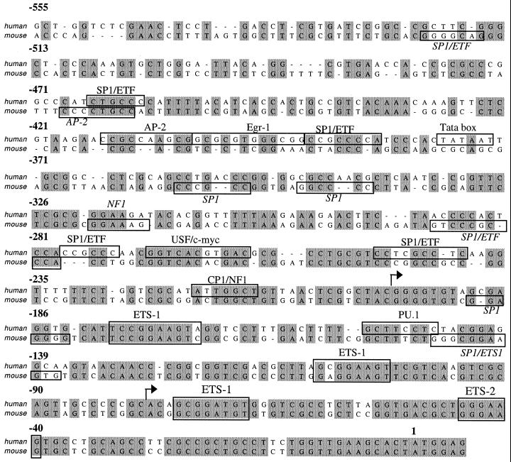
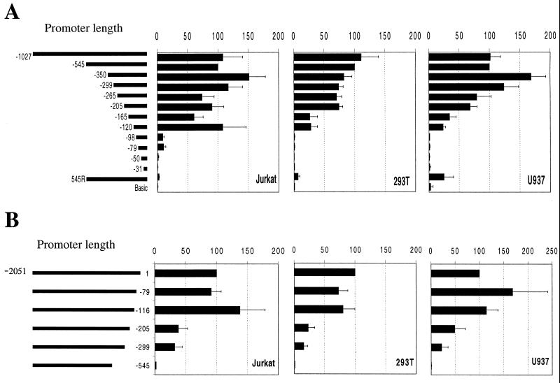
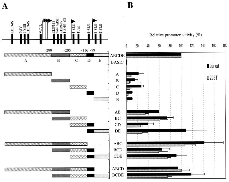
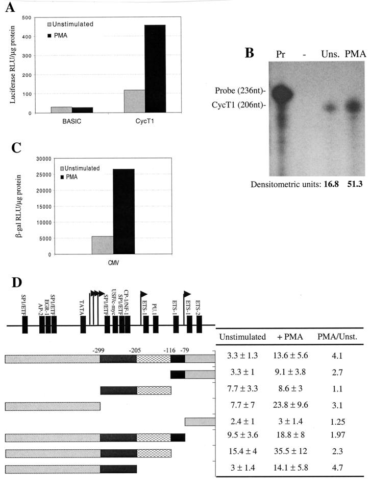
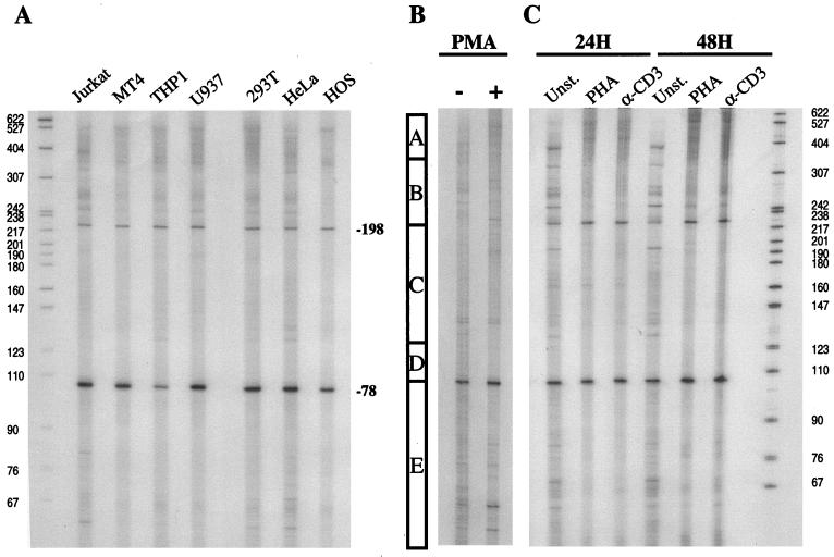
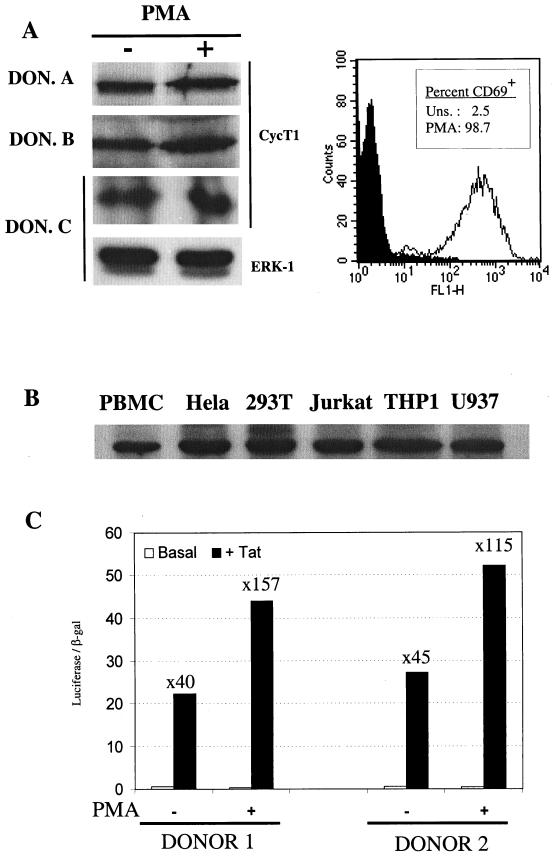
Cyclin T1 (CycT1), a component of positive-transcription-elongation factor-b (P-TEFb), is an essential cofactor for transcriptional activation by lentivirus Tat proteins. It is thought that low CycT1 expression levels restrict human immunodeficiency virus type 1 (HIV-1) expression levels and replication in resting CD4+ lymphocytes. In this study, we undertook a functional analysis of the cycT1 promoter to determine which, if any, promoter elements might be responsible for cellular activation state-dependent CycT1 expression. The cycT1 gene contains a complex promoter that exhibits an extreme degree of functional redundancy: five nonoverlapping fragments were found to exhibit significant promoter activity in immortalized cell lines, and these elements could interact in a synergistic or redundant manner to mediate cycT1 transcription. Reporter gene expression, mediated by the cycT1 promoter, was detectable in unstimulated transfected primary lymphocytes and multiple sites within the promoter could serve to initiate transcription. While utilization of these start sites was significantly altered by the application of exogenous stimuli to primary lymphocytes and two distinct promoter elements exhibited enhanced activity in the presence of phorbol ester, overall cycT1 transcription was only modestly enhanced in response to cell activation. These observations prompted a reexamination of CycT1 protein expression in primary lymphocytes. In fact, steady-state CycT1 expression is only slightly lower in unstimulated lymphocytes compared to phorbol ester-treated cells or a panel of immortalized cell lines. Importantly, CycT1 is expressed at sufficient levels in unstimulated primary cells to support robust Tat activity. These results strongly suggest that CycT1 expression levels in unstimulated primary lymphocytes do not profoundly limit HIV-1 gene expression or provide an adequate mechanistic explanation for proviral latency in vivo.


Figures

FIG. 1.
FIG. 1.
Conservation and divergence in putative transcription factor binding sites present in the human and murine cycT1 promoters. The sequence of the 545-nucleotide human cycT1 promoter is shown, aligned with the corresponding murine genomic sequence. Transcription factor binding sites, predicted by using MacVector (Oxford Molecular) or Transcription Element Search Software (TESS), are indicated by boxes. Arrows indicate major discrete sites of transcription initiation, as defined in Fig. 5.
FIG. 2.
FIG. 2.
Functional analysis of the cycT1 promoter in immortalized cell lines. Jurkat (T cells), 293T (epithelial), or U937 (monocytic) cell lines were transfected with pGL3-derived luciferase reporter plasmids. (A) The 5′ deletion series contains human genomic sequences from the CycT1 translation intiation site extending 5′ to the indicated nucleotide; 545R represents the complete 545-nucleotide promoter iserted into pGL3 in the reverse orientation. (B) The 3′ deletion series contains sequences from position −2051 relative to the CycT1 translation initiation site extending 3′ to the indicated nucleotide. In each case, cells were cotransfected with pBC12/CMV/lacZ. The luciferase and β-galactosidase activities were determined in cell lysates 48 h after transfection. Promoter activity is expressed as a percentage of the luciferase activity present in cells transfected with a reporter plasmid containing the 545-nucleotide promoter (determined, by this analysis, to contain the entire transcription promoting activity) and were normalized for minor variations in transfection efficiency, as determined from the β-galactosidase activity.
FIG. 3.
FIG. 3.
Combinatorial analysis of minimally active cycT1 promoter fragments in immortalized cell lines. (A) Minimal cycT1 promoter fragments, found to retain at least 10% of the intact promoter activity are termed modules A (−545 to −300), B (−299 to −206), C (−205 to −117), D (−116 to −80), and E (−79 to +1). These were analyzed individually or as all possible contiguous combinations of two, three, or four modules. (B) Luciferase activities resulting from transfection of Jurkat and 293T cells with reporter plasmids are expressed as a percentage of that obtained after transfection of a construct containing the intact 545-nucleotide cycT1 promoter.
FIG. 4.
FIG. 4.
Analysis of cycT1 promoter activity in primary lymphocytes. (A) Primary lymphocytes obtained from a normal donor were electroporated with pGL3-based plasmids either that lacked a promoter or contained the 545-nucleotide cycT1 promoter. (B) RNase protection analysis of endogenous cycT1 transcription with total RNA extracted from nontransfected, unsimulated (Uns) or PMA-treated primary lymphocytes. Lanes: Pr, undigested cycT1 probe; −, digested probe after hybridization with yeast tRNA. (C) Cells from the same donor as in panel A were transfected with pBC12/CMV/lacZ. For both panels A and C, cells were either left unstimulated or were treated with PMA after transfection. Luciferase or β-galactosidase activities in cell lysates were determined 24 h after electroporation and are representative of at least three independent experiments. (D) Primary lymphocytes were electroporated with reporter plasmids containing selected cycT1 promoter fragments, and cells either were left unstimulated or were treated with PMA. Results are presented as the fold increase in luciferase activity (± the standard deviation) over that obtained with a promoterless reporter plasmid and lymphocytes from three different donors.
FIG. 5.
FIG. 5.
Transcription start site utilization at the cycT1 promoter in immortalized cell lines and primary lymphocytes. (A) Total RNA was obtained from a panel of immortalized cell lines and subjected to primer extension analysis with an oligonucleotide proximal to the cycT1 translation start site (T1GP1; see Materials and Methods). Similar analyses were done with RNA extracted from primary lymphocytes that were either unstimulated or subjected to treatment with PMA for 24 h (B) or treatment with either PHA or αCD3 antibody for 24 and 48 h (C). The bar in panel B indicates the boundaries of the promoter modules A, B, C, and D as defined in Fig. 3.
FIG. 6.
FIG. 6.
CycT1 is expressed in unstimulated primary lymphocytes at levels sufficient to support Tat function. (A) Western blot analysis of CycT1 expression in primary lymphocytes obtained from three representative donors that either were unstimulated or were treated with PMA for 24 h. As a loading control, some blots were also probed with an anti-ERK-1 antibody. The panel to the right shows FACS analysis of unstimulated and PMA-treated lymphocytes with an anti-CD69 monoclonal antibody. (B) Comparative Western blot analysis of CycT1 expression levels in freshly isolated, unstimulated primary lymphocytes and a panel of immortalized cell lines known to support HIV-1 Tat function or virus replication. (C) Analysis of Tat function in unstimulated or PMA-treated primary lymphocytes. Data obtained with lymphocytes from two representative donors, electroporated with pHIV/luc, pBC12/CMV/lacZ, and either pcTat or pBC12/CMV is shown. The luciferase and β-galactosidase activities in cell lysates were determined 24 h after electroporation. For each experiment, the fold increase in luciferase expression in response to Tat is shown.

References

    1. Adams, M., L. Sharmeen, J. Kimpton, J. M. Romeo, J. V. Garcia, B. M. Peterlin, M. Groudine, and M. Emerman. 1994. Cellular latency in human immunodeficiency virus-infected individuals with high CD4 levels can be detected by the presence of promoter-proximal transcripts. Proc. Natl. Acad. Sci. USA 91:3862–3866. - PMC - PubMed
    1. Albrecht, T. R., L. H. Lund, and M. A. Garcia-Blanco. 2000. Canine cyclin T1 rescues equine infectious anemia virus tat trans-activation in human cells. Virology 268:7–11. - PubMed
    1. Alcami, J., T. Lain de Lera, L. Folgueira, M. A. Pedraza, J. M. Jacque, F. Bachelerie, A. R. Noriega, R. T. Hay, D. Harrich, R. B. Gaynor, et al. 1995. Absolute dependence on kappa B responsive elements for initiation and Tat-mediated amplification of HIV transcription in blood CD4 T lymphocytes. EMBO J. 14:1552–1560. - PMC - PubMed
    1. Antequera, F., and A. Bird. 1999. CpG islands as genomic footprints of promoters that are associated with replication origins. Curr. Biol. 9:R661–R667. - PubMed
    1. Barboric, M., R. Taube, N. Nekrep, K. Fujinaga, and B. M. Peterlin. 2000. Binding of Tat to TAR and recruitment of positive transcription elongation factor b occur independently in bovine immunodeficiency virus. J. Virol. 74:6039–6044. - PMC - PubMed

Publication types

MeSH terms