. 2002 Jan;22(1):1-11.
doi: 10.1128/MCB.22.1.1-11.2002.

Protein kinase A regulates sexual development and gluconeogenesis through phosphorylation of the Zn finger transcriptional activator Rst2p in fission yeast

Affiliations

Protein kinase A regulates sexual development and gluconeogenesis through phosphorylation of the Zn finger transcriptional activator Rst2p in fission yeast

Toru Higuchi et al. Mol Cell Biol. 2002 Jan.

Abstract

Protein kinase A (PKAi a cyclic AMP-dependent protein kinase) negatively regulates sexual development and gluconeogenesis in fission yeast by suppressing the transcription of ste11 required for the former and the transcription of fbp1 required for the latter. Here we show that Rst2p, a zinc finger protein that can bind to the upstream region of ste11 and fbp1 via the STREP motif, mediates the activity of PKA to transcription of these genes. A simple reporter system confirmed that PKA could cause its negative effect on transcription through the combination of Rst2p and STREP. Rst2p was phosphorylated by PKA in vitro at two consensus sequences on it. Substitution of the target threonine residues by alanine made the protein active even in the presence of high PKA activity. Rst2p underwent hyperphosphorylation in the medium lacking glucose, and PKA inhibited this hyperphosphorylation. Rst2p was mainly cytoplasmic under high PKA activity but was concentrated in the nucleus when this activity was lowered, suggesting that PKA might regulate ste11 and fbp1 negatively by excluding Rst2p from the nucleus. However, the shift of Rst2p localization was not perfect under physiological conditions, leaving the possibility that PKA inhibits Rst2p function in another way as well. Although the PKA-Rst2p-STREP pathway is apparently central to the regulation of ste11 and fbp1 transcription in accordance with nutritional conditions, some additional paths are likely to connect nitrogen to repression of ste11 and glucose to repression of fbp1. These paths may ensure the specificity between the type of nutrients in shortage and the type of genes to be expressed.


Figures

FIG. 1.
FIG. 1.
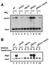
The rst2 gene function is required for transcription of the fbp1 gene. (A) Induction of fbp1 transcription by a medium shift. Cells, either rst2+ (JY333) or rst2Δ (JX233), were grown in MMR containing 8% glucose to a concentration of 5 × 10⁶ cells/ml and then shifted to MMD containing 3% glycerol and 0.1% glucose. Cells were then harvested at the indicated times after the shift, and the expression of fbp1 and leu1 as an internal control was examined by Northern blotting. RNA (5 μg) was loaded in each lane after denaturation by formamide. (B) Alignment of STREP sequences, recognized by Rst2p, in the upstream activation regions of ste11 and fbp1. The numbers refer to positions relative to the transcriptional start.
FIG. 2.
FIG. 2.
Effects of PKA-related mutations on expression of ste11 and fbp1. (A) To examine ste11 expression, cells of each genotype indicated (wild type [wt], JY333; rst2Δ, JX233; cgs1Δ, JX222; pka1Δ, JX384; pka1Δ rst2Δ, JX282) were grown in MM supplemented with ammonium chloride (+), and some of them were subjected to starvation of nitrogen for 2 h (−). Total RNA was extracted from each preparation and analyzed by Northern blotting. RNA (5 μg) was loaded in each lane after denaturation by formamide. Expression of ste11 and leu1 was detected by appropriate probes. (B) To examine fbp1 expression, cells of each strain (the same as in panel A) were grown in glucose-based MMR (R), and some of them were shifted to glycerol-based MMD (D) and incubated for 1 h. Expression of fbp1 was detected as described in panel A.
FIG. 3.
FIG. 3.
An Rst2p- and STREP-dependent reporter system. The lacZ-reporter plasmids used here are constructed as described in Materials and Methods. Various strains as indicated (as defined in Fig. 2) were transformed with a reporter plasmid carrying either the perfect or a mutated STREP sequence. Lysates were prepared from cells grown in MMR (open bars) or derepressed in MMD for 3 h (shaded bars). An assay of β-galactosidase activity was done as described in Materials and Methods. An average of at least three independent measurements, performed with stable transformants, is shown for each case.
FIG. 4.
FIG. 4.
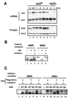
Rst2p activity is likely to be regulated posttranscriptionally. (A) Detection of rst2 mRNA and the gene product before and after a medium shift. To determine the level of rst2 mRNA, extracts were prepared from cells grown in MMD for the indicated duration, as detailed in Fig. 1. Poly(A) RNA was selected from each sample and subjected to gel electrophoresis after denaturation with formamide. We detected leu1 mRNA as an internal loading control (the time for exposure on X-ray film is much shorter than that for rst2 mRNA). For analysis of protein, boiled cell lysates were subjected to SDS-PAGE, followed by immunoblot analysis as described in Materials and Methods. (B) Phosphatase treatment of Rst2p. Protein precipitates prepared from JY333 cells either growing in MMR or incubated in MMD for 1 h were treated with calf intestinal alkaline phosphatase (CIP), with or without the addition of the inhibitor mix, and subjected to SDS-PAGE and immunoblot analysis. (C) Detection of wild-type and mutated Rst2p in PKA-related mutant cells under repressing or derepressing conditions. The M3 mutant form of Rst2p lacks phosphorylatable residues at the two putative PKA-target sites. Cells of JX233 (rst2Δ), JY333 (WT), JW373 (rst2-M3), JX222 (cgs1Δ), JW374 (cgs1Δ rst2-M3), JX384 (pka1Δ), and JW377 (pka1Δ rst2-M3) were grown in MMR. A portion of each culture was shifted to MMD and incubated for 1 h. Cell lysates were prepared from the cultures in MMR and MMD and then subjected to SDS-PAGE and immunoblot analysis.
FIG. 5.
FIG. 5.
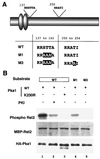
Phosphorylation of Rst2p by PKA in vitro. (A) Schematic diagram of Rst2p showing consensus sequences for PKA phosphorylation and a table of mutations introduced into bacterially expressed Rst2p used for in vitro phosphorylation assay. Zinc finger motifs are shown as ovals. (B) Audoradiogram showing phosphorylation of bacterially expressed Rst2p, fused with MBP. The engineered proteins were incubated with [γ-32P]ATP and PKA immunoprecipitated from fission yeast cells. A kinase-negative PKA variant (K230R) was used in lane 3. The PKA inhibitor 5-24 (PKI) was added to lane 2.
FIG. 6.
FIG. 6.
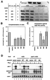
Unphosphorylatable mutations in the PKA target sites enhances the activity of Rst2p. (A) Iodine-staining assay of sporulation efficiency in various rst2 transformants. Cells of JX231 (h90 cgs1+ rst2Δ) and JW487 (h90 cgs1Δ rst2Δ) were transformed with pREP81-based plasmids carrying each allele as indicated on the left. They were incubated in patches on SSA plates for 4 days and stained with iodine vapor, which turns sporulated cells dark brown. JY450 cells transformed with the vector plasmid were used as a control. (B) JX231 transformants, shown in panel A, were examined microscopically for their conjugation efficiency. (C) JW487 transformants, shown in panel A, were examined microscopically for their conjugation efficiency. (D) Restoration of ste11 transcription in cgs1Δ cells by the rst2-M3 allele. Total RNA was prepared from heterothallic (h) cgs1+ rst2Δ (JX233) or cgs1Δ rst2Δ (JX261) cells transformed with a pREP81-based plasmid carrying either the rst2+ or the rst2-M3 allele. Cells were either growing exponentially in MM (+) or were subjected to nitrogen starvation for 4 h (−). RNA (5 μg) was loaded in each lane after denaturation by formamide and analyzed by Northern blotting. Expression of cam1 and plasmid-borne rst2 was measured as an internal control and for confirmation, respectively.
FIG. 7.
FIG. 7.
Effects of glucose on subcellular localization of Rst2p. (A) Immunostaining of Rst2-3HAp. Cells (JW367) grown in MMR (R panels) were harvested by filtration, transferred to MMD, and incubated for 1 h (D panels). They were stained with anti-HA antibody and counterstained with Hoechst 33342 to visualize the DNA. (B) Addition of glucose promotes exclusion of Rst2p from the nucleus. JW352 cells expressing Rst2p-GFP were adapted in MMD for an hour (no addition). Glucose was added to a portion of the culture to the final concentration of 4% (+Glucose). To block exportin-dependent nuclear export, leptomycin B (0.1 μg/ml) was added simultaneously to a portion (+Glucose +LMB). DNA was stained with Hoechst 33342. Bar, 10 μm. (C) Subcellular localization of Rst2p-GFP in PKA-related mutants. Cells of JW363 lacking PKA (pka1Δ) and cells of JW359 with constitutively active PKA (cgs1Δ), both of which were grown in MM, were observed for GFP fluorescence. Cells of JW355 lacking adenylyl cyclase were first grown in MM containing 2 mM cAMP and 5 mM caffeine to block phosphodiesterase (cyr1Δ + cAMP). They were filtered, transferred to cAMP-free MM, and incubated for 10 min (cyr1Δ). DNA was stained with Hoechst 33342. The presence of caffeine somehow weakened staining of DNA by Hoechst 33342. Bars, 10 μm.
FIG. 8.
FIG. 8.
Quantitative measurements of subcellular localization of the wild-type Rst2p and the M3 mutant under the repressing and derepressing conditions. Rst2p and Rst2-M3p, both tagged with HA, were expressed from the expression vector pREP81 in cells that were either cgs1+ rst2Δ (JX 233) or cgs1Δ rst2Δ (JX 261). Cells cultured in MMR or shifted to MMD (30 min) were stained with anti-HA antibody, and the percentage of cells showing clear nuclear accumulation of Rst2p was culculated for each sample. Bar, 10 μm.
FIG. 9.
FIG. 9.
Diagram of the regulatory network involving the PKA-Rst2p pathway for transcriptional activation of ste11 and fbp1. The pathway established in this study is shown in thick lines. Other possible paths are shown in thin lines.

Similar articles

Cited by

References

    1. Alfa, C., P. A. Fantes, J. Hyams, M. McLeod, and E. Warbrick. 1993. Experiments with fission yeast: a laboratory course manual. Cold Spring Harbor Laboratory Press, Cold Spring Harbor, N.Y.
    1. Bähler, J., J. Q. Wu, M. S. Longtine, N. G. Shah, A. McKenzie III, A. B. Steever, A. Wach, P. Philippsen, and J. R. Pringle. 1998. Heterologous modules for efficient and versatile PCR-based gene targeting in Schizosaccharomyces pombe. Yeast 14:943–951. - PubMed
    1. Byrne, S. M., and C. S. Hoffman. 1993. Six git genes encode a glucose-induced adenylate cyclase activation pathway in the fission yeast Schizosaccharomyces pombe. J. Cell Sci. 105:1095–1100. - PMC - PubMed
    1. Cherry, J. R., T. R. Johnson, C. Dollard, J. R. Shuster, and C. L. Denis. 1989. Cyclic AMP-dependent protein kinase phosphorylates and inactivates the yeast transcriptional activator ADR1. Cell 56:409–419. - PubMed
    1. Denis, C. L., and E. T. Young. 1983. Isolation and characterization of the positive regulatory gene ADR1 from Saccharomyces cerevisiae. Mol. Cell. Biol. 3:360–370. - PMC - PubMed

Publication types

MeSH terms

LinkOut - more resources