. 2002 Jan;22(1):12-22.
doi: 10.1128/MCB.22.1.12-22.2002.

c-Jun and hypoxia-inducible factor 1 functionally cooperate in hypoxia-induced gene transcription

Affiliations

c-Jun and hypoxia-inducible factor 1 functionally cooperate in hypoxia-induced gene transcription

Arántzazu Alfranca et al. Mol Cell Biol. 2002 Jan.

Abstract

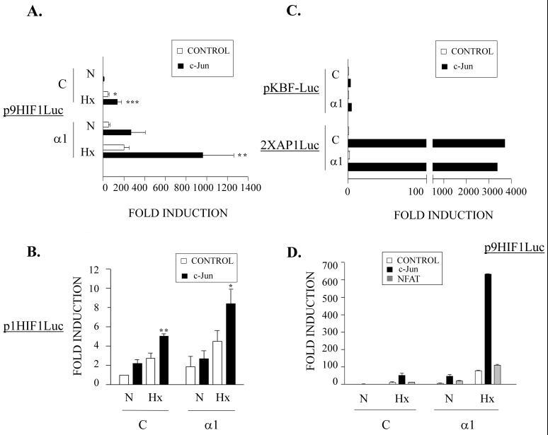
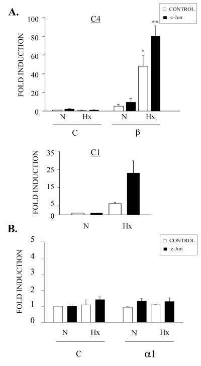
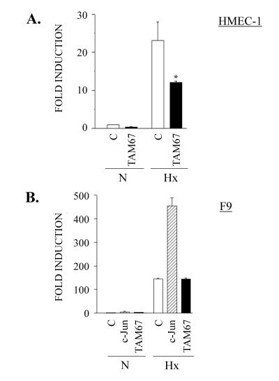
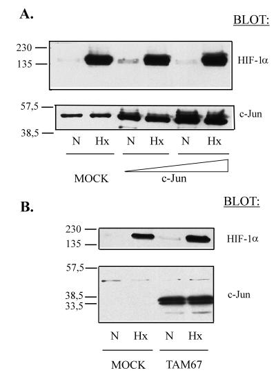
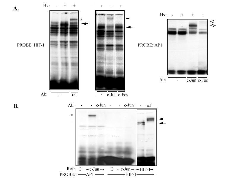
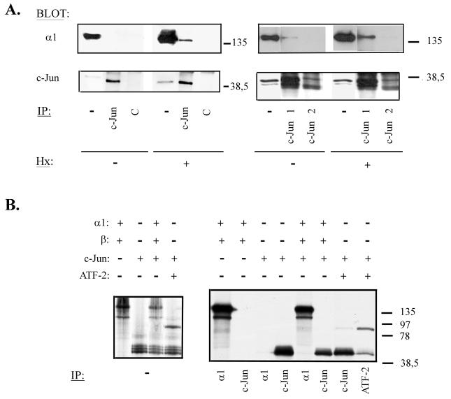
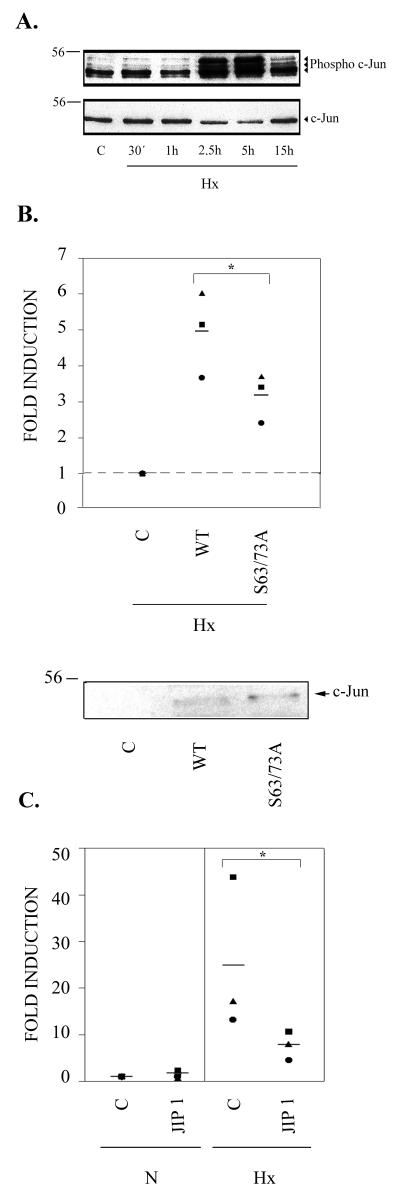
Under low-oxygen conditions, cells develop an adaptive program that leads to the induction of several genes, which are transcriptionally regulated by hypoxia-inducible factor 1 (HIF-1). On the other hand, there are other factors which modulate the HIF-1-mediated induction of some genes by binding to cis-acting motifs present in their promoters. Here, we show that c-Jun functionally cooperates with HIF-1 transcriptional activity in different cell types. Interestingly, a dominant-negative mutant of c-Jun which lacks its transactivation domain partially inhibits HIF-1-mediated transcription. This cooperative effect is not due to an increase in the nuclear amount of the HIF-1alpha subunit, nor does it require direct binding of c-Jun to DNA. c-Jun and HIF-1alpha are able to associate in vivo but not in vitro, suggesting that this interaction involves the participation of additional proteins and/or a posttranslational modification of these factors. In this context, hypoxia induces phosphorylation of c-Jun at Ser(63) in endothelial cells. This process is involved in its cooperative effect, since specific blockade of the JNK pathway and mutation of c-Jun at Ser(63) and Ser(73) impair its functional cooperation with HIF-1. The functional interplay between c-Jun and HIF-1 provides a novel insight into the regulation of some genes, such as the one for VEGF, which is a key regulator of tumor angiogenesis.


Figures

FIG. 1.
FIG. 1.
Cooperative effect of c-Jun on HIF-1 transcriptional activity. (A) c-Jun-defective F9 teratocarcinoma cells were transiently transfected with 0.5 μg of the p9HIF1Luc luciferase reporter plasmid together with 1 μg of pcDNA3α1 (α1) or the empty vector pcDNA3 (C) and pRSV c-Jun (solid bars) or control pUCRSV empty vector (open bars). After transfection, the cells were incubated in 1% O2 (Hx) or 21% O2 (N) for 12 h and then analyzed for luciferase activity. (B) F9 cells were transfected and processed as for panel A, but the luciferase reporter plasmid was p1HIF1Luc. The data in panels A and B are the means + standard errors of the mean of five independent experiments performed in duplicate. ∗, P < 0.05; ∗∗, P < 0.01; ∗∗∗, P < 0.001 compared to controls in normoxia. (C) F9 cells were transfected as described above, but the luciferase reporter plasmids were pKBF-luc and 2XAP1Luc. (D) F9 cells were transiently transfected with the p9HIF1Luc luciferase reporter plasmid (0.5 μg) together with 1 μg of pcDNA3α1 (α1) or control pcDNA3 (C) and expression vectors for c-Jun (solid bars), NFATc (shaded bars), or control empty vector (open bars) and processed as for panel A. The data are the means + standard deviations of a representative experiment out of three performed in duplicate.
FIG. 2.
FIG. 2.
Lack of c-Jun cooperative effect in the absence of a functional HIF-1 or a preserved HIF-1-response element. (A) (Top) HIF-1β-defective Hepa-1c4 mouse hepatoma cells (C4) were transfected with 0.25 μg of p9HIF1Luc reporter plasmid together with 0.5 μg of pARNT (β) or the empty vector pcDNA3 (C) and pRSV c-Jun (solid bars) or control pUCRSV empty vector (open bars). Sixteen hours after transfection, the cells were grown under normoxic (N) or hypoxic (Hx) conditions for 8 h, and luciferase activity was determined. The histogram shows the means + standard errors of the mean of three independent experiments performed in duplicate; ∗, P < 0.05, and ∗∗, P < 0.001 compared to control in normoxia. (Bottom) The Hepa-1c1c7 mouse hepatoma cell line (C1) was transfected with the p9HIF1Luc reporter plasmid together with pRSV c-Jun (solid bars) or control pUCRSV empty vector (open bars) and processed as C4 cells. The data are means + standard deviations of a representative experiment out of four performed in triplicate. (B) F9 cells were transfected with 0.5 μg of p1HIF1mLuc luciferase reporter plasmid (which contains a mutated HRE) together with 1 μg of pcDNA3 α1 (α1) or empty vector pcDNA3 (C) and pRSV c-Jun (solid bars) or control pUCRSV empty vector (open bars). After transfection, the cells were processed as for panel A. The data are means + standard errors of the mean of three independent experiments performed in duplicate.
FIG. 3.
FIG. 3.
Effect of c-Jun dominant-negative mutant on HIF-1-dependent transcription. (A) The HMEC-1 endothelial cell line was transiently transfected with 0.2 μg of p9HIF1Luc reporter plasmid together with 0.2 μg of pCMV TAM67 (TAM 67), or control vector pCMV (C). Sixteen hours after transfection, the cells were either left untreated (N) or grown in 1% O2 for 8 h (Hx) and analyzed for luciferase activity. The data are means + standard errors of the mean of four independent experiments performed in duplicate. ∗, P < 0.05 compared to control in hypoxia. (B) F9 cells were transiently transfected with 0.5 μg of p9HIF1Luc together with 1 μg of pRSVc-Jun (c-Jun), pCMV TAM67, or control empty vector. After transfection, the cells were incubated under normoxic (N) or hypoxic (Hx) conditions for 12 h, and then luciferase activity was measured. The data are means plus standard errors of the mean of three independent experiments performed in duplicate.
FIG. 4.
FIG. 4.
Effect of c-Jun or its dominant-negative mutant on HIF-1α expression. (A) COS-7 cells were transfected with control empty vector (MOCK) or increasing amounts of c-Jun expression vector (c-Jun). After transfection, the cells were grown under normoxic (N) or hypoxic (Hx) conditions for 5 h. Nuclear extracts were obtained and subjected to immunoblotting (20 μg per lane) with an anti-HIF-1α antibody (top) or an anti-c-Jun antibody (bottom). (B) COS-7 cells were transfected with 8 μg of control empty vector (MOCK) or pCMV TAM67 (TAM67) and processed as for panel A. Molecular weight markers are shown on the left.
FIG. 5.
FIG. 5.
c-Jun binding to VEGF 5′ UTR HIF-1 consensus sequence in endothelial cells. (A) Electrophoretic mobility shift assay of nuclear extracts obtained from primary endothelial cells either untreated or subjected to hypoxia (Hx) for 4 h; these extracts were incubated (3 μg per lane) with a 32P-labeled probe which contains the HIF-1 DNA binding consensus sequence of the VEGF 5′ UTR (HIF-1; 0.5 ng per lane), showing a hypoxia-inducible DNA-protein complex (arrows). In some cases, nuclear extracts were incubated with specific antibodies (Ab) against the HIF-1α subunit (α1) (left) or c-Jun or c-Fos (middle) before the probe was added. The autoradiographs show supershifted complexes with anti-HIF-1α (asterisk) and c-Jun (arrowhead) antibodies, whereas anti-c-Fos had no effect; (right) electrophoretic mobility shift assay performed as described above but with a 32P-labeled probe containing an AP-1 consensus sequence from the CD11c promoter (AP-1) used as a positive control. Supershifted complexes can be observed with both anti-c-Jun (open arrow) and anti c-Fos (open arrowhead) antibodies. (B) In vitro-translated (Ret.) pcDNA3 (C), c-Jun, or HIF-1 (α1 plus β subunits) was incubated with the same labeled probes as for panel A (1 ng per lane). Specific complexes with AP-1 (asterisk) and HIF-1 (arrow) probes could be observed, which could be identified, respectively, as c-Jun and the HIF-1 α subunit (arrowhead) with specific antibodies (Ab) against these factors. +, present; −, absent.
FIG. 6.
FIG. 6.
Association between c-Jun and HIF-1α. (A) COS-7 cells were cotransfected with 8 μg of pcDNA3α1 and pRSV c-Jun (left) or pCMV TAM67 (right) expression vector. After 40 h, the cells were incubated in normoxia or hypoxia (Hx) for 4 h, and nuclear extracts were obtained. These extracts were immunoprecipitated (IP) with polyclonal antibodies against the DNA binding domain (c-Jun and c-Jun 1) or the transactivation domain (c-Jun 2) of c-Jun or a control antibody (C) and immunoblotted with anti-HIF-1α (top) and anti-c-Jun (bottom) antibodies; as a control for protein expression, aliquots of the lysates (representing 1/10 of each immunoprecipitation reaction) (−) were also subjected to Western blotting with antibodies against HIF-1α and c-Jun. (B) HIF-1α (α1), c-Jun, HIF-1β (β), and ATF-2 were in vitro translated or cotranslated in the presence of [35S]methionine and immunoprecipitated with antibodies against HIF-1α (α1), c-Jun, or ATF-2. The immunocomplexes were analyzed by SDS-PAGE (right); the autoradiograph on the left shows 1/10 of each translation reaction as a control. +, present; −, absent. Molecular weight markers are shown on the right.
FIG. 7.
FIG. 7.
Involvement of JNK pathway in functional cooperation between c-Jun and HIF-1. (A) Western blot analysis of phosphorylated c-Jun in endothelial cells under hypoxic conditions. (Top) Nuclear extracts were obtained from HUVEC grown in 1% O2 (Hx) for 30 min (30′) and 1, 2.5, 5, and 15 h or under normoxic conditions (C) and subjected to immunoblotting with an antibody which specifically recognizes c-Jun phosphorylated at Ser63. (Bottom) The same membrane was stripped and reblotted with an anti-c-Jun antibody to show equivalent amounts of protein in each lane. (B) (Top) F9 cells were transfected with 0.5 μg of p9HIF1Luc luciferase reporter plasmid, together with 1 μg of pRSVc-Jun (WT), pRSVc-Jun S63A-S73A (S63/73A), or pUCRSV control vector (C); after transfection, the cells were grown under hypoxic conditions (Hx) for 16 h, and luciferase activity was determined. Data from three separate experiments are shown, normalized to control in hypoxia (set as 1); the bars represent the means of the different values for each experimental condition. ∗, P < 0.05. (Bottom) F9 cells were transfected as described above, and nuclear extracts were obtained and subjected to immunoblotting (20 μg per lane) with a specific antibody against c-Jun. Molecular weight markers are shown on the left. (C) The HMEC-1 endothelial cell line was transiently transfected with 0.2 μg of p9HIF1Luc reporter plasmid together with 0.2 μg of JIP1 or control vector (C). Sixteen hours after transfection, the cells were either left untreated (N) or grown in 1% O2 for 8 h (Hx) and analyzed for luciferase activity. Data from three separate experiments are shown, normalized to control in normoxia; the bars represent the means of the different values for each experimental condition. ∗, P < 0.05.

References

    1. Aragones, J., D. R. Jones, S. Martin, M. A. San Juan, A. Alfranca, F. Vidal, A. Vara, I. Merida, and M. O. Landazuri. 2001. Evidence for the involvement of diacylglycerol kinase in the activation of hypoxia-inducible transcription factor-1 by low oxygen tension. J. Biol. Chem. 276:10548–10555. - PubMed
    1. Arany, Z., L. E. Huang, R. Eckner, S. Bhattacharya, C. Jiang, M. A. Goldberg, H. F. Bunn, and D. M. Livingston. 1996. An essential role for p300/CBP in the cellular response to hypoxia. Proc. Natl. Acad. Sci. USA 93:12969–12973. - PMC - PubMed
    1. Ausserer, W. A., B. Bourrat-Floeck, C. J. Green, K. R. Laderoute, and R. M. Sutherland. 1994. Regulation of c-jun expression during hypoxic and low-glucose stress. Mol. Cell. Biol 14:5032–5042. - PMC - PubMed
    1. Bandyopadhyay, R. S., M. Phelan, and D. V. Faller. 1995. Hypoxia induces AP-1-regulated genes and AP-1 transcription factor binding in human endothelial and other cell types. Biochim. Biophys. Acta 1264:72–78. - PubMed
    1. Bannister, A. J., T. Oehler, D. Wilhelm, P. Angel, and T. Kouzarides. 1995. Stimulation of c-Jun activity by CBP: c-Jun residues Ser63/73 are required for CBP induced stimulation in vivo and CBP binding in vitro. Oncogene 11:2509–2514. - PubMed

Publication types

MeSH terms