. 2002 Jan;22(1):41-56.
doi: 10.1128/MCB.22.1.41-56.2002.

Cytoplasmic death signal triggered by SRC-mediated phosphorylation of the adenovirus E4orf4 protein

Affiliations

Cytoplasmic death signal triggered by SRC-mediated phosphorylation of the adenovirus E4orf4 protein

Marie-Claude Gingras et al. Mol Cell Biol. 2002 Jan.

Abstract

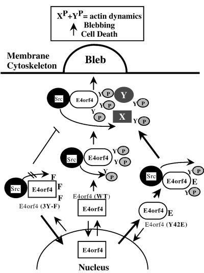
In transformed cells, the adenovirus E4orf4 death factor works in part by inducing a Src-mediated cytoplasmic apoptotic signal leading to caspase-independent membrane blebbing and cell death. Here we show that Src-family kinases modulate E4orf4 phosphorylation on tyrosine residues. Mutation of tyrosines 26, 42, and 59 to phenylalanines inhibited Src-induced phosphorylation of E4orf4 in vivo and in vitro but had no effect on the molecular association of E4orf4 with Src. However, in contrast to wild-type E4orf4, the nonphosphorylatable E4orf4 mutant was unable to modulate Src-dependent phosphorylation and was deficient in recruiting a subset of tyrosine-phosphorylated proteins. Indeed, the Src substrates cortactin and p62dok were found to associate with wild-type E4orf4 but not with the nonphosphorylatable E4orf4. Importantly, the nonphosphorylatable mutant E4orf4 was preferentially distributed in the cell nucleus, was unable to induce membrane blebbing, and had a highly impaired killing activity. Conversely, an activated form of E4orf4 was obtained by mutation of tyrosine 42 to glutamic acid. This pseudophosphorylated mutant E4orf4 was enriched in the cytoplasm and plasma membrane, showed increased binding to phosphotyrosine-containing proteins, and induced a dramatic blebbing phenotype associated with increased cell death. Altogether, our findings strongly suggest that Src-mediated phosphorylation of adenovirus type 2 E4orf4 is critical to promoting its cytoplasmic and membrane localization and is required for the transduction of E4orf4-Src-dependent induction of membrane blebbing. We propose that E4orf4 acts in part by uncoupling Src-dependent signals to drive the formation of a signaling complex that triggers a cytoplasmic death signal.


Figures

FIG. 1.
FIG. 1.
Modulation of Ad2 E4orf4 tyrosine phosphorylation by Src kinase activity. (A) 293T cells were transfected with empty vector (EV) or Flag-E4orf4 with or without c-src, kinase-deficient c-src(K295M), or v-src. Twenty-four hours after transfection, immunoprecipitations were performed with anti-Flag antibody. E4orf4 immune complexes were analyzed by Western blotting using anti-Flag and antiphosphotyrosine (P-Tyr; PY20) antibodies. (B) Twenty-four hours after transfection of 293T cells with the indicated constructs, cells were metabolically labeled with 32P, and when indicated, an inhibitor of tyrosine phosphatases (Na3VO4, 0.1 mM) or a selective Src kinase inhibitor (PP2, 50 μM) was added during the labeling period. Cells were then processed for immunoprecipitation, and phosphorylation levels were detected by autoradiography. The levels of immunoprecipitated E4orf4 were determined by Western blotting using anti-Flag antibody, and phosphorylation levels were quantified with NIH Image and corrected for E4orf4 levels in immune complexes. R.U., relative units; DMSO, dimethyl sulfoxide. (C) In vitro kinase assays were performed with either precipitated v-src or kinase-deficient c-src(K295R) from transfected 293T cells, incubated with either purified GST or GST-E4orf4. Labeled reaction mixtures were resolved by SDS-PAGE and visualized by autoradiography ([γ-32P]ATP) or analyzed by Western blotting after electrotransfer onto nitrocellulose using antiphosphotyrosine antibody (P-Tyr).
FIG. 2.
FIG. 2.
Mutation of tyrosines 26, 42, and 59 to phenylalanines inhibits Src-mediated phosphorylation of Ad2 E4orf4. (A) Various E4orf4 proteins were obtained by mutation of the indicated tyrosine residue(s) to phenylalanine by site-directed mutagenesis. The Flag-E4orf4 constructs were transfected into 293T cells, and 24 h after transfection, E4orf4 expression levels were analyzed by Western blotting of equal amounts of total cell lysates using anti-Flag antibody. Ponceau staining of the gel is shown as a loading control. EV, empty vector. (B) 293T cells were transfected with empty vector (EV), wild-type Flag-E4orf4 (WT), or mutant Flag-E4orf4 bearing Y-F substitution(s) of the indicated tyrosine residue(s), together with v-src or activated c-src(Y527F). Twenty-four hours after transfection, equal amounts of cell extracts were processed for immunoprecipitation with anti-Flag antibody. E4orf4 immune complexes were resolved by SDS-PAGE, and tyrosine-phosphorylated E4orf4 (P-Tyr) was detected by Western blotting with antiphosphotyrosine antibody (PY20). The levels of Flag-E4orf4 in immune complexes (IP), and levels of transfected v-src in total lysates (TL) were analyzed by Western blotting with anti-Flag M2 and anti-Src (Ab-1), respectively. The phosphotyrosine levels of E4orf4 mutants were quantified from scanned enhanced chemiluminescence-derived images by densitometric analysis with NIH Image and were corrected for the amount of precipitated Flag-E4orf4 proteins. The results are expressed as percent tyrosine-phosphorylated E4orf4 relative to wild-type (WT) E4orf4 and are the means ± SE of five independent experiments. (C) In vitro kinase assays were performed with precipitated c-src(Y527F) from transfected 293T cells incubated with either purified GST, wild-type GST-E4orf4, or mutant GST-E4orf4(3Y-F). Labeled reaction mixtures were resolved by SDS-PAGE and visualized by autoradiography ([γ-32P]ATP).
FIG. 3.
FIG. 3.
Src-mediated phosphorylation of Ad2 E4orf4 is not required for molecular association of E4orf4 with Src. (A) Pull-down assays were performed with lysates of 293T cells transfected with either wild-type Flag-E4orf4 (WT), Flag-E4orf4(3Y-F), polyomavirus middle-T antigen, or Flag-GFP (left panels) or cotransfected with activated c-src(Y527F) and either Flag-E4orf4, Flag-E4orf4(3Y-F), or Myc-FAK (right panels). Cell lysates were incubated with 10 μg of purified GST or the indicated GST-c-src fusion proteins bound to glutathione-Sepharose. The bound material was analyzed by Western blotting with anti-Flag M2, anti-middle-T antigen, or anti-c-myc to detect the amounts of absorbed E4orf4 or GFP, middle-T antigen, and FAK, respectively. The input lanes (TL) represent 2% of the total extracts. (B) 293T cells were transfected with kinase-deficient c-src(K295R) together with either vector alone (EV), wild-type Flag-E4orf4, or Flag-E4orf4(3Y-F). Twenty-four hours after transfection, immunoprecipitations were performed with anti-Flag M2, and equal amounts of immune complexes were analyzed by Western blotting using anti-Flag M2 and anti-Src (SRC2) to reveal E4orf4 proteins and coprecipitated kinase-deficient c-src, respectively. The amounts of transfected c-src(K295R) in total cell lysates (TL) are shown.
FIG. 4.
FIG. 4.
Modulation of Src-dependent phosphorylation by Ad2 E4orf4 requires tyrosine phosphorylation of E4orf4. (A) 293T cells were transfected with activated c-src alone (EV) or together with either wild-type E4orf4 (WT) or nonphosphorylatable E4orf4(3Y-F). Equal amounts of total cell extracts 24 h after transfection were analyzed by Western blotting using antiphosphotyrosine (P-Tyr, RC20-HRP), anti-Src (SRC2), or anti-Flag M2. Arrows indicate proteins whose Src-dependent phosphorylation is upregulated in cells expressing wild-type E4orf4 but not the nonphosphorylatable E4orf4(3Y-F). (B) 293T cells were transfected as for panel A, and 24 h after transfection, cells were lysed in modified RIPA buffer. Equal amounts of cell lysates were processed for immunoprecipitation of E4orf4 proteins using anti-Flag M2, and immune complexes were analyzed by Western blotting using anti-Flag M2, anti-Src (SRC2), and antiphosphotyrosine (RC20-HRP) to reveal E4orf4 proteins, activated c-src, and phosphotyrosine proteins, respectively. Antiphosphotyrosine blots were stripped and reprobed with either anti-phospho-Src (Y416), anti-Dok-1 (M-276), or anticortactin (4F11). Levels of transfected c-src(Y527F) and endogenous p62dok and cortactin (p80) in total lysates (TL) are shown. (C) 293T cells were transfected with vector alone (EV), wild-type Flag-E4orf4 (WT), or mutant Flag-E4orf4(3YF) and immunoprecipitations were performed using anti-Flag M2. Immune complexes were analyzed by Western blotting with anti-Flag M2, SRC2, M-276, and 4F11. Protein levels in total lysates prior to immunoprecipitation are shown (TL).
FIG. 5.
FIG. 5.
The tyrosine phosphorylation of Ad2 E4orf4 promotes its accumulation in the cytoplasm and its translocation to the plasma membrane. (A) 293T cells were transfected either with wild-type Flag-E4orf4 (a and a′), nonphosphorylatable Flag-E4orf4(3Y-F) (b and b′), or pseudophosphorylated Flag-E4orf4(Y42E) (c and c′), and immunostaining of E4orf4 proteins was performed 24 h after transfection by using rabbit anti-E4orf4. Specimens were analyzed by fluorescence confocal microscopy. Arrows are designating typical staining: c, cytoplasm mainly; n, nucleus mainly; m, clear plasma membrane accumulation. With these criteria, the numbers of cells that presented clear accumulation of E4orf4 in the different cell compartments were determined and are presented as means ± SE of four independent counts performed on different populations of transfected cells (>1,000 cells). (B) 293T cells were transfected with wild-type Flag-E4orf4 or Flag-E4orf4(3Y-F), and cell fractionation was performed 24 h after transfection. Equal proportions of P1, P2, and S fractions were analyzed by Western blotting using anti-Flag M2, anti-SP1, and anti-ERK. The relative amounts of E4orf4 proteins were quantified from scanned enhanced chemiluminescence-derived images by densitometric analysis with NIH Image and are expressed as the percentage of E4orf4 in each fraction relative to the total amount of transfected E4orf4. (C) 293T cells were cotransfected with wild-type c-src and vector alone (EV) or together with Flag-E4orf4 (WT) or the pseudophosphorylated mutant Flag-E4orf4(Y42E). Twenty-four hours after transfection, immunoprecipitations (IP) were performed with anti-Flag M2, and equal amounts of immune complexes were analyzed by Western blotting with anti-Flag-M2 and antiphosphotyrosine (RC20-HRP). The antiphosphotyrosine blot was stripped and reprobed with anti-phospho-Src(Y416) to reveal the amount of active autophosphorylated c-src in immune complexes of E4orf4. The amounts of transfected wild-type c-src in total lysates (TL) were detected by immunoblotting with anti-Src (SRC2).
FIG. 6.
FIG. 6.
Tyrosine phosphorylation of Ad2 E4orf4 is required for E4orf4-dependent induction of membrane blebbing. (A) The morphology of 293T cells cotransfected with Flag-GFP and the vector alone or together with the indicated Flag-E4orf4 constructs, at a plasmid DNA ratio of 1:20, was analyzed by fluorescence confocal microscopy 12 h after transfection. Representative GFP-positive cells coexpressing various E4orf4 proteins are shown, and arrows indicate membrane blebs typically observed in wild-type-E4orf4-expressing cells, compared to cells expressing equivalent amounts of the pseudophosphorylated E4orf4(Y42E). Percentages of blebbing cells were determined by counting GFP-positive cells presenting more than one bleb per cell 24 h after transfection and are expressed relative to the total number of GFP-positive cells. The data are representative of at least four independent experiments in which a minimum of 300 cells per condition were evaluated (means ± SE). After morphological analysis, cells were lysed in SDS sample buffer, and expression levels of the transfected proteins (Flag-E4orf4 and GFP proteins) were determined by Western blotting analysis of equal amounts of cell lysates, using anti-Flag M2 and anti-Src (SRC2) to detect endogenous Src proteins as loading controls. (B) In vivo time lapse analyses of 293T blebbing cells transfected with GFP together with either wild-type E4orf4 or pseudophosphorylated E4orf4(Y42E) were performed by taking pictures of the same cells every 5 s over a period of 2.5 min, 12 to 24 h after transfection. Representative series of pictures are presented for cells expressing either wild-type (WT) E4orf4 or E4orf4(Y42E). Arrows indicate blebbing events (either protrusion or retraction of a bleb), and the numbers of events per cell per min are shown as means ± SE of the blebbing movement estimated in at least 40 cells expressing each of the E4orf4 proteins. (C) 293T transfected with GFP together with either wild-type or pseudophosphorylated E4orf4, with and without c-src, were analyzed by fluorescence microscopy. The occurrence of uropod-like structures was determined by counting the number of giant protrusions (arrows pointing to cell 1) in each GFP-positive cell population 24 h after transfection, relative to the total number of GFP-positive cells. The data are means ± SE of three independent experiments in which at least 300 GFP-positive cells were analyzed. Western analysis of Flag-E4orf4 expression levels and Src (endogenous and transfected) is shown, and Ponceau staining of the gel is presented as a loading control.
FIG. 7.
FIG. 7.
The killing activity of Ad2 E4orf4 is regulated by its tyrosine phosphorylation level. (A) 293T cells were plated on fibronectin-coated slides and transfected with either vector only (EV), wild-type E4orf4, nonphosphorylatable E4orf4(3Y-F), or pseudophosphorylated E4orf4(Y42E), using equivalent amounts of plasmid DNA. At the indicated times after transfection, cells were fixed and immunostaining of E4orf4 proteins was performed, coupled to DNA staining with DAPI. Specimens were analyzed by fluorescence microscopy, and representative phenotypes are presented. The percent apoptotic nuclei was obtained by counting at least 300 E4orf4-positive cells, and data are expressed as the number of cells presenting nuclear condensation (arrows) relative to the total number of E4orf4-positive cells. The data are representative of at least four independent experiments (means ± SE). (B) 293T cells were transfected with pGKpuro together with either the vector only (EV), wild-type E4orf4, E4orf4(3Y-F), or E4orf4 (Y42E), using a plasmid DNA ratio of 1:20, and cell survival assays were performed as described in Material and Methods. Aliquots of transfected cells were kept for Western blot analyses of expression levels 24 h after transfection, before selection of transfected cells. Percentages of cells surviving were obtained by counting the number of resulting colonies and are expressed relative to the total number of colonies obtained in cells transfected with the vector only (control). Data are representative of three independent experiments (means plus SE).
FIG. 8.
FIG. 8.
The proapoptotic activity of Ad2 E4orf4 is regulated by tyrosine phosphorylation in human tumor H1299 cells. (A) Plasmid DNA encoding either Flag-GFP (50 ng/μl), wild-type E4orf4 fusion protein E4orf4-GFP (100 ng/μl), nonphosphorylatable E4orf4(3Y-F)-GFP (100 ng/μl), or pseudophosphorylated E4orf4(Y42E)-GFP (100 ng/μl) was microinjected into the nucleus of H1299 cells. Two hours after injection, the subcellular distribution of E4orf4-GFP proteins and their effects on cell morphology were analyzed by fluorescence microscopy in vivo, and representative effects are shown. The subcellular localization of E4orf4-GFP proteins was evaluated 2 and 3 h after injection by counting the number of cells expressing E4orf4 either mainly in the nucleus (n), in the nucleus and the cytoplasm (n/c), or mainly in the cytoplasm (c). The data are means ± SE and are representative of two independent experiments in which at least 50 positive cells were evaluated in each condition. (B) The blebbing-inducing activity of the various E4orf4-GFP proteins was evaluated by counting the number of GFP-positive blebbing cells in vivo 2 h after injection. The killing activity of the various E4orf4-GFP proteins was determined 7 h after microinjection following cell fixation and DNA staining with DAPI. Data are means ± SE of three independent experiments in which at least 50 positive cells were evaluated for each condition.
FIG. 9.
FIG. 9.
Working model. Tyrosine phosphorylation of Ad2 E4orf4 orchestrates the formation of a blebbing-inducing signaling complex and is required to initiate a Src-dependent cytoplasmic death signal. The data indicated that tyrosine phosphorylation of Ad2 E4orf4 is critical for the stable accumulation of E4orf4 in the cytoplasm, presumably by inducing a change in E4orf4 conformation. Based on the data, Src-mediated phosphorylation of E4orf4 is also believed to contribute to the recruitment of substrates of Src (proteins X, Y, and possibly cortactin and p62dok) involved in the modulation of actin dynamics by providing SH2-binding sites or by triggering a change in Src conformation that would promote its binding to specific substrates. Accordingly, Ad2 E4orf4-induced dysregulation of Src kinases may lead to the formation of a signaling complex promoting Src-dependent morphogenic events. Such an imbalance in Src signals is proposed to trigger a cytoplasmic death signal, leading to membrane blebbing and cell death. Symbols: arrows, stimulation; broken arrows, inhibition; squares, hypophosphorylated E4orf4; ovals, hyperphosphorylated E4orf4; YP, phosphotyrosines; XP and YP, tyrosine-phosphorylated substrates of Src.

Similar articles

Cited by

References

    1. Abram, C. L., and S. A. Courtneidge. 2000. Src family tyrosine kinases and growth factor signaling. Exp. Cell Res. 254:1–13. - PubMed
    1. Alexandropoulos, K., and D. Baltimore. 1996. Coordinate activation of c-Src by SH3- and SH2-binding sites on a novel p130Cas-related protein, Sin. Genes Dev. 10:1341–1355. - PubMed
    1. Ashkenazi, A., and V. M. Dixit. 1999. Apoptosis control by death and decoy receptors. Curr. Opin. Cell Biol. 11:255–260. - PubMed
    1. Ashkenazi, A., and V. M. Dixit. 1998. Death receptors: signaling and modulation. Science 281:1305–1308. - PubMed
    1. Bishop, A. L., and A. Hall. 2000. Rho GTPases and their effector proteins. Biochem. J. 348(Pt. 2):241–255. - PMC - PubMed

Publication types

MeSH terms

LinkOut - more resources