. 2002 Jan;22(1):69-77.
doi: 10.1128/MCB.22.1.69-77.2002.

Assembly of the RAG1/RAG2 synaptic complex

Affiliations

Assembly of the RAG1/RAG2 synaptic complex

Cynthia L Mundy et al. Mol Cell Biol. 2002 Jan.

Abstract

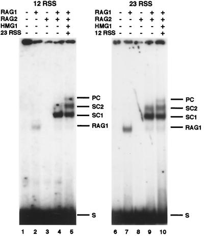
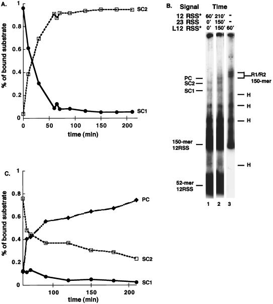
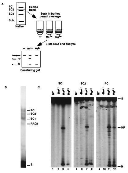
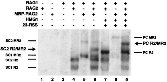
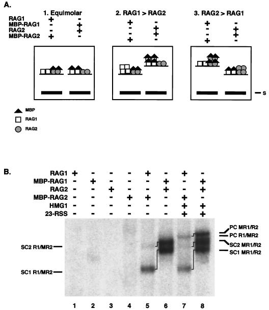
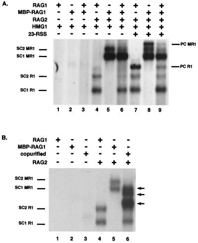
Assembly of antigen receptor genes by V(D)J recombination requires the site-specific recognition of two distinct DNA elements differing in the length of the spacer DNA that separates two conserved recognition motifs. Under appropriate conditions, V(D)J cleavage by the purified RAG1/RAG2 recombinase is similarly restricted. Double-strand breakage occurs only when these proteins are bound to a pair of complementary signals in a synaptic complex. We examine here the binding of the RAG proteins to signal sequences and find that the full complement of proteins required for synapsis of two signals and coupled cleavage can assemble on a single signal. This complex, composed of a dimer of RAG2 and at least a trimer of RAG1, remains inactive for double-strand break formation until a second complementary signal is provided. Thus, binding of the second signal activates the complex, possibly by inducing a conformational change. If synaptic complexes are formed similarly in vivo, one signal of a recombining pair may be the preferred site for RAG1/RAG2 assembly.


Figures

FIG. 1.
FIG. 1.
Detection of multiple complexes of RAG1 and RAG2 bound to recombination signal sequences. Complexes formed on a labeled 12RSS (left panel) or labeled 23RSS (right panel) substrate are shown. The presence (+) or absence (−) of RAG1, RAG2, HMG1, and a complementary unlabeled RSS is indicated. The positions of the two SCs, SC1 and SC2, and the PC are as indicated. A complex containing RAG1 only (RAG1), which appears to a variable extent in these assays, is also indicated at right. S, substrate.
FIG. 2.
FIG. 2.
Time course of SC and PC assembly. (A) Time course for the assembly of SC1 and SC2 on a 12RSS. RAG1, RAG2, and HMG1 were incubated with a 32P labeled 12RSS for the indicated times, and the complexes were separated on a 4.5% polyacrylamide gel. The percentage of each complex formed at a given time relative to the total amount of bound substrate as determined by PhosphorImager analysis of the gel is shown. (B) RAG/RSS complexes do not continue to form after 1 h of incubation. All reactions contain RAG1, RAG2, and HMG1. The presence of radiolabeled (✽) or unlabeled (no asterisk) substrates is indicated. The length of time the substrate was present in the binding reaction is indicated. The total length of incubation was 60 min (lanes 1 and 3) or 210 min (lane 2). For complete details, see Results. L12RSS = 150-mer substrate. (C) Time course for the assembly of PC. RAG1, RAG2, and HMG1 were incubated with a 12-signal for 60 min. At 60 min, an unlabeled 23-signal was added to the reaction mixture, and binding was allowed to proceed for the times indicated prior to analysis. The gel was analyzed on a PhosphorImager as for panel A, and a graphic representation of the data is shown. Labeling is as described in Fig. 1 and 2A.
FIG. 3.
FIG. 3.
Functional analysis of individual RAG-RSS complexes. (A) Diagram of the assay. A representative analysis of the SC2 complex is shown. (B) Representative native gel lane from which individual complexes were excised. (C) Analyses of cleavage products formed by SC1 (left), SC2 (middle), and PC (right) are shown. Each slice was subject either to no additional treatment after gel excision (NT) or to incubation in reaction buffer lacking metal ion (−Me2+), or to incubation with Mn2+ or Mg2+ as indicated. N, nick; H, hairpin; S, substrate. Other labeling is as described in Fig. 1.
FIG. 4.
FIG. 4.
Stoichiometry of RAG2 in RAG-RSS complexes. The presence (+) or absence (−) of RAG1, RAG2, MBP-RAG2, HMG1, and an unlabeled 23-signal is indicated. Positions of SC1 and SC2 derived from either RAG2 (R2), MBP-RAG2 (MR2), or both (R2/MR2, see arrow) in conjunction with RAG1 are indicated on the left. Positions of each form of PC are indicated on the right, with the R2/MR2 marked by an arrow.
FIG. 5.
FIG. 5.
Relative content of RAG1 and RAG2 in SCs and PC. (A) Predicted relative mobilities of MBP-RAG1/RAG2 and RAG1/MBP-RAG2 complexes. The predicted relative mobilities of the RAG1/RAG2 complexes formed when the RAG proteins are present in equal numbers (left panel), when RAG1 is present in molar excess (middle), or when RAG2 is present in molar excess (right panel) are shown. (B) Actual relative mobilities of RAG1/RAG2 complexes. The presence (+) or absence (−) of RAG1, MBP-RAG1, RAG2, MBP-RAG-2, HMG1, and an unlabeled 23-signal is indicated. The positions of SC1 and SC2 and PC formed from the different combinations are indicated. Lines between bands mark the same complex formed from different combinations of protein.
FIG. 6.
FIG. 6.
Purified RAG1 does not reassort during formation of SC1, SC2, or PC. (A) Gel shift analysis of complexes formed after incubation with core and MBP-tagged RAG1. All reactions contain a labeled 52-mer-12RSS. The presence (+) or absence (−) of RAG1, MBP-RAG1, RAG2, HMG1, and an unlabeled 23-signal is indicated. The positions of SC1 and SC2 derived from either RAG1 (R1) or MBP-RAG1 (MR1) in conjunction with RAG2 are indicated on the left. The positions of the PCs are indicated on the right. Lanes 1 to 6 and lanes 7 to 9 are derived from the same exposure of the same gel. (B) Core RAG1 and MBP-RAG1 are capable of forming heteromeric complexes when coexpressed. The presence (+) or absence (−) of RAG1, MBP-RAG1, coexpressed and copurified RAG1/MBP-RAG1 (copurified), and RAG2 is indicated. The positions of SC1 and SC2 derived from RAG1 or MBP-RAG1 in conjunction with RAG2 are indicated on the left. The arrows on the right indicate the positions of heteromeric forms isolated from copurification of RAG1 and MBP-RAG1.

References

    1. Akamatsu, Y., and M. A. Oettinger. 1998. Distinct roles of RAG1 and RAG2 in binding the V(D)J recombination signal sequences. Mol. Cell. Biol. 18:4670–4678 - PMC - PubMed
    1. Aldaz, H., E. Schuster, and T. A. Baker. 1996. The interwoven architecture of the Mu transposase couples DNA synapsis to catalysis. Cell 85:257–269. - PubMed
    1. Asante-Appiah, E., and A. M. Skalka. 1997. A metal-induced conformational change and activation of HIV-1 integrase. J. Biol. Chem. 272:16196–16205. - PubMed
    1. Bailin, T., X. Mo, and M. J. Sadofsky. 1999. A RAG1 and RAG2 tetramer complex is active in cleavage in V(D)J recombination. Mol. Cell. Biol. 19:4664–4671 - PMC - PubMed
    1. Baker, T. A., and K. Mizuuchi. 1992. DNA-promoted assembly of the active tetramer of the Mu transposase. Genes Dev. 6:2221–2232 - PubMed

Publication types

LinkOut - more resources