. 2002 Jan;22(1):78-93.
doi: 10.1128/MCB.22.1.78-93.2002.

Novel function of the cyclin A binding site of E2F in regulating p53-induced apoptosis in response to DNA damage

Affiliations

Novel function of the cyclin A binding site of E2F in regulating p53-induced apoptosis in response to DNA damage

Jung-Kuang Hsieh et al. Mol Cell Biol. 2002 Jan.

Abstract

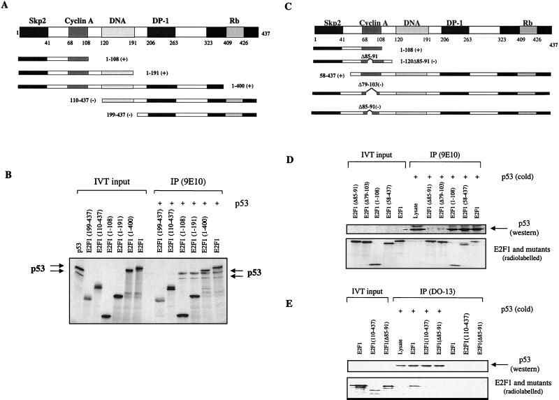
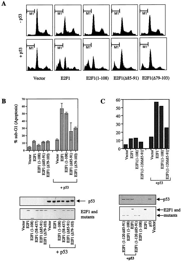
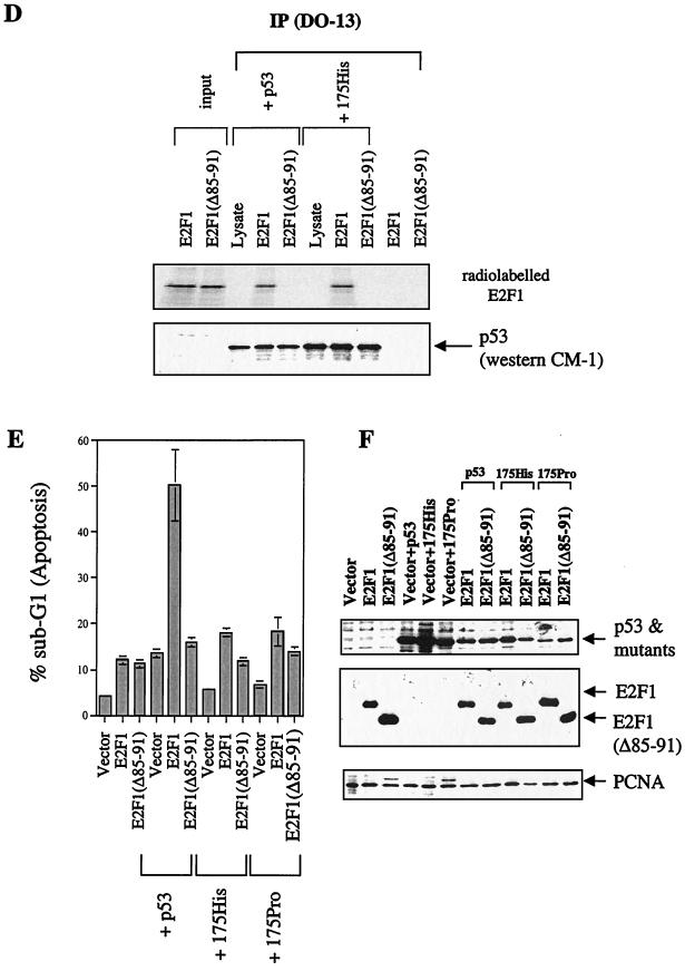
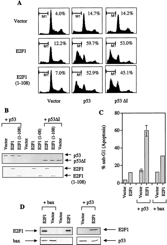
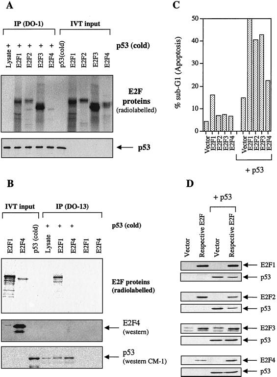
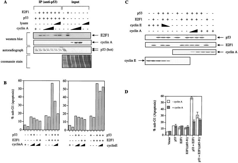
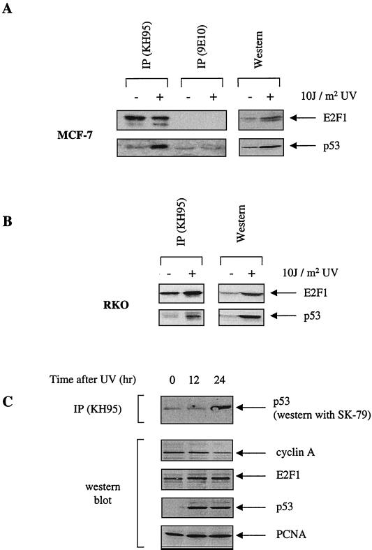
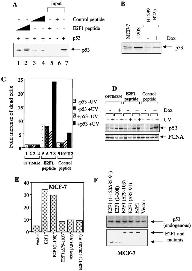
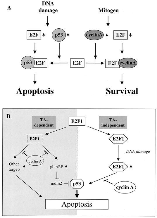
We demonstrate here that the E2F1 induced by DNA damage can bind to and promote the apoptotic function of p53 via the cyclin A binding site of E2F1. This function of E2F1 does not require its DP-1 binding, DNA binding, or transcriptional activity and is independent of mdm2. All the cyclin A binding E2F family members can interact and cooperate with p53 to induce apoptosis. This suggests a novel role for E2F in regulating apoptosis in response to DNA damage. Cyclin A, but not cyclin E, prevents E2F1 from interacting and cooperating with p53 to induce apoptosis. However, in response to DNA damage, cyclin A levels decrease, with a concomitant increase in E2F1-p53 complex formation. These results suggest that the binding of E2F1 to p53 can specifically stimulate the apoptotic function of p53 in response to DNA damage.


Figures

FIG. 1.
FIG. 1.
p53 binds to the cyclin A binding domain of the E2F1 protein. (A and C) Mapping was done using a series of E2F1 truncation mutants. The numbers refer to the residues the peptides contain, while the prefix Δ denotes the residues that are deleted. The E2F1 mutants that bind p53 are denoted by (+), while nonbinding mutants are indicated by (−). (B) Both in vitro-translated [35S]methionine-labeled E2F1 and p53 were used. The antibody 9E10 was used to immunoprecipitate 9E10-tagged E2F1 with the coimmunoprecipitation of radiolabeled p53. The presence of p53 and E2F1 mutants was detected by autoradiography. (D and E) Only in vitro-translated [35S]methionine-labeled E2F1 was used. The in vitro-translated p53 was unlabeled, and its presence was detected by Western blotting using rabbit polyclonal antibody SK-79 (D) or CM-1 (E). For panel E, an anti-p53 antibody (DO-13) was used to immunoprecipitate p53 and the corresponding binding of E2F1 was detected by autoradiography.
FIG. 2.
FIG. 2.
Only E2F1 mutants that bind to p53 can cooperate with p53 to induce apoptosis. (A to C) FACS analysis showing identical-scale histogram plots (A) or bar graphs (B and C) of transiently transfected Saos-2 cells in the absence or presence of p53 (3 μg/10-cm dish) with E2F1 and its mutants (5 μg/10-cm dish) as indicated. The lower portions of panels B and C show the expression level of p53, E2F1, and mutants. The transfected p53 protein was detected with DO-1. (D) Results from a binding assay using E2F1 and E2F1(Δ85–91) and in vitro-translated p53 and p53 mutant 175his. (E) Cells undergoing apoptosis when p53 (3 μg) or the p53 mutants 175his and 175pro (5 μg/dish) were transfected with E2F1 (3 μg) or E2F1(Δ85–91) (3 μg) in Saos-2 cells. (F) Expression of p53, E2F1, and mutant. PCNA was used as a loading control.
FIG. 2.
FIG. 2.
Only E2F1 mutants that bind to p53 can cooperate with p53 to induce apoptosis. (A to C) FACS analysis showing identical-scale histogram plots (A) or bar graphs (B and C) of transiently transfected Saos-2 cells in the absence or presence of p53 (3 μg/10-cm dish) with E2F1 and its mutants (5 μg/10-cm dish) as indicated. The lower portions of panels B and C show the expression level of p53, E2F1, and mutants. The transfected p53 protein was detected with DO-1. (D) Results from a binding assay using E2F1 and E2F1(Δ85–91) and in vitro-translated p53 and p53 mutant 175his. (E) Cells undergoing apoptosis when p53 (3 μg) or the p53 mutants 175his and 175pro (5 μg/dish) were transfected with E2F1 (3 μg) or E2F1(Δ85–91) (3 μg) in Saos-2 cells. (F) Expression of p53, E2F1, and mutant. PCNA was used as a loading control.
FIG. 3.
FIG. 3.
E2F1 can specifically cooperate with p53 to induce apoptosis independent of mdm2. (A) Cells undergoing apoptosis as determined by FACS analysis of Saos-2 cells were transfected with 3 μg of p53 or p53ΔI or 5 μg of E2F1 or E2F1(1–108). The numbers at the top right of each panel refer to the percentage in sub-G1 (labeled M1; indicative of apoptosis). (C) Bar graph showing the amount of apoptosis as determined by FACS analysis of Saos-2 cells transfected with 5 μg of E2F1 or E2F1(1–108) in the presence of 3 μg of p53 or 1 μg of Bax. (B and D) Immunoblotting to show the expression level of p53, E2F1, and mutants, of p53ΔI (B) and p53, and of E2F1 and Bax (D). The transfected p53 (and p53ΔI in panel B) were detected with the anti-p53 antibody DO-13, and the Bax protein (D) was detected using N-20.
FIG. 4.
FIG. 4.
Only E2F members containing the cyclin A binding domain (E2F1, E2F2, and E2F3) bind to and activate p53 to induce apoptosis. (A) In vitro binding assay showing the results of radiolabeled E2F1, E2F2, E2F3, and E2F4 and unlabeled p53 [referred to as p53 (cold)] immunoprecipitated with anti-p53 mouse monoclonal antibody DO-1. The presence of [35S]methionine-labeled E2Fs was detected by autoradiography, while the unlabeled p53 protein was detected using rabbit polyclonal antibody SK-79. (B) Immunoprecipitation using radiolabeled E2F1 and E2F4 with unlabeled p53 and immunoprecipitated by a anti-p53 antibody (DO-13). Since the radioactive E2F4 signal is rather weak, a blot showing that E2F4 is expressed but not immunoprecipitated under these conditions is included. (C) The percentage of apoptotic cells as determined by FACS of Saos-2 cells transfected with E2F1-4 (5 μg each/10-cm dish) in the absence or presence of p53 (3 μg/10-dish). (D) Immunoblotting showing the expression of the various E2F proteins using the respective antibodies (9E10 epitope-tagged E2F1 [9E10], E2F2 [C-20], E2F3 [N-20], and E2F4 [C-108]) and p53 using the antibody DO-1.
FIG. 5.
FIG. 5.
Cyclin A, but not cyclin E, competitively inhibits the cooperation of E2F1- and p53-induced apoptosis. (A) In vitro competition assay using baculovirus-expressed cyclin A (2, 5, or 10 μg) to compete out the binding of E2F1 to p53. Vector-infected baculovirus lysate was used as a control. The polyclonal anti-p53 (DO-13) was used to immunoprecipitate radiolabeled p53. (B) Apoptosis induced in Saos-2 cells transfected with E2F1 (5 μg/10-cm dish), p53 (3 μg/10-cm dish), and increasing concentrations of cyclin A or cyclin E (0, 5, and 10 μg/10-cm dish) as indicated. (C) Immunoblotting was used to show the expression of cyclin A using rabbit polyclonal antibody (H-432), cyclin E using rabbit polyclonal antibody (M-20), E2F1 using C-20 (anti-E2F1 polyclonal antibody), and p53 using DO-1. (D) Percentage of apoptotic cells as determined by FACS analysis of Saos-2 cells transfected with E2F1 and mutants (5 μg/10-cm dish) and p53 (3 μg/10-cm dish) in the absence or presence of cyclin A (10 μg/10-cm dish).
FIG. 6.
FIG. 6.
UV-induced endogenous E2F1 and p53 can complex in vivo. (A and B) MCF-7 cells (A) and RKO cells (B) were UV irradiated with 10 J m−2 (+) or left untreated (−) and then incubated for 16 h. The left parts of each panel show the results of the immunoprecipitation using the respective lysates with anti-E2F1 (KH95) and control antibody (9E10) (panel A only). The right parts of each panel show the corresponding Western blot of the cell lysate. In both cases, the rabbit polyclonal antibodies used were C-20 (for E2F1) and SK-79 (for p53). (C) Time course for U20S cells treated with UV. Immunoprecipitation using anti-E2F1 (KH95) and probed for p53 using the polyclonal antibody indicated is shown in the top part. The lower four parts show the results of a Western blot analysis using the corresponding lysate with endogenous levels of E2F1 (detected by C-20), p53 (detected by DO-1), and cyclin A (detected by H-432) shown at the indicated times after UV. PCNA (detected by PC-10) is used to show equal loading.
FIG. 7.
FIG. 7.
E2F1 can cooperate with endogenous p53 to induce apoptosis. p53 binding peptide of E2F1 can specifically enhance the apoptotic function of p53 in response to treatment with UV. (A) Immunoprecipitation data using biotin-conjugated anti-FITC to immunoprecipitate increasing concentrations of FITC-labeled E2F1 peptide and FITC-labeled control peptide. The presence of [35S]methionine-labeled p53 was detected using autoradiography. (B) Comparison of the levels of endogenous p53 in MCF7 and U20S and the p53 in the H1299 B225 p53-inducible cell line induced by 2 μg of doxycycline (Dox) per ml. The presence of p53 was detected using the mouse monoclonal antibody DO-1. (C) The p53-inducible cell line H1299 B225 was incubated in the absence (−p53) or presence (+p53) of 2 μg of doxycycline per ml untreated (−UV) or UV irradiated at 10 J m−2 (+UV) in medium only (Optimem) or 100 μM E2F1 peptide or the control peptide in Optimem as detailed in Materials and Methods. To take into account the p53-independent mechanism of cell killing exhibited by the peptide and the various effects of treatment, the relative fold increase in the number of dead cells is calculated and presented as described in Materials and Methods. (D) The corresponding Western blot where p53 was detected using DO-13 and the PC-10 supernatant used to detect PCNA. (E) Apoptosis was determined by FACS when E2F1 and mutants (10 μg) were transfected in wild-type expressing MCF7 cells. (F) Expression levels of transfected E2F1 and mutants and endogenous p53 detected using 9E10 and DO-1, respectively.
FIG. 8.
FIG. 8.
(A) Method by which cyclin A may influence the apoptotic function of p53 via E2F1. (B) Summary of the various mechanisms by which E2F1 and p53 can cooperate to induce apoptosis under different conditions. The left half shows transactivation-dependent mechanisms, while the right half depicts the transactivation-independent mechanism we propose here.

Similar articles

Cited by

References

    1. Bandara, L. R., V. M. Buck, M. Zamanian, L. H. Johnston, and N. La Thangue. 1993. Functional synergy between DP1 and E2F1 in the cell cycle-regulating transcription factor DRTF/E2F. EMBO J. 12:4317–4324. - PMC - PubMed
    1. Bates, S., A. Phillips, P. Clark, F. Stott, G. Peters, R. Ludwig, and K. Vousden. 1998. p14ARF links the tumour suppressors RB and p53. Nature 395:124–125. - PubMed
    1. Blattner, C., A. Sparks, and D. Lane. 1999. Transcription factor E2F-1 is upregulated in response to DNA damage in a manner analogous to that of p53. Mol. Cell. Biol. 19:3704–3713. - PMC - PubMed
    1. Chen, Y. N., S. K. Sharma, T. M. Ramsey, L. Jiang, M. S. Martin, K. Baker, P. D. B. Adams, and W. G. J. Kaelin. 1999. Selective killing of transformed cells by cyclin/cyclin-dependent kinase 2 antagonists. Proc. Natl. Acad. Sci. U-S-A 96:4325–4329. - PMC - PubMed
    1. Clarke, A. R., C. A. Purdie, D. J. Harrison, R. G. Morris, C. C. Bird, M. L. Hooper, and A. H. Wyllie. 1993. Thymocyte apoptosis induced by p53-dependent and independent pathways. Nature 362:849–852. - PubMed

Publication types

MeSH terms