. 2002 Jan;22(1):148-60.
doi: 10.1128/MCB.22.1.148-160.2002.

SRm160 splicing coactivator promotes transcript 3'-end cleavage

Affiliations

SRm160 splicing coactivator promotes transcript 3'-end cleavage

Susan McCracken et al. Mol Cell Biol. 2002 Jan.

Abstract

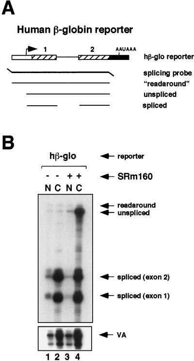
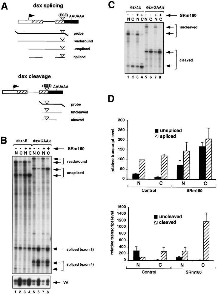
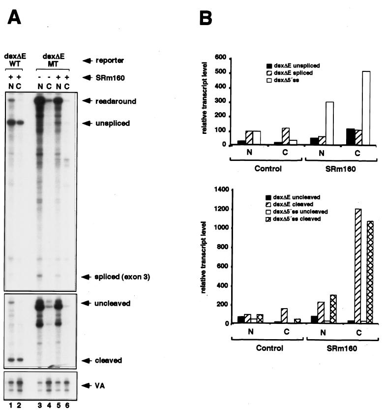
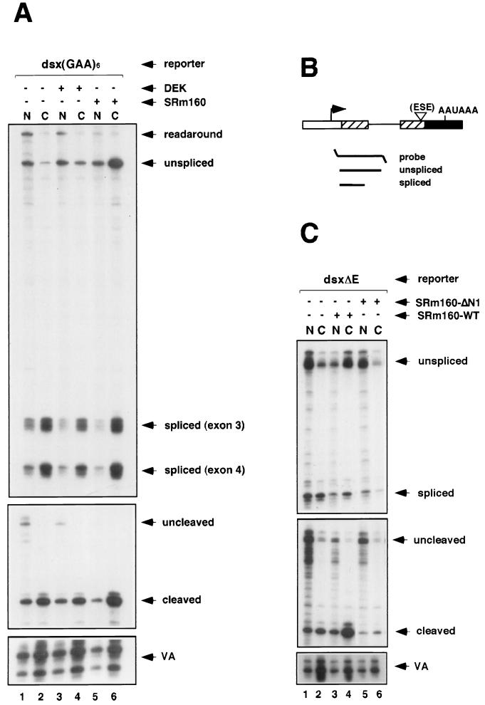
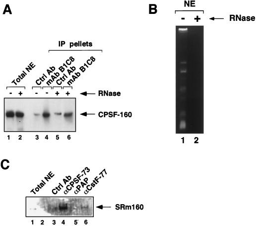
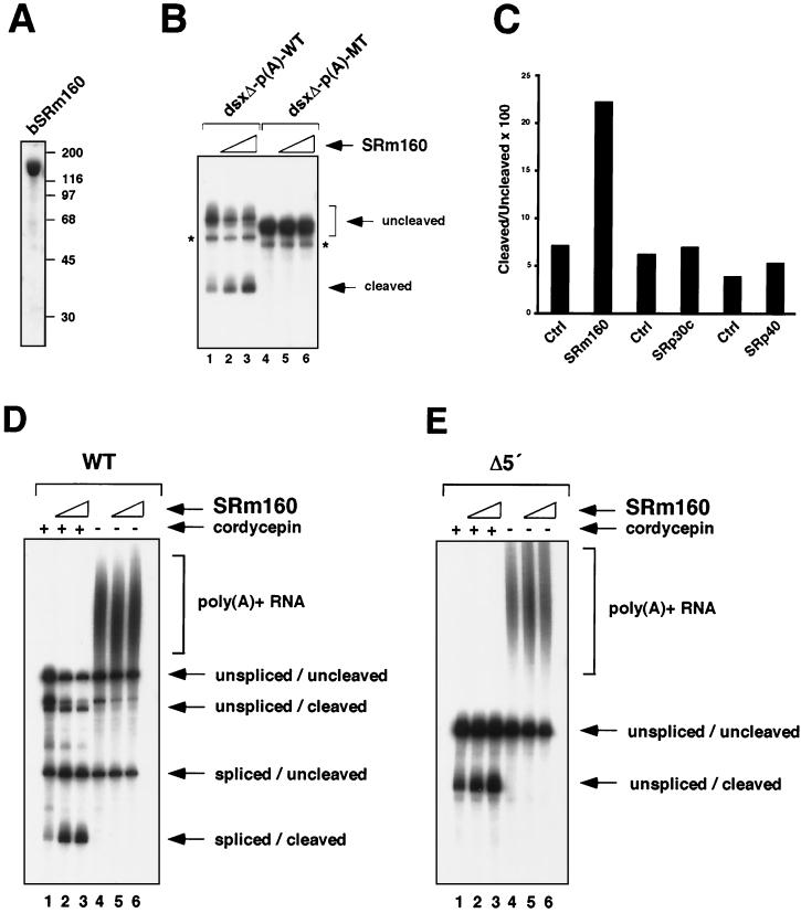
Individual steps in the processing of pre-mRNA, including 5'-end cap formation, splicing, and 3'-end processing (cleavage and polyadenylation) are highly integrated and can influence one another. In addition, prior splicing can influence downstream steps in gene expression, including export of mRNA from the nucleus. However, the factors and mechanisms coordinating these steps in the maturation of pre-mRNA transcripts are not well understood. In the present study we demonstrate that SRm160 (for serine/arginine repeat-related nuclear matrix protein of 160 kDa), a coactivator of constitutive and exon enhancer-dependent splicing, participates in 3'-end formation. Increased levels of SRm160 promoted the 3'-end cleavage of transcripts both in vivo and in vitro. Remarkably, at high levels in vivo SRm160 activated the 3'-end cleavage and cytoplasmic accumulation of unspliced pre-mRNAs, thereby uncoupling the requirement for splicing to promote the 3'-end formation and nuclear release of these transcripts. Consistent with a role in 3'-end formation coupled to splicing, SRm160 was found to associate specifically with the cleavage polyadenylation specificity factor and to stimulate the 3'-end cleavage of splicing-active pre-mRNAs more efficiently than that of splicing-inactive pre-mRNAs in vitro. The results provide evidence for a role for SRm160 in mRNA 3'-end formation and suggest that the level of this splicing coactivator is important for the proper coordination of pre-mRNA processing events.


Figures

FIG. 1.
FIG. 1.
Expression of high levels of SRm160 in vivo results in the cytoplasmic accumulation of unspliced RNA. (A) Schematic representation of the RNase protection probe used to analyze splicing of transcripts from a reporter consisting of hβ-glo exons 1 and 2 with the intervening intron. The predicted RNase protection products are shown below the probe (for sizes refer to Materials and Methods and supplementary information located at http://www.utoronto.ca/intron/supp_info). (B) Human 293 cells were transiently transfected with the hβ-glo pre-mRNA reporter and a pol III reporter (pSPVA) as an internal control (lanes 1 to 4) together with a control, empty expression vector (pcDNA3-Flag) (lanes 1 and 2) or an expression vector for Flag epitope-tagged SRm160 (pcDNA3-fSRm160) (lanes 3 and 4). Proportional amounts of RNA isolated from the nuclear (N) and cytoplasmic (C) fractions were analyzed by RNase protection using the probe illustrated in panel A.
FIG. 2.
FIG. 2.
Increased expression of SRm160 promotes 3′-end cleavage in vivo. (A) Schematic representation of the RNase protection probes used to analyze splicing and 3′-end cleavage in pre-mRNAs transcribed from reporters derived from exons 3 and 4 of the Drosophila doublesex gene (dsx). The dsx reporters contained either no ESE (dsxΔE) or a six-GAA repeat ESE [dsx(GAA)6]. The predicted RNase protection products are shown below each probe (for sizes refer to Materials and Methods and supplementary information available at http://www.utoronto.ca/intron/supp_info). (B and C) Human 293 cells were transiently transfected with the dsxΔE (lanes 1 to 4) or dsx(GAA)6 reporter (lanes 5 to 8) together with a control expression vector containing no insert (pcDNA3-Flag) (lanes 1 and 2 and 5 and 6) or an expression vector for Flag epitope-tagged SRm160 (pcDNA3-fSRm160) (lanes 3 and 4 and 7 and 8); the pol III reporter (pSPVA) was cotransfected in each case as an internal control. Proportional amounts of RNA isolated from the nuclear (N) and cytoplasmic (C) fractions of the transfected cells were analyzed by RNase protection using either the splicing protection probe (B) or the 3′-end protection probe (C). The identity of each RNA species is indicated. Note that exon 4 of the dsxΔE pre-mRNA, migrating near the bottom of the gel in panel B, is less strongly detected by the [32P]UTP-labeled RNase protection probe due to the low A content of this exon. It was, however, readily detected following a longer exposure of the gel (data not shown). (D) Quantification of the effect of SRm160 expression on the yields of different RNA species transcribed from the dsxΔE reporter. RNA isolated from transfected cells was analyzed by RNase protection as described above, and the amounts of unspliced, spliced, uncleaved, and cleaved RNAs from three independent experiments were quantified. The values were normalized for both VA signal and U content. For the purpose of averaging different experiment values, the nuclear spliced RNA and the nuclear cleaved RNA were normalized to a value of 100.
FIG. 3.
FIG. 3.
Mutation of the polyadenylation signal, but not deletion of the 5′ splice site, prevents cleavage and cytoplasmic accumulation of unspliced RNA by SRm160. (A) 293 cells were transiently transfected with the dsxΔE reporter containing either a wild-type (dsxΔE-WT) (lanes 1 and 2) or mutant (dsxΔE-MT) (lanes 3 to 6) polyadenylation signal (see the text). The cells were cotransfected with a control, empty expression vector (pcDNA3-Flag) (lanes 3 and 4) or an expression vector for Flag epitope-tagged SRm160 (pcDNA3-fSRm160) (lanes 1 and 2 and 5 and 6) and with pSPVA as an internal control. Proportional amounts of RNA isolated from the nuclear (N) and cytoplasmic (C) fractions were analyzed by RNase protection using the splicing and cleavage probes shown in Fig. 2A. (B) Quantification of the effect of SRm160 expression on the nuclear and cytoplasmic levels of RNAs transcribed from a splicing-inactive dsx reporter, which lacks a functional 5′ splice site (dsxΔ5′ss), and the dsxΔE reporter. RNA isolated from transfected cells was analyzed by RNase protection as described in the legend to Fig. 2A, and the amounts of the unspliced, spliced, uncleaved, and cleaved RNAs were quantified as described in the legend to Fig. 2 (refer to supplementary information at the web site cited above for details of the dsxΔ5′ss reporter and protection probe).
FIG. 4.
FIG. 4.
Specificity of the cleavage-stimulatory and transcript export activities of SRm160. (A) Human 293 cells were transiently transfected with the dsx(GAA)6 reporter together with a control expression vector containing no insert (pcDNA3-Flag) (lanes 1 and 2), an expression vector for HA epitope-tagged DEK (pcDNA3-DEK) (lanes 3 and 4), or an expression vector for Flag epitope-tagged SRm160 (pcDNA3-fSRm160) (lanes 5 and 6); the pol III reporter (pSPVA) was cotransfected in each case as an internal control. Proportional amounts of RNA isolated from the nuclear (N) and cytoplasmic (C) fractions of the transfected cells were analyzed by RNase protection using either a splicing protection probe or the 3′-end protection probe (refer to the legend to Fig. 2). The identity of each RNA species is indicated. (B) Schematic representation of the short splicing RNase protection probe used to analyze splicing of transcripts from the dsxΔE reporter. The predicted RNase protection products are shown below each probe (for sizes refer to Materials and Methods and supplementary information available at http://www.utoronto.ca/intron/supp_info). (C) Human 293 cells were transiently transfected with the dsxΔE reporter together with a control expression vector containing no insert (pcDNA3-Flag) (lanes 1 and 2), an expression vector for Flag epitope-tagged SRm160 (pcDNA3-fSRm160) (lanes 3 and 4), or an expression vector for Flag epitope-tagged SRm160 deleted from amino acids 1 to 155 (pcDNA3-fSRm160ΔN1) (lanes 5 and 6); the pol III reporter (pSPVA) was cotransfected in each case as an internal control. Proportional amounts of RNA isolated from the nuclear (N) and cytoplasmic (C) fractions of the transfected cells were analyzed by RNase protection using either the short splicing protection probe or the 3′-end protection probe. The identity of each RNA species is indicated.
FIG. 5.
FIG. 5.
SRm160 associates with the 3′-end cleavage machinery. (A) Immunoprecipitates (IP) were collected from HeLa nuclear extract (NE) by using the SRm160-specific MAb (MAb-B1C8) (lanes 4 and 6) and excess levels of a control antibody (rabbit anti-mouse immunoglobulin; Ctrl Ab) (lanes 3 and 5). The immunoprecipitates were separated on a 7.5% sodium dodecyl sulfate-polyacrylamide gel and immunoblotted with an affinity-purified antibody specific for the 160-kDa subunit of the human CPSF (CPSF-160). Total nuclear extract, separated in lanes 1 and 2, represents 7% of the amount of extract used in each immunoprecipitation. Nuclear extract was preincubated in the presence (lanes 2, 5, and 6) or absence (lanes 1, 3, and 4) of RNase prior to immunoprecipitation. (B) Analysis of the RNA content of the nuclear extract used for immunoprecipitation shown in panel A. RNA isolated from nuclear extract pretreated with (lane 2) or without (lane 1) RNase was analyzed on a 10% denaturing acrylamide gel stained with ethidium bromide. (C) Immunoprecipitates were collected from RNase-pretreated HeLa nuclear extract by using rabbit polyclonal antibodies to the 73-kDa subunit of human CPSF (CPSF-73) (lane 4), PAP (lane 5), CstF-77 (lane 6), and a control antibody (rabbit anti-glutathione S-transferase) (lane 3). The immunoprecipitates were separated on a 7.5% sodium dodecyl sulfate- polyacrylamide gel and immunoblotted with MAb-B1C8. Total nuclear extract, separated in lane 1, represents ∼0.5% of the amount of extract used in each immunoprecipitation.
FIG. 6.
FIG. 6.
Highly purified, recombinant SRm160 preferentially stimulates cleavage of spliced substrates in vitro. (A) Analysis of recombinant, baculovirus-expressed SRm160 (bSRm160) by sodium dodecyl sulfate gel electrophoresis and Coomassie blue staining (see Materials and Methods for purification details). bSRm160 (4.5 μg) was loaded on the gel shown. (B) bSRm160 stimulates the 3′-end cleavage of a dsx substrate lacking functional splice sites in vitro. Reactions were performed with substrates derived from the 3′ half of dsx exon 4 (no ESE present) containing either a wild-type SV40 late poly(A) site [dsxΔ-p(A)-WT] (lanes 1 to 3) or a mutant poly(A) site [dsxΔ-p(A)-MT] (lanes 4 to 6) in the presence (lanes 2 and 3 and 5 and 6) or absence (lanes 1 and 4) of bSRm160. bSRm160 (165 ng) was added to the reaction shown in lanes 2 and 5, and 330 ng of bSRm160 was added to the reaction shown in lanes 3 and 6. The reactions were performed with added 5′-cordycepin triphosphate, as described in Materials and Methods. Duplicate reactions performed in the absence of 5′-cordycepin triphosphate confirmed the identity of the 3′-end cleaved bands (data not shown). Asterisks indicate a nonspecific degradation product not related to cleavage. (C) Specificity of the 3′-end stimulatory activity of SRm160. Approximately equal amounts (176 ng) of bSRm160, bSRp30c, and bSRp40, as assessed by Bradford assay, were added to 3′-end cleavage reaction mixtures incubated with the dsxΔ-p(A)-WT substrate. The reaction products were analyzed by electrophoresis on a denaturing gel and quantified by using a Molecular Dynamics PhosphorImager and ImageQuant software. (D and E) In vitro splicing and cleavage reactions containing a wild-type (WT) (D) or a 5′-splice site-deleted (Δ5′) (E) adenovirus pre-mRNA substrate, with an SV40 late polyadenylation signal (MSXVL) (35), were performed in the presence (lanes 2 and 3 and 5 and 6) or absence (lanes 1 and 4) of bSRm160. bSRm160 (110 ng) was added to the reaction shown in lanes 2 and 5, and 220 ng of bSRm160 was added to the reaction shown in lanes 3 and 6. The reactions were performed with (lanes 1 to 3) or without (lanes 4 to 6) added 5′-cordycepin triphosphate in order to distinguish the 3′-end cleaved products (see the text).

Similar articles

Cited by

References

    1. Bauren, G., S. Belikov, and L. Wieslander. 1998. Transcriptional termination in the Balbiani ring 1 gene is closely coupled to 3′-end formation and excision of the 3′-terminal intron. Genes Dev 12:2759–2769. - PMC - PubMed
    1. Birse, C. E., L. Minvielle-Sebastia, B. A. Lee, W. Keller, and N. J. Proudfoot. 1998. Coupling termination of transcription to messenger RNA maturation in yeast. Science 280:298–301. - PubMed
    1. Blencowe, B. J., G. Bauren, A. G. Eldridge, R. Issner, J. A. Nickerson, E. Rosonina, and P. A. Sharp. 2000. The SRm160/300 splicing coactivator subunits. RNA 6:111–120. - PMC - PubMed
    1. Blencowe, B. J., A. L. Bowman, S. McCracken, and E. Rosonina. 1999. SR-related proteins and the processing of messenger RNA precursors. Biochem. Cell Biol. 77:277–291. - PubMed
    1. Blencowe, B. J., R. Issner, J. A. Nickerson, and P. A. Sharp. 1998. A coactivator of pre-mRNA splicing. Genes Dev. 12:996–1009. - PMC - PubMed

Publication types

MeSH terms