. 2002 Jan;22(1):171-81.
doi: 10.1128/MCB.22.1.171-181.2002.

Absence of the CAAX endoprotease Rce1: effects on cell growth and transformation

Affiliations

Absence of the CAAX endoprotease Rce1: effects on cell growth and transformation

Martin O Bergo et al. Mol Cell Biol. 2002 Jan.

Abstract

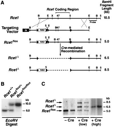
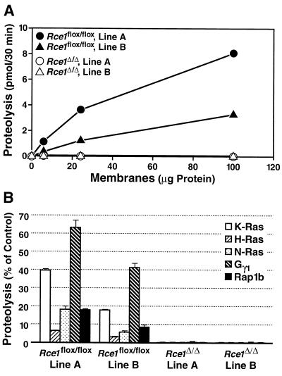
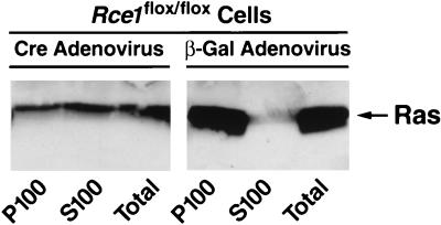
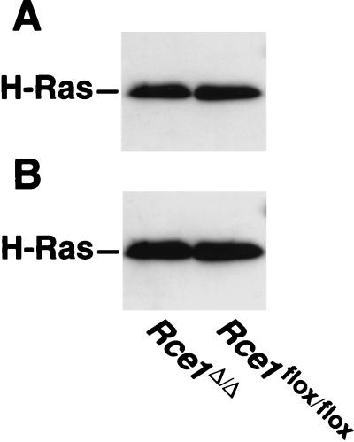
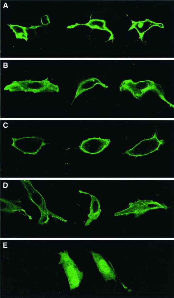
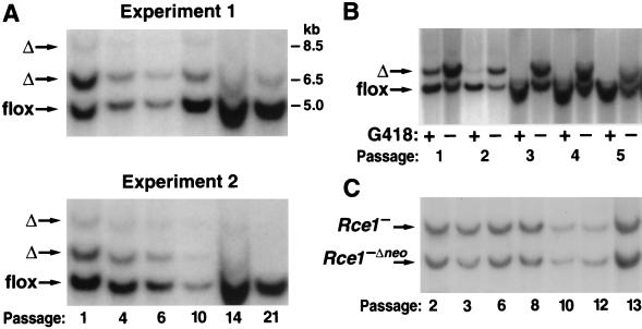
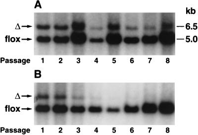
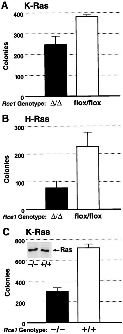
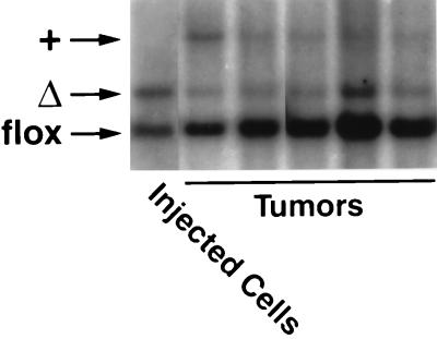
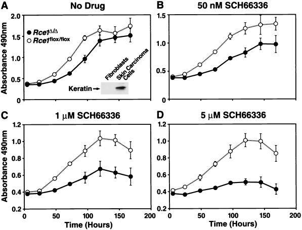
After isoprenylation, the Ras proteins and other CAAX proteins undergo two additional enzymatic modifications-endoproteolytic release of the last three amino acids of the protein by the protease Rce1 and methylation of the carboxyl-terminal isoprenylcysteine by the methyltransferase Icmt. This postisoprenylation processing is thought to be important for the association of Ras proteins with membranes. Blocking postisoprenylation processing, by inhibiting Rce1, has been suggested as a potential approach for retarding cell growth and blocking cellular transformation. The objective of this study was to develop a cell culture system for addressing these issues. We generated mice with a conditional Rce1 allele (Rce1(flox)) and produced Rce1(flox/flox) fibroblasts. Cre-mediated excision of Rce1 (thereby producing Rce1(Delta/Delta) fibroblasts) eliminated Ras endoproteolytic processing and methylation and caused a partial mislocalization of truncated K-Ras and H-Ras fusion proteins within cells. Rce1(Delta/Delta) fibroblasts grew more slowly than Rce1(flox/flox) fibroblasts. The excision of Rce1 also reduced Ras-induced transformation, as judged by the growth of colonies in soft agar. The excision of Rce1 from a Rce1(flox/flox) skin carcinoma cell line also significantly retarded the growth of cells, and this effect was exaggerated by cotreatment of the cells with a farnesyltransferase inhibitor. These studies support the idea that interference with postisoprenylation processing retards cell growth, limits Ras-induced transformation, and sensitizes tumor cells to a farnesyltransferase inhibitor.


Figures

FIG. 1.
FIG. 1.
Production of mice with a conditional Rce1 allele. (A) Sequence-replacement strategy to introduce loxP sites 5′ and 3′ of the Rce1 protein-coding sequences. Approximately 1 in 200 G418- and FIAU-resistant ES cell clones were targeted and contained all of the loxP sites. (B) Southern blot identification of Rce1+/+, Rce1flox/+, and Rce1flox/flox mice with EcoRV-cleaved genomic DNA and a 3′ flanking probe. (C) Southern blot identification of Cre-mediated recombination events, with BamHI-cleaved genomic DNA and the same 3′ flanking probe. Shown are examples of Rce1flox/flox cells infected with small (106 PFU) or large (108 PFU) amounts of AdRSVCre.
FIG. 2.
FIG. 2.
Rce1 activity in two different immortalized lines (lines A and B) of Rce1flox/flox and Rce1Δ/Δ fibroblasts. (A) Concentration dependence of protease activity in membrane fractions from fibroblasts. Fibroblast membranes (0 to 100 μg) were incubated with 2.0 μM farnesyl-K-Ras for 30 min at 37°C. (B) Endoprotease activity in membranes from Rce1flox/flox and Rce1Δ/Δ fibroblasts against farnesyl-K-Ras, farnesyl-H-Ras, farnesyl-N-Ras, farnesyl-Gγ1, and geranylgeranyl-Rap1b. Protease activity is expressed as a percentage of the total CAAX protein turnover.
FIG. 3.
FIG. 3.
Intracellular localization of Ras proteins in immortalized fibroblasts. Cre adenovirus fully converted Rce1flox/flox into Rce1Δ/Δ fibroblasts. Fibroblasts were fractionated into cytosolic (S100) and membrane (P100) fractions; Ras proteins were immunoprecipitated and analyzed on Western blots of sodium dodecyl sulfate-polyacrylamide gels with antibody Ab-4. Virtually identical results were observed with independently derived lines of Rce1flox/flox and Rce1Δ/Δ fibroblasts (not shown). Also, very similar results were obtained when the experiments were performed with K-Ras-specific antibodies (data not shown).
FIG. 4.
FIG. 4.
Palmitoylation of Ras proteins in H-Ras-transfected Rce1flox/flox and Rce1Δ/Δ fibroblasts. (A) Autoradiograph showing the incorporation of [3H]palmitate into immunoprecipitated Ras proteins. Cells were metabolically labeled with [3H]palmitate according to the procedures of Mumby and Buss (29a), and the Ras proteins were immunoprecipitated with a different Ras-specific antibody (25). (B) Western blot of the same immunoprecipitated proteins with a Ras-specific antibody. Note that the Ras proteins from Rce1flox/flox and Rce1Δ/Δ fibroblasts differ very slightly in their electrophoretic mobilities. A slower electrophoretic mobility of Ras proteins in Rce1-deficient cells has been noted previously (25).
FIG. 5.
FIG. 5.
Confocal microscope images demonstrating the intracellular localization of EGFP-tK-Ras and EGFP-tH-Ras fusions in Rce1flox/flox and Rce1Δ/Δ fibroblasts. Confocal images of Rce1flox/flox fibroblasts (A and C) and Rce1Δ/Δ fibroblasts (B and D) transfected with either EGFP-tH-Ras (A and B) or EGFP-tK-Ras (C and D). (E) Confocal image demonstrating the intracellular localization of an EGFP-tH-Ras fusion in cells treated with a farnesyltransferase inhibitor (SCH66336, 1.0 μM). The genotype of the cell on the left is Rce1flox/flox; the genotype of the cell on the right is Rce1Δ/Δ. Inhibition of isoprenylation with SCH66336 was associated with the presence of the fusion protein in the nucleus. In viewing many transfected cells under the microscope, we do not believe that the Rce1 gene excision caused different degrees of mislocalization with the EGFP-tH-Ras and EGFP-tK-Ras fusion proteins.
FIG. 6.
FIG. 6.
Southern blot assessment of the ratio of Rce1flox and Rce1Δ alleles during the growth of mixed cultures of Rce1flox/flox and Rce1Δ/Δ fibroblasts. Mixed cultures of Rce1flox/flox and Rce1Δ/Δ cells were obtained by infecting immortalized Rce1flox/flox fibroblasts with AdRSVnlacZ and AdRSVCre as described in Materials and Methods. (A) Southern blots showing the ratio of Rce1Δ and Rce1flox alleles at different passages in two independent experiments. Very similar results were obtained with six other experiments involving fibroblasts from two different mouse embryos. (B) Southern blot demonstrating the rapid disappearance of the Rce1Δ band from a mixed culture of Rce1flox/flox and Rce1Δ/Δ cells in the presence of G418. (C) Southern blot showing Rce1 and Rce1−Δneo bands during the growth of mixed cultures of Rce1−/− and Rce1−Δneo/−Δneo cells.
FIG. 7.
FIG. 7.
Southern blot demonstrating the effect of a farnesyltransferase inhibitor on the growth of Rce1flox/flox and Rce1Δ/Δ fibroblasts. Rce1flox/flox and Rce1Δ/Δ fibroblasts in the presence and absence of a farnesyltransferase inhibitor. Approximately equal numbers of Rce1flox/flox and Rce1Δ/Δ fibroblasts were mixed and grown in the absence (A) and presence (B) of a farnesyltransferase inhibitor (SCH66336, 5.0 μM). Southern blots were performed to assess the ratio of Rce1flox and Rce1Δ alleles at different passages.
FIG. 8.
FIG. 8.
Growth of Rce1Δ/Δ and Rce1flox/flox fibroblasts in the presence and absence of a farnesyltransferase inhibitor. Equal numbers of Rce1flox/flox or Rce1Δ/Δ fibroblasts were seeded separately onto 96-well plates in medium containing vehicle (dimethyl sulfoxide) alone or vehicle plus different concentrations of SCH66336 (12 wells for each concentration of drug). (A) No SCH66336. (B) SCH66336 at 5 μM. Cell growth was assessed with the CellTiter 96 AQueous One Solution cell proliferation assay (Promega). Gradually increasing differences in the growth rates of Rce1flox/flox or Rce1Δ/Δ fibroblasts were noted with 50 nM SCH66336 and with 1 μM SCH66336 (not shown).
FIG. 9.
FIG. 9.
Comparison of the ability of K-Ras-transformed immortalized Rce1Δ/Δ and Rce1flox/flox fibroblasts to form colonies in soft agar. Rce1flox/flox fibroblasts were infected with a K-Ras retrovirus and then treated with either AdRSVnlacZ or AdRSVCre. (A) The ability of K-Ras-transformed Rce1Δ/Δ and Rce1flox/flox fibroblasts to grow in soft agar (P = 0.029). Similar results were observed in three other independent experiments (P = 0.005, P = 0.09, and P = 0.008). (B) The ability of H-Ras-transformed Rce1Δ/Δ and Rce1flox/flox fibroblasts to grow in soft agar (P = 0.002). (C) Comparison of the ability of primary Rce1−/− and Rce1+/+ fibroblasts, matched for K-Ras expression (note Western blot insert), to form colonies in soft agar after transformation with E1A and K-Ras (P = 0.003). Similar results were observed in two other experiments (P < 0.001 and P < 0.001).
FIG. 10.
FIG. 10.
Southern blot demonstrating the relative ability of mixed cultures of K-Ras-transfected Rce1flox/flox and Rce1Δ/Δ fibroblasts to form tumors in nude mice. Mixed cultures of the fibroblasts were obtained by infecting K-Ras-transfected Rce1flox/flox fibroblasts with AdRSVnlacZ and AdRSVCre as described in Materials and Methods. Cells were grown for several passages, and then a total of 2 × 10⁶ cells were injected into nude mice (n = 10). A Southern blot showed the ratio of Rce1Δ and Rce1flox alleles in the injected cells and in the tumors generated from the cells. As judged by a phosphorimager, the Rce1flox/Rce1Δ ratio in the injected cells was 1.99; in the tumors, the ratio was 11.8 ± 2.9 (mean ± SD).
FIG. 11.
FIG. 11.
Growth of Rce1Δ/Δ and Rce1flox/flox skin carcinoma cells in the presence and absence of a farnesyltransferase inhibitor. Equal numbers of Rce1flox/flox skin carcinoma cells (cells treated with AdRSVnlacZ) and Rce1Δ/Δ cells (treated with AdRSVnlacZ and AdRSVCre) were seeded separately onto 96-well plates in medium containing vehicle (dimethyl sulfoxide) alone or vehicle plus different concentrations of SCH66336 (12 wells for each concentration of drug). (A) No SCH66336. The insert shows a Western blot documenting the expression of keratin in the skin carcinoma cells. (B) SCH66336 at 50 nM. (C) SCH66336 at 1 μM. (D) SCH66336 at 5 μM. Cell growth was assessed with the CellTiter 96 AQueous One Solution cell proliferation assay (Promega). This experiment was representative of three separate experiments.

References

    1. Ashby, M. N., and J. Rine. 1995. Ras and a-factor converting enzyme. Methods Enzymol. 250:235–251. - PubMed
    1. Bergo, M. O., G. K. Leung, P. Ambroziak, J. C. Otto, P. J. Casey, and S. G. Young. 2000. Targeted inactivation of the isoprenylcysteine carboxyl methyltransferase gene causes mislocalization of K-Ras in mammalian cells. J. Biol. Chem. 275:17605–17610. - PubMed
    1. Boyartchuk, V. L., M. N. Ashby, and J. Rine. 1997. Modulation of Ras and a-factor function by carboxyl-terminal proteolysis. Science 275:1796–1800. - PubMed
    1. Brown, M. S., and J. L. Goldstein. 1993. Protein prenylation. Mad bet for Rab. Nature 366:14–15. - PubMed
    1. Chakravarti, D., P. Mailander, J. Franzen, S. Higginbotham, E. L. Cavalieri, and E. G. Rogan. 1998. Detection of dibenzol[a,l]pyrene-induced H-ras codon 61 mutant genes in preneoplastic SENCAR mouse skin using a new PCR-RFLP method. Oncogene 16:3203–3210. - PubMed

Publication types

MeSH terms