. 2002 Jan;22(1):196-206.
doi: 10.1128/MCB.22.1.196-206.2002.

ARF function does not require p53 stabilization or Mdm2 relocalization

Affiliations

ARF function does not require p53 stabilization or Mdm2 relocalization

Chandrashekhar Korgaonkar et al. Mol Cell Biol. 2002 Jan.

Abstract

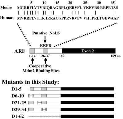
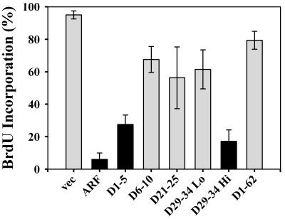
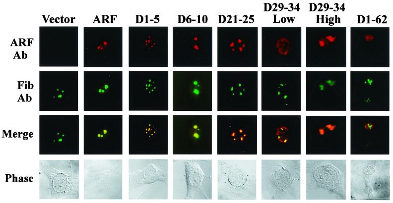
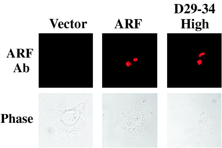
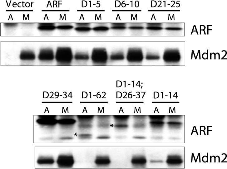
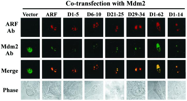
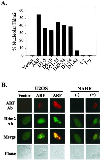
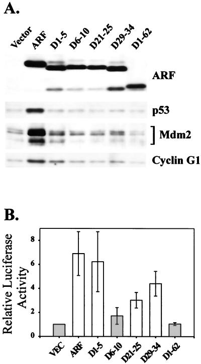
It is generally accepted that the ARF tumor suppressor induces p53-dependent growth arrest by sequestering the p53 antagonist Mdm2 in the nucleolus. Previous mutagenic studies of murine ARF suggested that residues 1 through 14 and 26 through 37 were critical for Mdm2 binding, while the latter domain also governed ARF nucleolar localization. We show that mouse ARF residues 6 to 10 and 21 to 25 are required for ARF-induced growth arrest whereas residues 1 to 5 and 29 to 34 are dispensable. Deletion of the putative nucleolar localization signal (31)RRPR(34) did not prevent nucleolar localization. Surprisingly, unlike wild-type ARF, growth-inhibitory mutants D1-5 and D29-34 failed to stabilize p53 yet induced its transcriptional activation in reporter assays. This suggests that p53 stabilization is not essential for ARF-mediated activation of p53. Like wild-type ARF, both mutants also exhibited p53-independent function since they were able to arrest p53/Mdm2-null cells. Notably, other mutants lacking conserved residues 6 to 10 or 21 to 25 were unable to suppress growth in p53-positive cells despite nucleolar localization and the ability to import Mdm2. Those observations stood in apparent contrast to the ability of wild-type ARF to block growth in some cells without relocalizing endogenous Mdm2 to nucleoli. Together, these data show a lack of correlation between ARF activity and Mdm2 relocalization, suggesting that additional events other than Mdm2 import are required for ARF function.


Figures

FIG. 1.
FIG. 1.
Schematic representations of mouse ARF and its mutants. (Top) Alignment of mouse and human ARF protein sequences between amino-terminal residues 1 and 37. Identical amino acids are indicated by bars. (Middle) Structure of the mouse ARF protein, showing the positions of the two reported Mdm2 binding domains (shaded) and the predicted NoLS, RRPR. (Bottom) Structures of mouse ARF deletion mutants analyzed in this study. aa, amino acids.
FIG. 2.
FIG. 2.
Analysis of cell cycle distributions in fibroblasts expressing ARF mutants. ARF-null NIH 3T3-D1 cells were infected with bicistronic retroviruses encoding the cell surface protein, CD8 (vector), or CD8 plus wild-type mouse ARF or the different ARF deletion mutants, as indicated. The DNA content of successfully infected (CD8-positive) cells was analyzed 2 days after infection by dual-color flow cytometry. (A) Histograms from a representative experiment showing G1 and G2/M populations shaded in gray and S-phase cells highlighted in black. The percentage of cells in S phase is noted within each histogram. (B) The relative percent S phase for cells expressing ARF or its mutants was calculated relative to the vector control. Each value represents the mean and its standard deviation from at least three independent experiments.
FIG. 3.
FIG. 3.
Growth-suppressive activities of ARF mutants in BrdU incorporation assays. NIH 3T3 cells infected with retroviruses encoding empty vector, ARF, or ARF mutants, as indicated, were pulsed with BrdU for 24 h 1 day after infection. Cells were fixed and stained with antibodies to ARF and BrdU, and assayed by immunofluorescence on a confocal microscrope. The percentage of ARF-positive cells that were BrdU-positive was scored in three independent experiments (at least 100 cells per cell type counted per experiment). Error bars indicate standard deviations.
FIG. 4.
FIG. 4.
Subnuclear localization of ARF mutants. ARF-null NIH 3T3 cells infected with vector, ARF, or the indicated ARF mutant retroviruses were examined by immunofluorescence after staining with antibodies (Ab) to ARF (red, top row) and the nucleolar marker protein fibrillarin (Fib) (green, second row). Colocalization between ARF and fibrillarin was revealed in the merged images (yellow, third row). Individual cells were visualized by phase-contrast microscopy (bottom row). The localization pattern of D29-34 in cells expressing moderate to high levels of the protein (D29-34 High) is compared with that in cells expressing low levels (D29-34 Low). Although not shown, the localization of the D29-34 mutant was rigorously examined in horizontal optical slices of 0.3 μm obtained by confocal microscopy.
FIG. 5.
FIG. 5.
Localization of ARF or its mutants is not altered in the absence of Mdm2. ARF/p53/Mdm2-null MEFs were infected with retroviruses encoding vector, ARF, or the deletion mutant D29-34. Cells were examined by immunofluorescence after staining with antibodies (Ab) to ARF (red, top row). Nucleoli within individual cells are visible in the phase-contrast images (bottom row). A representative picture of cells expressing moderate to high levels of D29-34 (70% of cells) is shown.
FIG. 6.
FIG. 6.
Inactive ARF deletion mutants, D6-10 and D21-25, bind to Mdm2 in vivo. U2OS cells were transfected with expression constructs encoding wild-type Mdm2 plus empty vector, wild-type ARF, or the indicated ARF mutants. Complexes between ARF and Mdm2 were identified by immunoprecipitation with antibodies to Mdm2 (M) and ARF (A), followed by Western blotting with both antibodies, as indicated to the right of each blot. The inactive ARF mutants which lack Mdm2 binding capability (D1-62 and D1-14;D26-37) are highlighted by asterisks.
FIG. 7.
FIG. 7.
Inactive ARF deletion mutants, D6-10 and D21-25, import exogenous Mdm2 into nucleoli. NIH 3T3 cells were transfected with an expression vector encoding Mdm2 plus empty vector, ARF, or the indicated ARF mutants. The localization of ARF (red, top row) and Mdm2 (green, second row) was assayed by antibody (Ab) staining and confocal microscopy. Colocalization between ARF and Mdm2 was observed (merged images, third row) in the nucleoli of all cells for all mutants tested, except D1-62, which remained nucleoplasmic with Mdm2. Nucleolar localization was confirmed in parallel studies examining ARF with fibrillarin (data not shown).
FIG. 8.
FIG. 8.
Lack of correlation between ARF activity and Hdm2 import. Human U2OS cells were transfected with empty vector, full-length ARF, or ARF mutant plasmids. Hdm2 and ARF localization were determined by immunofluorescence 2 days later. (A) Graphical representation of the efficiency of Hdm2 nucleolar import by different forms of ARF (data averaged from two or more experiments). (B) Localization of exogenous wild-type ARF (red, top row) and endogenous Hdm2 (green, second row), either in U2OS cells transfected with vector or ARF plasmids or in NARF cells that were treated with (+) IPTG for 2 days or left untreated (−). Although not shown, IPTG treatment caused complete G1 and G2 phase growth arrest of NARF cells, as reported previously (34, 55). Ab, antibody.
FIG. 9.
FIG. 9.
Growth-inhibitory ARF mutants, D1-5 and D29-34, activate but do not stabilize p53. (A) NIH 3T3 cells infected with the indicated retroviruses were harvested 2 days after infection and lysed, and equivalent amounts of total cellular protein (50 μg per lane) were analyzed by Western blotting. The expression of ARF, p53, and two p53 transcriptional targets, Mdm2 and cyclin G1, was examined. (B) NIH 3T3 cells stably expressing a p53 luciferase reporter construct were infected with the indicated retroviruses, and luciferase assays were performed 2 days after infection. Relative p53-dependent luciferase activities were determined by normalizing luciferase readings to the percentage of cells expressing ARF or its mutants (determined by immunofluorescence). Standard deviations are shown for three independent experiments.

References

    1. Agarwal, M. L., W. R. Taylor, M. V. Chernov, O. B. Chernova, and G. R. Stark. 1998. The p53 network. J. Biol. Chem. 273:1–4. - PubMed
    1. Chen, C., and H. Okayama. 1987. High-efficiency transformation of mammalian cells by plasmid DNA. Mol. Cell. Biol. 7:2745–2752. - PMC - PubMed
    1. Chernov, M. V., C. V. Ramana, V. V. Adler, and G. R. Stark. 1998. Stabilization and activation of p53 are regulated independently by different phosphorylation events. Proc. Natl. Acad. Sci. USA 95:2284–2289. - PMC - PubMed
    1. Chin, L., J. Pomerantz, and R. A. DePinho. 1998. The INK4a/ARF tumor suppressor: one gene—two products—two pathways. Trends Biochem. Sci. 23:291–296. - PubMed
    1. Cong, F., X. Zou, K. Hinrichs, K. Calame, and S. P. Goff. 1999. Inhibition of v-Abl transformation by p53 and p19ARF. Oncogene 18:7731–7739. - PubMed

Publication types

MeSH terms