. 2002 Jan;22(1):231-44.
doi: 10.1128/MCB.22.1.231-244.2002.

Gab3, a new DOS/Gab family member, facilitates macrophage differentiation

Affiliations

Gab3, a new DOS/Gab family member, facilitates macrophage differentiation

Ingrid Wolf et al. Mol Cell Biol. 2002 Jan.

Abstract

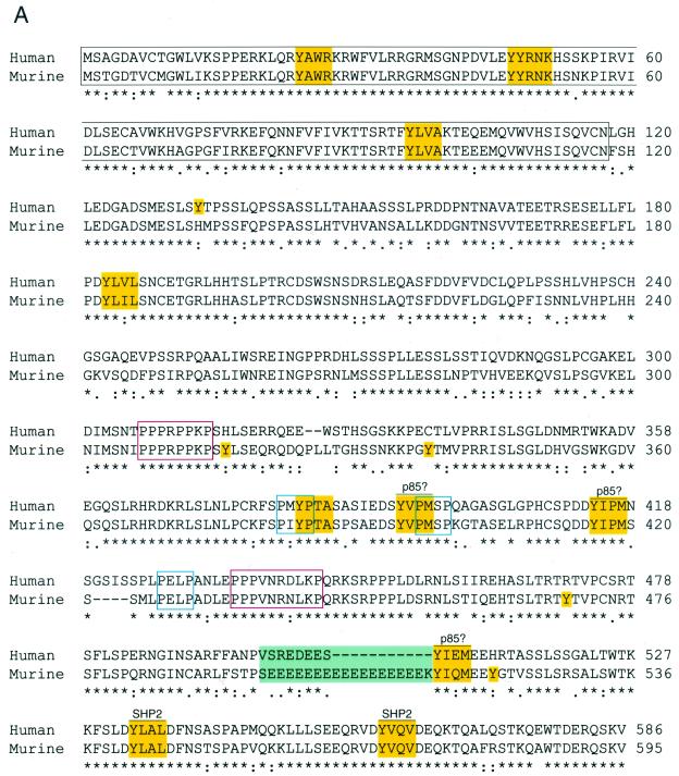
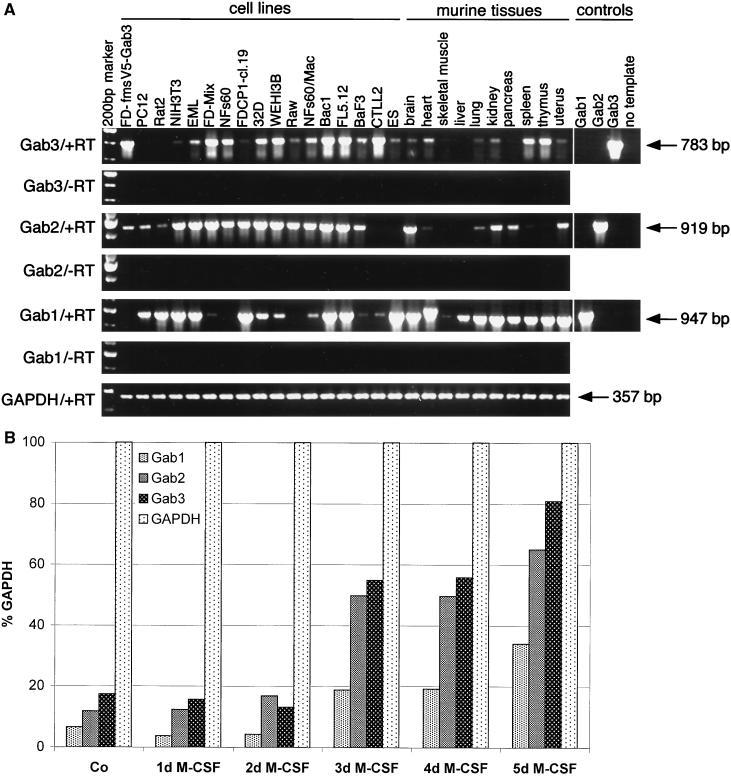
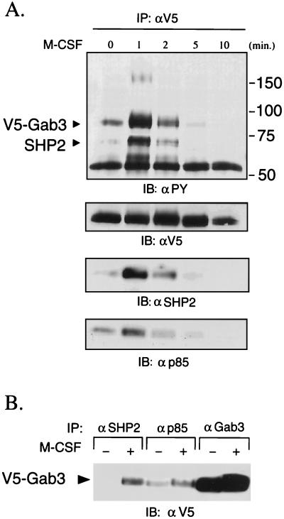
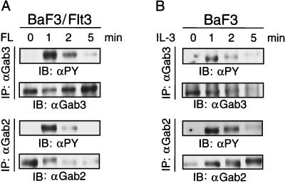
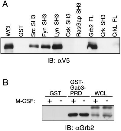
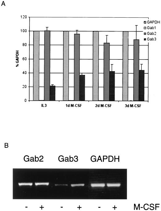
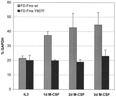
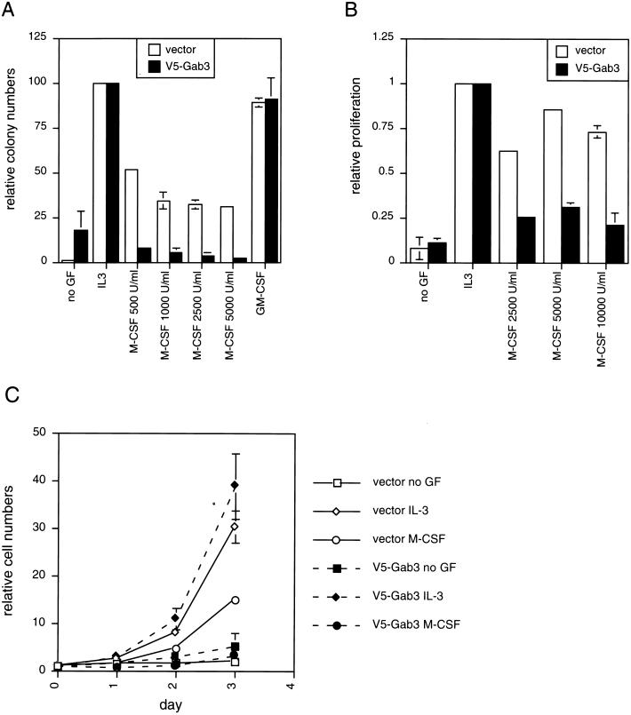
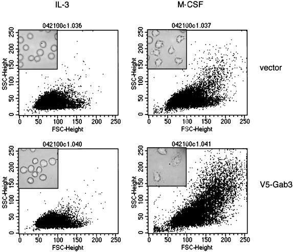
Using the FDC-P1 cell line expressing the exogenous macrophage colony-stimulating factor (M-CSF) receptor, Fms, we have analyzed the role of a new mammalian DOS/Gab-related signaling protein, called Gab3, in macrophage cell development of the mouse. Gab3 contains an amino-terminal pleckstrin homology domain, multiple potential sites for tyrosine phosphorylation and SH2 domain binding, and two major polyproline motifs potentially interacting with SH3 domains. Among the growing family of Gab proteins, Gab3 exhibits a unique and overlapping pattern of expression in tissues of the mouse compared with Gab1 and Gab2. Gab3 is more restricted to the hematopoietic tissues such as spleen and thymus but is detectable at progressively lower levels within heart, kidney, uterus, and brain. Like Gab2, Gab3 is tyrosine phosphorylated after M-CSF receptor stimulation and associates transiently with the SH2 domain-containing proteins p85 and SHP2. Overexpression of exogenous Gab3 in FD-Fms cells dramatically accelerates macrophage differentiation upon M-CSF stimulation. Unlike Gab2, which shows a constant mRNA expression level after M-CSF stimulation, Gab3 expression is initially absent or low in abundance in FD cells expressing the wild-type Fms, but Gab3 mRNA levels are increased upon M-CSF stimulation. Moreover, M-CSF stimulation of FD-FmsY807F cells (which grow but do not differentiate) fails to increase Gab3 expression. These results suggest that Gab3 is important for macrophage differentiation and that differentiation requires the early phosphorylation of Gab2 followed by induction and subsequent phosphorylation of Gab3.


Figures

FIG. 1.
FIG. 1.
Amino acid sequence and domain structure of the human and murine Gab3 proteins. (A) The sequences of the 595-amino acid murine Gab3 protein and the 586-amino acid human Gab3 protein deduced from the nucleotide open reading frames of each cDNA are shown in the single-letter amino acid designations. The PH domain is outlined in black, and tyrosine-containing motifs with potential for interacting with SH2 domains when phosphorylated are highlighted in yellow (as are single, unmatched tyrosine [Y] residues). Several PXXP motifs with potential for SH3 domain interactions are shown outlined in blue. The red outlined sequences represent highly conserved regions contained in all three Gab proteins, and the green rectangle contains a polyglutamic acid stretch. The consensus line below the amino acid sequences utilizes an asterisk for complete identity, a colon for a conserved substitution, and a period for a semiconserved substitution. (B) Diagram showing the relative structural details of Gab1, Gab2, and Gab3. The amino-terminal PH domains are highlighted in black, the location of tyrosine amino acids are shown along the length of each protein (Y), and PXXP amino acid motifs are designated in black with a “P” below the relevant location. The position of the major conserved polyproline-rich sequences are designated by open red rectangles on each Gab protein. The scale above the C terminus of Gab1 indicates the length of 50 amino acids. (C) Amino acid sequence alignment of the Gab1, Gab2, and Gab3 PH domains with tyrosine residues highlighted (yellow).
FIG. 2.
FIG. 2.
RT-PCR analysis of Gab1, Gab2, and Gab3 mRNA expression in cell lines and tissues of the mouse (and rat). (A) The designations Gab3/+RT and Gab3/−RT indicate PCRs using primers specific for Gab3 and conducted with or without prior reverse transcription, respectively. Similar reactions were performed for detecting Gab2, Gab1, and GAPDH as positive control. Additional positive and negative RT-PCR experiments demonstrated the specificity of each primer set for its corresponding Gab protein. These reactions are shown on the right-hand end (controls), and the nucleotide length for the expected products is adjacent to each arrow. The cell lines are PC12, Rat2 (rat cell lines; all others are mouse derived); NIH 3T3 (fibroblast); EML, FD-Mix (pluripotent, hematopoietic); NFS60, FDC-P1 cl 19, 32D, WEHI 3B, Raw, NFS60/Mac, Bac1 (myeloid); FL5.12, BaF3 (B cells); CTLL2 (T cells); ES (129Sv-derived Ak7 stem cells). (B) RT-PCR analysis of Gab1, Gab2, and Gab3 in primary BM cells cultivated in the presence of M-CSF. BM cells were collected and cultured in the presence of 1,000 U of M-CSF per ml for 5 days. Total RNA was isolated before the addition of M-CSF (Co) and at the time points indicated. RT-PCRs were performed using specific primers for Gab1, Gab2, Gab3, or GAPDH. Quantification of the ethidium bromide-stained DNA bands in gels was performed with ImageQuant software. Gab1, Gab2, and Gab3 signals were normalized to the GAPDH signal and are shown as percentages of GAPDH signals. One representative example of three independent experiments is shown.
FIG. 3.
FIG. 3.
Gab3 is tyrosine phosphorylated by M-CSF and associates with SHP2 and p85/PI3K. (A) FD-Fms-expressing mGab3V5 cells were starved, stimulated with 2,500 U of M-CSF per ml, and lysed at the times indicated. The Gab3 proteins were immunoprecipitated (IP) with anti-V5 antibody and the immune complex was separated by polyacrylamide gel electrophoresis. After electrophoretic transfer of proteins onto nitrocellulose filters, phosphotyrosine (PY)-containing proteins were detected by immunoblotting (IB) with the 4G10 monoclonal antibody. Likewise, equal levels of V5-tagged Gab3 are shown by immunoblotting with antibody to the V5 epitope. The transient M-CSF-dependent association of mGab3V5 with SHP2 and p85 is detected by immunoblotting the anti-V5 IP with the respective antibodies shown in the bottom two panels. The migration positions of Gab3 and SHP2 are shown in the top left panel, and the positions of molecular weight marker proteins are on the right. (B) Reciprocal co-IPs showing the association of SHP2 and p85 with V5-Gab3. Cell lysates from quiescent (−) or M-CSF (2,500 U/μl)-stimulated (+) FD-Fms cells expressing mGab3V5 were immunoprecipitated with antibodies as indicated. The immunocomplexes were separated by sodium dodecyl sulfate-7.5% polyacrylamide gel electrophoresis, transferred onto nitrocellulose filters, and immunoblotted with anti-V5 antibody.
FIG. 4.
FIG. 4.
Tyrosine phosphorylation of Gab3 in response to activation by various ligand-receptor systems. (A) BaF3 cells expressing the Flt3 receptor were stimulated with FL for the times indicated and the endogenous Gab3 or Gab2 protein was immunoprecipitated from aliquots of the cell lysate with antibody specific to each protein. After gel electrophoresis and transfer to nitrocellulose, the filters were immunoblotted with antibody to either phosphotyrosine (IB: αPY), Gab2 (IB: αGab2), or Gab3 (IB: αGab3). (B) BaF3 cells were stimulated with IL-3 and the tyrosine phosphorylation of Gab3 and Gab2 was tested as described above.
FIG. 5.
FIG. 5.
Potential interactions of Gab3 with various SH3 domains measured by GST pull-down assays. (A) Lysates from unstimulated FD-Fms(Gab3V5) cells were reacted with GST fused to SH3 domains from the various proteins identified above the figure and the presence of the V5-tagged Gab3 protein identified by immunoblotting. The left-hand lane marked WCL (whole-cell lysate) shows the position of mGab3V5. (B) The PRD of the Gab3 protein was fused to GST and used in pull-down experiments to demonstrate an interaction with Grb2. Immunoblotting with antibody to Grb2 (IB: αGrb2) identified Grb2 in the whole-cell lysates of the FD-Fms(Gab3V5) cells and the complex with GST-Gab3-PRD but not with GST alone. Where indicated, M-CSF was present in excess for 5 min at room temperature.
FIG. 6.
FIG. 6.
Expression of Gab protein mRNA in FD-Fms and NFS60-Fms cells. (A) RT-PCR analysis of Gab1, Gab2, and Gab3 in FD-Fms UW cells. FD-Fms UW cells were either cultured in IL-3 or stimulated with 2,500 U of M-CSF per ml, and total RNA was isolated at the time points indicated. Quantification of the PCR products was performed as described in the legend for Fig. 2B. Standard deviations of three independent experiments are indicated. (B) RT-PCR analysis of Gab2 and Gab3 in NFS60-Fms cells. NFS60-Fms cells were either cultured in IL-3 (−) or stimulated with 2,500 U of M-CSF per ml (+) for 3 days. Total RNA was isolated and RT-PCRs were performed using specific primers for Gab2, Gab3, or GAPDH. A representative ethidium bromide-stained gel of PCR products from three independent experiments is shown.
FIG. 7.
FIG. 7.
Comparison of Gab3 signals in FD-Fms WT and Y807F cells. FD-Fms WT and FD-FmsY807F cells were either cultured in IL-3 or stimulated with 2,500 U of M-CSF per ml and total RNA was isolated at the time points indicated. RT-PCRs and quantification of the PCR products were performed as described in the legend to Fig. 2B. Data represent mean values of three independent experiments and standard deviations are indicated.
FIG. 8.
FIG. 8.
Biological effects of mGab3V5 overexpression on growth of FD-Fms cells. (A) Soft agar colony assays were performed on FD-Fms cells expressing either the empty vector or the vector expressing the mGab3V5. The agar assay conditions included either no growth factor, IL-3, GM-CSF, or increasing M-CSF concentrations from 500 to 5,000 U/ml. (B) An MTT proliferation assay of the vector- or mGab3V5-FD-Fms cells was performed for the IL-3- and M-CSF-stimulated cells. (C) Cell growth in liquid suspension culture was measured by cell counts at 24-h intervals after ligand addition. The vector- or Gab3-expressing FD-Fms cells were grown without ligand or with either IL-3 or M-CSF as indicated in the legend to the right of the figure.
FIG. 9.
FIG. 9.
mGab3V5 overexpression in FD-Fms cells facilitates M-CSF-induced morphological differentiation. FD-Fms(vector) and FD-Fms(mGab3V5) cells were grown in liquid culture for 2 days with the indicated growth factor (IL-3 or 2,500-U/ml M-CSF) and analyzed by flow cytometry for forward and side scatter, which reflect the size and intracellular architecture, respectively. The inset within each plot shows a phase-contrast photographic image of the corresponding cells and culture conditions.

Similar articles

Cited by

References

    1. Allard, J. D., H. C. Chang, R. Herbst, H. McNeill, and M. A. Simon. 1996. The SH2-containing tyrosine phosphatase corkscrew is required during signaling by sevenless, Ras1 and Raf. Development 122:1137–1146. - PubMed
    1. Bardelli, A., P. Longati, D. Gramaglia, M. C. Stella, and P. M. Comoglio. 1997. Gab1 coupling to the HGF/Met receptor multifunctional docking site requires binding of Grb2 and correlates with the transforming potential. Oncogene 15:3103–3111. - PubMed
    1. Bausenwein, B. S., M. Schmidt, B. Mielke, and T. Raabe. 2000. In vivo functional analysis of the daughter of sevenless protein in receptor tyrosine kinase signaling. Mech. Dev. 90:205–215. - PubMed
    1. Bourette, R. P., G. M. Myles, K. Carlberg, and L. R. Rohrschneider. 1995. Uncoupling of the proliferation and differentiation signals mediated by the murine macrophage colony-stimulating factor receptor expressed in myeloid FDC-P1 cells. Cell Growth Differ. 6:631–645. - PubMed
    1. Bourette, R. P., G. M. Myles, J.-L. Choi, and L. R. Rohrschneider. 1997. Sequential activation of phosphatidylinositol 3-kinase and phospholipase C-γ2 by the M-CSF receptor is necessary for differentiation signaling. EMBO J. 16:5880–5893. - PMC - PubMed

Publication types

MeSH terms

Substances

Associated data