. 2002 Jan;22(1):286-97.
doi: 10.1128/MCB.22.1.286-297.2002.

Yeast hnRNP K-like genes are involved in regulation of the telomeric position effect and telomere length

Affiliations

Yeast hnRNP K-like genes are involved in regulation of the telomeric position effect and telomere length

Oleg Denisenko et al. Mol Cell Biol. 2002 Jan.

Abstract

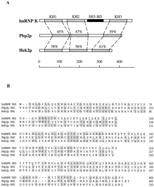
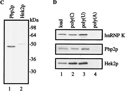
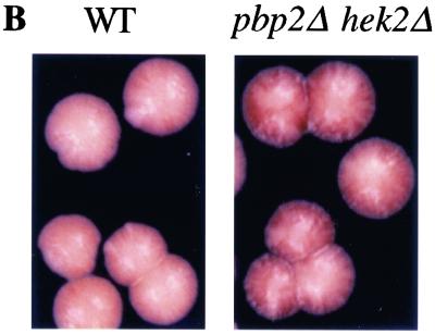
Mammalian heterogeneous nuclear ribonucleoprotein K (hnRNP K) is an RNA- and DNA-binding protein implicated in the regulation of gene expression processes. To better understand its function, we studied two Saccharomyces cerevisiae homologues of the human hnRNP K, PBP2 and HEK2 (heterogeneous nuclear RNP K-like gene). pbp2Delta and hek2Delta mutations inhibited expression of a marker gene that was inserted near telomere but not at internal chromosomal locations. The telomere proximal to the ectopic marker gene became longer, while most of the other telomeres were not altered in the double mutant cells. We provide evidence that telomere elongation might be the primary event that causes enhanced silencing of an adjacent reporter gene. The telomere lengthening could, in part, be explained by the inhibitory effect of hek2Delta mutation on the telomeric rapid deletion pathway. Hek2p was detected in a complex with chromosome regions proximal to the affected telomere, suggesting a direct involvement of this protein in telomere maintenance. These results identify a role for hnRNP K-like genes in the structural and functional organization of telomeric chromatin in yeast.


Figures

FIG. 1
FIG. 1
Comparison of the human K protein and the yeast homologues Pbp2p and Hek2p. (A) Similarity between the deduced amino acid sequences of the human hnRNP K protein and S. cerevisiae Pbp2p (YBR233w) and Hek2p (YBL032w). KH1 to -3, KH domains (percentage of similarity is shown); and SH3-BD, SH3-binding domain. The scale depicts length in amino acid residues. (B) results of KH domain alignment. Identical positions are shaded. (C) Murine K protein and yeast PBP2 and HEK2 cDNAs were transcribed and translated in vitro. 35S-labeled Pbp2p and Hek2p translational products were analyzed by SDS-electrophoresis and autoradiography. (D) RNA- and protein-binding specificity of K protein, Pbp2p, and Hek2p. 35S-labeled translational products were incubated with agarose beads bearing different homopolynucleotides in a buffer containing 150 mM KCl. After incubation the beads were washed with the same buffer, and bound proteins were eluted with SDS sample buffer and analyzed by SDS-gel electrophoresis and autoradiography. Load, 20% of the sample used in the binding reaction.
FIG. 1
FIG. 1
Comparison of the human K protein and the yeast homologues Pbp2p and Hek2p. (A) Similarity between the deduced amino acid sequences of the human hnRNP K protein and S. cerevisiae Pbp2p (YBR233w) and Hek2p (YBL032w). KH1 to -3, KH domains (percentage of similarity is shown); and SH3-BD, SH3-binding domain. The scale depicts length in amino acid residues. (B) results of KH domain alignment. Identical positions are shaded. (C) Murine K protein and yeast PBP2 and HEK2 cDNAs were transcribed and translated in vitro. 35S-labeled Pbp2p and Hek2p translational products were analyzed by SDS-electrophoresis and autoradiography. (D) RNA- and protein-binding specificity of K protein, Pbp2p, and Hek2p. 35S-labeled translational products were incubated with agarose beads bearing different homopolynucleotides in a buffer containing 150 mM KCl. After incubation the beads were washed with the same buffer, and bound proteins were eluted with SDS sample buffer and analyzed by SDS-gel electrophoresis and autoradiography. Load, 20% of the sample used in the binding reaction.
FIG. 2
FIG. 2
Enhanced TPE in pbp2Δ hek2Δ strains. (A) PBP2 HEK2 (UCC509 [44]), pbp2Δ (DY14), hek2Δ (DY15), and pbp2Δ hek2Δ (DY16) strains were grown on yeast-peptone-dextrose agar plates for 2 days; separate colonies were grown in yeast-peptone-dextrose liquid medium. Tenfold serial dilutions of overnight cultures were plated on either complete or 5-FOA-supplemented agar media. The graph represents the fraction of cells forming colonies on 5-FOA medium compared with cells forming colonies on nonselective medium, from at least four independent experiments. (B) Subtelomeric ADE2 gene expression in PBP2 HEK2 and pbp2Δ hek2Δ strains. Wild-type (PBP2 HEK2, strain UCC3505) and mutant (pbp2Δ hek2Δ, strain DY28) cells with the ADE2 gene introduced near the VR telomere (52) were grown on yeast-peptone-dextrose agar for 3 days at 30°C, and then the dish was kept at 4°C for 1 week to develop the color. (C) Growth of wild-type and hek mutant strains containing URA3 inserted into the HML locus on media either lacking uracil or containing 5-FOA. Tenfold dilutions of wild-type (WT; strain UCC3515 [53]), pbp2Δ (strain DY115), hek2Δ (strain DY116), pbp2Δ hek2Δ (strain DY117), and ppr1Δ (strain UCC4565) strains were spotted onto complete medium (Complete), medium lacking uracil (−URA), or medium containing 5-FOA. The experiment was repeated three times, and the results of one representative experiment are shown. (D) Level of URA3 mRNA expression in pbp2Δ hek2Δ strains. Individual colonies were grown to mid-log phase in yeast-peptone-dextrose, cells were harvested by centrifugation, and total RNA was extracted by the phenol/glass bead method. RNA samples (5 μg each) were examined by Northern blot analysis with fragments of URA3 (upper panel) or ACT1 (lower panel) genes used as probes. The position of URA3, ura3-52, and ACT1 transcripts is shown. Strains used: 1 to 3, UCC509 (PBP2 HEK2); 4 to 6, DY16 (pbp2Δ hek2Δ); 7, UCC511 (PBP2 HEK2 [44]); 8, DY19 (pbp2Δ hek2Δ); and 9, YPH250 (PBP2 HEK2). Numbers above the panels represent frequencies of 5-FOA-resistant cells. N/A, not applicable. The chromosomal constructs used in these experiments are shown below.
FIG. 2
FIG. 2
Enhanced TPE in pbp2Δ hek2Δ strains. (A) PBP2 HEK2 (UCC509 [44]), pbp2Δ (DY14), hek2Δ (DY15), and pbp2Δ hek2Δ (DY16) strains were grown on yeast-peptone-dextrose agar plates for 2 days; separate colonies were grown in yeast-peptone-dextrose liquid medium. Tenfold serial dilutions of overnight cultures were plated on either complete or 5-FOA-supplemented agar media. The graph represents the fraction of cells forming colonies on 5-FOA medium compared with cells forming colonies on nonselective medium, from at least four independent experiments. (B) Subtelomeric ADE2 gene expression in PBP2 HEK2 and pbp2Δ hek2Δ strains. Wild-type (PBP2 HEK2, strain UCC3505) and mutant (pbp2Δ hek2Δ, strain DY28) cells with the ADE2 gene introduced near the VR telomere (52) were grown on yeast-peptone-dextrose agar for 3 days at 30°C, and then the dish was kept at 4°C for 1 week to develop the color. (C) Growth of wild-type and hek mutant strains containing URA3 inserted into the HML locus on media either lacking uracil or containing 5-FOA. Tenfold dilutions of wild-type (WT; strain UCC3515 [53]), pbp2Δ (strain DY115), hek2Δ (strain DY116), pbp2Δ hek2Δ (strain DY117), and ppr1Δ (strain UCC4565) strains were spotted onto complete medium (Complete), medium lacking uracil (−URA), or medium containing 5-FOA. The experiment was repeated three times, and the results of one representative experiment are shown. (D) Level of URA3 mRNA expression in pbp2Δ hek2Δ strains. Individual colonies were grown to mid-log phase in yeast-peptone-dextrose, cells were harvested by centrifugation, and total RNA was extracted by the phenol/glass bead method. RNA samples (5 μg each) were examined by Northern blot analysis with fragments of URA3 (upper panel) or ACT1 (lower panel) genes used as probes. The position of URA3, ura3-52, and ACT1 transcripts is shown. Strains used: 1 to 3, UCC509 (PBP2 HEK2); 4 to 6, DY16 (pbp2Δ hek2Δ); 7, UCC511 (PBP2 HEK2 [44]); 8, DY19 (pbp2Δ hek2Δ); and 9, YPH250 (PBP2 HEK2). Numbers above the panels represent frequencies of 5-FOA-resistant cells. N/A, not applicable. The chromosomal constructs used in these experiments are shown below.
FIG. 3
FIG. 3
The effect of PBP2 and HEK2 disruption on the length of telomeres. (A) UCC519 (PBP2 HEK2 [44]) and DY25 (pbp2Δ hek2Δ) strains were grown on yeast-peptone-dextrose agar media; separate colonies were grown overnight in the same liquid media. Cells were collected by centrifugation; DNA was extracted by the phenol/glass bead method (Materials and Methods). One microgram of DNA was digested with either HindIII (upper panel, lanes 1 and 2), HindIII and BamHI (lanes 3 and 4), or XhoI (lower panel, lanes 1 and 2). The products were analyzed by the Southern method with either VR (upper panel, lanes 1 through 4) or Y′ (lower panel, lanes 1 and 2) probes. Arrowhead and arrow mark the position of DNA fragments bearing the telomeric repeat either proximal to the URA3 gene (VR) or corresponding to the Y′-type chromosomal ends (Y′) respectively. WT, wild type; mut, mutant. (B) UCC513 (PBP2 HEK2 [44]) and DY22 (PBP2 hek2Δ) strains were grown and analyzed as described for panel A. DNA was digested with XhoI (lanes 1 and 2) and was analyzed by the Southern method with the Y′ probe. For lanes 3 and 4, genomic DNA was cut with SmaI (this site is introduced with URA3 to VR. The arrowhead marks DNA fragment corresponding to Y′ probe. All other chromosomal ends recognized by the Y′ probe do not contain a SmaI site within 25 kb adjacent to telomeres); the 11-kb zone was purified from gel, cut with XhoI, and then analyzed by the Southern method with the Y′ probe. WT, PBP2 HEK2 cells; mut, PBP2 hek2Δ cells; asterisk, position of DNA fragment specific to the sample purified from hek2Δ cells. (C) DNA samples shown in panel B, lanes 1 and 2, were cut with XhoI and analyzed by Southern blotting with a probe specific to the TG1-3 repeat. Position of fragments corresponding to the X- and Y′-type chromosomal ends is marked. The asterisk indicates the position of DNA fragment specific to the sample purified from hek2Δ (mut) cells. (D) Chromosomal constructs and probes (bold lines) used to measure telomere length. The diffuse dark end represents the telomeric (TG1-3)n repeat and is not drawn to scale.
FIG. 4
FIG. 4
RT-PCR analysis of transcription of the subtelomeric region in pbp2Δ hek2Δ cells. After DNase treatment, RNA samples (1 μg each, same as shown in Fig. 2D) were reverse transcribed with random hexanucleotide mixture as a primer and then amplified by PCR with primers specific to ACT1, URA3, and the chromosomal region localized between the URA3 gene and the neighboring TG1-3 repeat (INT). −RT, no RT added, DNA, 1 μg of total DNA was used as a template in PCR. Strains used: 1 and 2, UCC509 (WT, PBP2 HEK2); and 3 and 4, DY16 (mut, pbp2Δ hek2Δ).
FIG. 5
FIG. 5
Telomere length in 5-FOA-resistant cells. (A) The UCC523 strain (PBP2 HEK2 ppr1 [44]) that was used in these experiments contains the URA3 gene at the end of the VR chromosome. Cells were grown on either complete medium (Complete, lanes 1 to 3) or complete medium supplemented with 5-FOA (5FOAR, lanes 4 to 6), and the individual colonies were then grown overnight in corresponding liquid media. DNA purified from the overnight cultures was cut with HindIII (upper panel) or XhoI (lower panel) and was analyzed by the Southern method with VR (upper panel) or Y′ (lower panel) probes. Arrowheads mark fragments corresponding to the probes. (B) Cells (UCC523, PBP2 HEK2 ppr1) were grown on either complete medium (Complete, lane 1) or complete medium supplemented with 5-FOA. Individual colonies from the 5-FOA plate were then consecutively passed on two plates without selection. Several colonies from the final plate were grown overnight in liquid medium (After 5FOA, lanes 2 to 5). To assess silencing of subtelomeric URA3, overnight cultures were plated as 10-fold serial dilutions of either complete medium without (lower panel, Complete) or with (middle panel, 5FOA) 5-FOA. DNA purified from the same cultures was cut with HindIII and was analyzed by the Southern method with VR probe (upper panel) to estimate the length of the VR telomere. (C) Wild-type (WT) (UCC509, PBP2 HEK2 PPR1 RAD52) and rad52Δ mutant (DY1000, PBP2 HEK2 PPR1 rad52Δ) strains were grown on either complete (Complete, lanes 1 and 5) or 5-FOA-supplemented (5FOAR, lanes 2 to 4 and 6 to 8) media, and the length of the VR telomere was analyzed as done for panel A. In contrast to RAD52 colonies, rad52Δ colonies were small and grew slowly on 5-FOA media. Chromosomal constructs and VR probe (bold line) are shown below.
FIG. 6
FIG. 6
TPE and telomere lengthening depend on the distance between the ectopic URA3 and TG-repeat region. Strains (UCC519, UCC521, and UCC523, all PBP2 HEK2 ppr1 [44]) with URA3 introduced to the end of chromosome VR at distances 1.1, 2.4, and 5.2 kb from the TG1-3 repeat region were grown on either complete medium or complete medium supplemented with 5-FOA. The efficiency of URA3 silencing was measured as described in the legend to Fig. 2. The lengthening of the VR telomere in 5-FOAR cells was measured as the difference between the length of this telomere found in the cells growing in complete medium and the length found in 5-FOAR cells (VR telomere length was measured as described in the legend to Fig. 5). Results of two independent experiments with at least three individual colonies per point are shown.
FIG. 7
FIG. 7
Effect of SIR3 overexpression on length of telomeres. (A) UCC509 strain (PBP2 HEK2 PPR1) transformed with either pVP plasmid (2μm LEU2) (Vector) or the same plasmid containing the SIR3 gene (SIR3) was grown on agar plates lacking Leu (−Leu). Individual colonies were grown overnight in liquid −Leu medium, cells were counted, and 10-fold serial dilutions were plated on either −Leu or 5-FOA, −Leu agar plates. (B) DNA purified from the overnight cultures was cut with HindIII and analyzed by the Southern method with VR probe. Total, cells were grown on −Leu agar plates; and 5FOA-resistant, cells were grown on −Leu agar plates supplemented with 5-FOA. At least three colonies of each strain were analyzed; each lane corresponds to DNA purified from one colony. Chromosomal construct and VR-specific probe (bold line) are shown below gel.
FIG. 8
FIG. 8
Effect of hek mutations on TRD. UCC509 (WT, PBP2 HEK2), DY14 (pbp2Δ HEK2), DY15 (PBP2 hek2Δ), and DY16 (pbp2Δ hek2Δ) strains carrying URA3 near the VR telomere were grown on yeast-peptone-dextrose agar plates (NS, lanes 1 and 10 in the upper panel and 1 and 9 in the lower panel) and then transferred on 5-FOA-supplemented plates. Individual 5-FOAR colonies (5FOAR, lanes 2 and 11 in the upper panel and 2 and 10 in the lower panel) were consecutively passed on three yeast-peptone-dextrose plates without selection (s1 to s3, lanes 3 to 9 and 12 to 18 in the upper panel and lanes 3 to 8 and 11 to 16 in the lower panel), with each round consisting of ∼25 generations of growth. Individual colonies from each plate were grown overnight in 2-ml cultures, DNA was isolated from each culture, and the length of the URA3-marked VR telomere was assayed as described in the legend to Fig. 3. The chromosomal construct and the position of the HindIII site used to cut DNA and the VR-specific probe (bold line) are shown below.
FIG. 9
FIG. 9
In vivo association of Hek2p with the VR subtelomeric region. (A) Whole-cell extracts were prepared from formaldehyde cross-linked strains, and chromatin was sonicated to an average DNA size of 0.5 to 1.0 kb. Immunoprecipitations (IP) were performed with anti-HA antibody 12CA5 (αHA). Precipitated DNA was analyzed by PCR with primers specific to the VR chromosomal (Chr.) end, shown schematically in the upper panel, and by primers designed to the Y′ locus or TY1 element. Thirty PCR cycles were carried out for the VR set of primers and 25 cycles for TY1 and the Y′ set of primers. PCR products were resolved in 1% agarose gels and stained with ethidium bromide. The DNA size standard is shown in lane 1. PCR products of immunoprecipitated DNA are shown in lanes 2 to 6. PCR products from the respective input extracts are shown in lanes 7 to 11. A threefold dilution of DNA sample from lane 11 was used in PCR, and the products are shown in lane 12. The following strains were used: UCC509 (wild type, lanes 2 and 7), DY119 (wild type, HEK2-3HA, lanes 3 and 4 and 8 and 9), DY122 (sir3Δ, HEK2-3HA, lanes 5 and 10), and AYH2.45 (wild type, SIR3HA [56], lanes 6 and 11). (B) Western blot analysis of HEK2-3HA expression. UCC509 (HEK2), DY119 (HEK2HA), and DY122 (HEK2HA sir3Δ) strains were grown exponentially. Proteins were extracted with SDS loading buffer from equal number of cells, separated by SDS-gel electrophoresis, transferred on polyvinylidene difluoride membrane, and stained with anti-HA antibody 12CA5.

Similar articles

Cited by

References

    1. Aparicio, O. M., B. L. Billington, and D. E. Gottschling. 1991. Modifiers of position effect are shared between telomeric and silent mating-type loci in S. cerevisiae. Cell 66:1279–1287. - PubMed
    1. Baber, J. L., D. Libutti, D. Levens, and N. Tjandra. 1999. High precision solution structure of the C-terminal KH domain of heterogeneous nuclear ribonucleoprotein K, a c-myc transcription factor. J. Mol. Biol. 289:949–962. - PubMed
    1. Bomsztyk, K., I. Van Seuningen, H. Suzuki, O. Denisenko, and J. Ostrowski. 1997. Diverse molecular interactions of the hnRNP K protein. FEBS Lett. 403:113–115. - PubMed
    1. Brachmann, C. B., A. Davies, G. J. Cost, E. Caputo, J. Li, P. Hieter, and J. D. Boeke. 1998. Designer deletion strains derived from Saccharomyces cerevisiae S288C: a useful set of strains and plasmids for PCR-mediated gene disruption and other applications. Yeast 14:115–132. - PubMed
    1. Burd, C. G., and G. Dreyfuss. 1994. Conserved structures and diversity of functions of RNA-binding proteins. Science 265:615–621. - PubMed

Publication types

MeSH terms

Substances