. 2002 Jan;22(1):357-69.
doi: 10.1128/MCB.22.1.357-369.2002.

Identification and characterization of a tissue-specific coactivator, GT198, that interacts with the DNA-binding domains of nuclear receptors

Affiliations

Identification and characterization of a tissue-specific coactivator, GT198, that interacts with the DNA-binding domains of nuclear receptors

Lan Ko et al. Mol Cell Biol. 2002 Jan.

Abstract

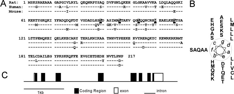
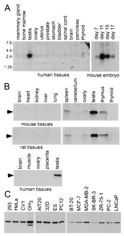
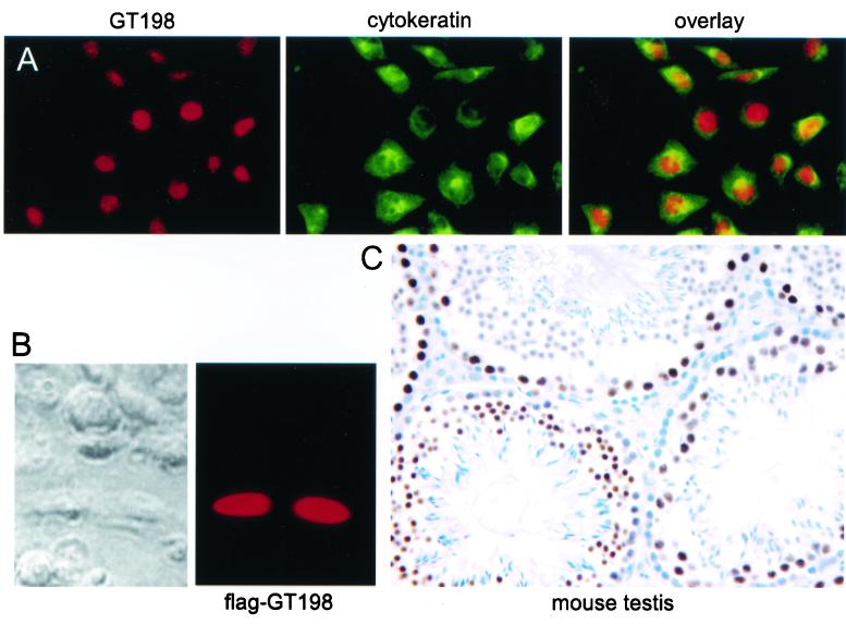
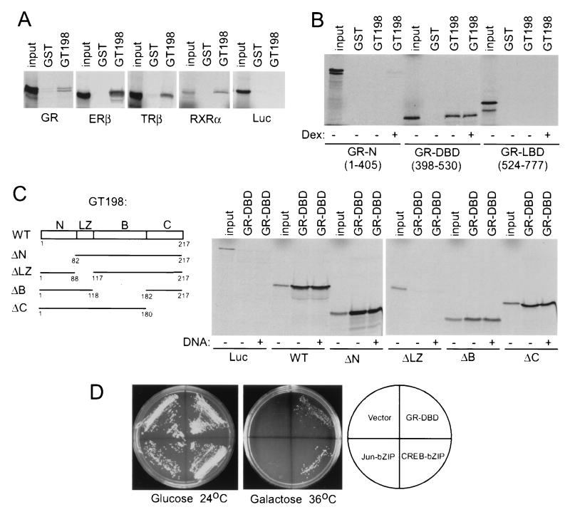
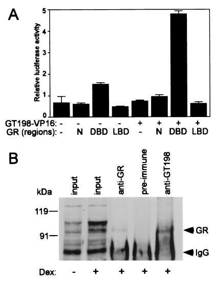
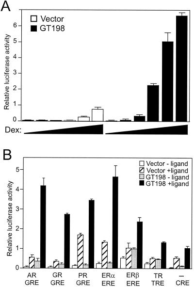
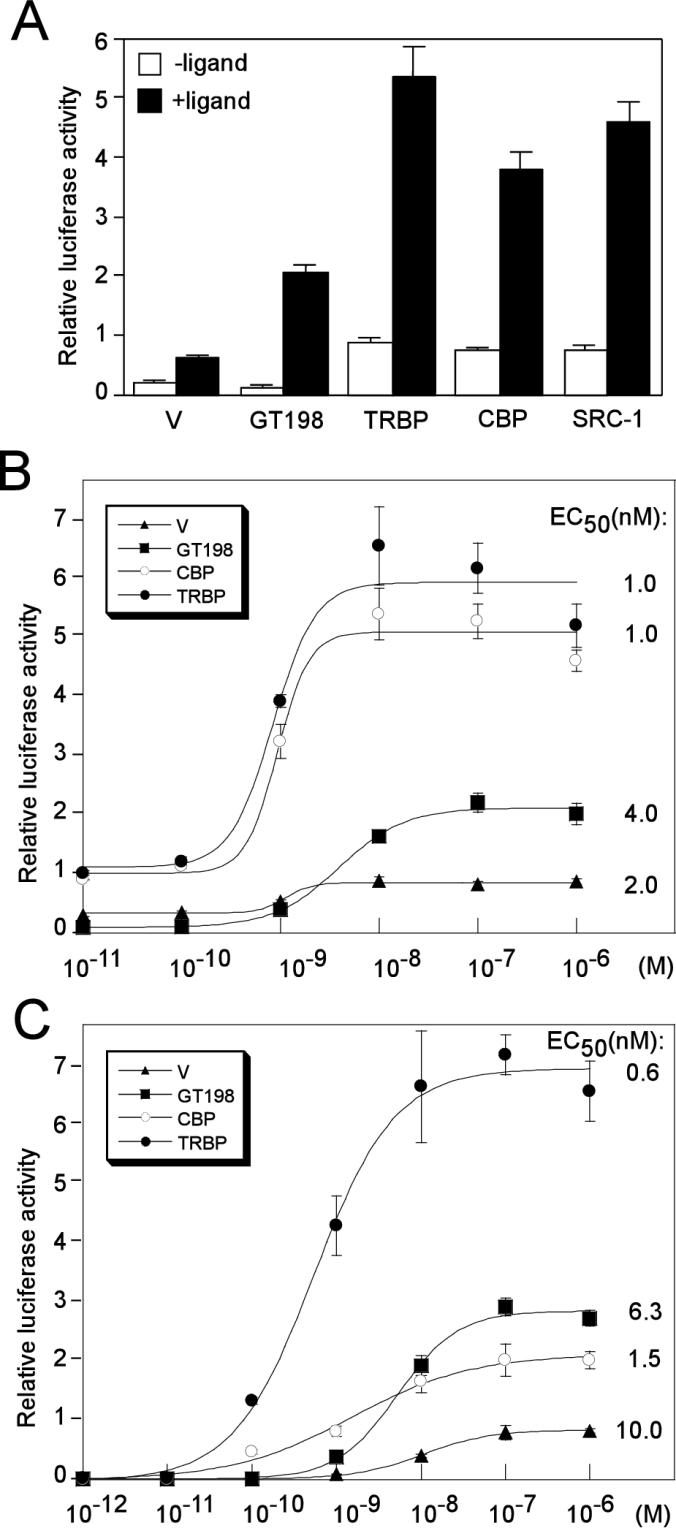
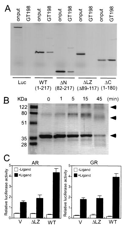
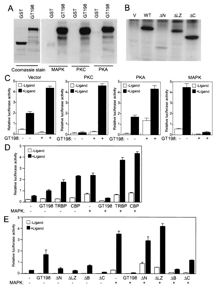
Gene activation mediated by nuclear receptors is regulated in a tissue-specific manner and requires interactions between nuclear receptors and their cofactors. Here, we identified and characterized a tissue-specific coactivator, GT198, that interacts with the DNA-binding domains of nuclear receptors. GT198 was originally described as a genomic transcript that mapped to the human breast cancer susceptibility locus 17q12-q21 with unknown function. We show that GT198 exhibits a tissue-specific expression pattern in which its mRNA is elevated in testis, spleen, thymus, pituitary cells, and several cancer cell lines. GT198 is a 217-amino-acid nuclear protein that contains a leucine zipper required for its dimerization. In vitro binding and yeast two-hybrid assays indicated that GT198 interacted with nuclear receptors through their DNA-binding domains. GT198 potently stimulated transcription mediated by estrogen receptor alpha and beta, thyroid hormone receptor beta1, androgen receptor, glucocorticoid receptor, and progesterone receptor. However, the action of GT198 was distinguishable from that of the ligand-binding domain-interacting nuclear receptor coactivators, such as TRBP, CBP, and SRC-1, with respect to basal activation and hormone sensitivity. Furthermore, protein kinase A, protein kinase C, and mitogen-activated protein kinase can phosphorylate GT198 in vitro, and cotransfection of these kinases regulated the transcriptional activity of GT198. These data suggest that GT198 is a tissue-specific, kinase-regulated nuclear receptor coactivator that interacts with the DNA-binding domains of nuclear receptors.


Figures

FIG. 1.
FIG. 1.
Coactivator GT198. (A) Alignment of amino acid sequences of rat GT198 with human GT198 (44) and mouse GT198 (TBP-IP) (50). Amino acid variations between species are indicated. Identical amino acids (−) are indicated. The heptad repeats of leucine and methionine residues are shaded. (B) An α-helical wheel diagram for the leucine dimerization domain of rat GT198 (aa 86 to 117). Aligned leucines are at the d position, and hydrophobic residues are at the a position. (C) Gene structure of mouse GT198 on chromosome 11. Introns are shown as lines and exons are depicted as boxes. Coding regions are shown in black and noncoding regions are in white.
FIG. 2.
FIG. 2.
Endogenous expression pattern of GT198. (A) Northern analysis of mRNA isolated from human tissues and mouse embryo at different stages. Blots were probed with rat GT198 cDNA. Human tissues examined are as shown on top of the panel. Mouse embryos were from day 7 to day 17, as indicated. (B) Western analysis using polyclonal anti-GT198 antibody. Results shown are for whole-cell lysates (50 μg/lane) from mouse, rat, and human tissues. (C) Western analysis with selected cell lines. Nuclear extract (50 μg) isolated from various cell lines was used (left and middle panels). Results for samples from whole-cell lysates of human cancer cells are shown in the panel on the right. ES, embryonic stem cells.
FIG. 3.
FIG. 3.
GT198 is a nuclear protein. (A) HeLa cells were methanol fixed and double stained with affinity-purified polyclonal anti-GT198 (left panel, red) and monoclonal anticytokeratin (middle panel, green), which was used as a cytoplasmic control. Secondary antibodies were anti-rabbit Cy3- and anti-mouse FITC-conjugated antibodies. An overlay of the two panels is shown on the right. (B) HeLa cells transfected with Flag-tagged GT198. Transfected cells were methanol fixed. Nuclear staining of GT198 was visualized by fluorescence with anti-Flag antibody (M2) and Cy3-conjugated secondary antibody. A phase-contrast view of the same field is shown at the left. (C) Immunohistochemical staining of the seminiferous tubules from mouse testis. The section was stained with affinity-purified anti-GT198 (1:100) and counterstained with hematoxylin.
FIG. 4.
FIG. 4.
GT198 interacts with DBD of nuclear receptors. (A) GST-GT198 fusion proteins were incubated with in vitro-translated, 35S-labeled full-length nuclear receptors (GR, ERβ, TRβ1, and RXRα). Bound proteins were resolved by SDS-PAGE and detected by autoradiography. GST alone was used as a control. Each nuclear receptor and luciferase (Luc) used as a negative control are indicated at the bottom. (B) The N-terminal region, DBD, and LBD of GR were in vitro translated and tested for binding GT198 in the presence (+) or absence (−) of 1 μM dexamethasone (Dex), as indicated. (C) Schematic representation of the deletion constructs of GT198 (left panel). Numbers indicate amino acids. The wild-type (WT) and deletion mutants of GT198 were in vitro translated and tested for the binding of GST-GR-DBD in the presence or absence of 30 ng of GRE-containing double-stranded DNA/ml as indicated. Input was 10%. (D) GR-DBD interacted with GT198 in a yeast two-hybrid assay. Full-length rat GT198 in pYes2 vector was cotransfected with bait plasmids, including GR-DBD, CREB-bZIP, c-Jun-bZIP, and pADNS empty vector, as indicated. Positive clones were detected at 36°C on a galactose plate. The same clones grown at 24°C on glucose plates were used as controls.
FIG. 5.
FIG. 5.
GT198 interacts with GR in vivo. (A) GT198 interacts with GR-DBD in a mammalian one-hybrid assay. CV-1 cells were transfected in 24-well plates with GRE-luciferase reporter (100 ng) and the following GR fragments: N (aa 1 to 405), DBD (aa 398 to 530), LBD (aa 524 to 777), or a vector control (100 ng). GT198-VP16 or control vector (100 ng) was also cotransfected. Luciferase activity was measured and is shown as the mean of triplicate transfections ± standard error. (B) Coimmunoprecipitation of endogenous GT198 and GR from HeLa cells. HeLa cells were treated with 100 nM dexamethasone (Dex) for 16 h. Nuclear extracts were isolated and incubated with anti-GT198 antibody, which was captured by protein A beads. Anti-GR antibody was used as a positive control and preimmune serum was used as a negative control. Bound GR was detected by immunoblotting with anti-GR antibody. Input of nuclear extracts (30 μg) from induced or uninduced cells is shown on the left. Arrows indicate GR and IgG.
FIG. 6.
FIG. 6.
GT198 is a nuclear receptor coactivator. (A) CV-1 cells were cotransfected in 24-well plates with MMTV luciferase reporter (100 ng), GR (10 ng), and GT198 (200 ng), or empty vector as control. Cells were grown in the presence of increasing amounts of dexamethasone (Dex) for GR (0, 10−11, 10−10, 10−9, 10−8, and 10−7 M). (B) CV-1 cells were transfected with the indicated luciferase reporters containing different minimal enhancer elements (2XGRE, 2XERE, F2 TRE, and 5XCRE) (100 ng). GT198 (200 ng), AR, GR, PR, ERα, ERβ, or TRβ1 (10 ng) and inducer plasmid containing the catalytic subunit of PKA (20 ng) for CRE were also cotransfected. For nuclear receptors, each cognate ligand (100 nM) was added to the medium 24 h after the transfection (mibolerone for AR, dexamethasone for GR, progesterone for PR, 17β-estradiol for ERα or ERβ, and T3 for TRβ1). Ligand-dependent activities are shown as the means of triplicate transfections ± standard errors.
FIG. 7.
FIG. 7.
Coactivator GT198 is functionally distinguishable from TRBP, CBP, and SRC-1. (A) Transcriptional activity of GT198 was compared with that of TRBP, CBP, and SRC-1 together with a vector control (V) (200 ng). CV-1 cells on 24-well plates were cotransfected with a GRE reporter (100 ng) and AR (10 ng) and treated with or without 100 nM mibolerone. (B) Transfections similar to those shown in panel A were performed with a GRE-luciferase reporter and AR, except that increasing amounts of ligand (mibolerone) were used (10−11, 10−10, 10−9, 10−8, 10−7, and 10−6 M). EC50 values are indicated at the right of each curve. Standard errors of EC50s were less than 5% (KaleidaGraph). (C) CV-1 cells were transfected as for panel B, except a MMTV-luc reporter and GR were used in the presence of indicated amounts of the ligand dexamethasone (10−12, 10−11, 10−10, 10−9, 10−8, 10−7, and 10−6 M). EC50 values are indicated on the right. Data shown are means of triplicate transfections ± standard errors. Vector control (filled triangle), GT198 (filled square), CBP (open circle), and TRBP (filled circle) are shown.
FIG. 8.
FIG. 8.
GT198 is a functional dimer. (A) Deletion of its leucine zipper abolished GT198 dimerization in vitro. Recombinant GST-GT198 fusion protein was incubated with 35S-labeled GT198 deletion fragments to detect homodimerization. Bound proteins were resolved by SDS-PAGE and detected by autoradiography. Luciferase (Luc) was used as a negative control. GT198 wild-type (WT) and mutants were as follows: WT (aa 1 to 217), N-terminal deletion (ΔN; aa 82 to 217), leucine zipper domain deletion (ΔLZ; Δ89–117) and C-terminal deletion (ΔC; aa 1 to 180). (B) Cross-linking of GT198. His-tagged recombinant GT198 (0.1 μg/μl) was treated with 0.15 mM glutaraldehyde for increasing amounts of time as indicated. Proteins were resolved by SDS-PAGE and detected by Coomassie blue staining. Arrowheads indicate the monomer, dimer, and polymer of GT198. (C) CV-1 cells were cotransfected with a GRE reporter (100 ng) and AR or GR (10 ng), along with wild-type GT198 (WT) or GT198 with the deletion at leucine zipper domain (ΔLZ; Δ89–117) or a pcDNA3 vector (V) as control. Ligand-dependent luciferase activities stimulated by 100 nM mibolerone or dexamethasone are shown as means of triplicate transfections ± standard errors.
FIG. 9.
FIG. 9.
GT198 is phosphorylated and regulated by PKA, PKC, and MAPK. (A) GST-GT198 was phosphorylated in vitro by PKA, PKC, and MAPK as described in Materials and Methods. After washing, phosphorylated GST-GT198 proteins were resolved by SDS-PAGE and detected by autoradiography. GST alone served as a negative control. Coomassie staining of GT198 and GST proteins, shown in the left panel, was to confirm that equal amounts of protein were used. (B) 293 cells were transfected with Flag-tagged GT198 and its deletion mutants. Abbreviations are as shown in the legend to Fig. 8A. Cells were Pi labeled, and GT198 proteins were immunoprecipitated with anti-Flag M2 antibody beads (Kodak). After washing, the bound protein was separated by SDS-PAGE and visualized by autoradiography. (C) Kinase stimulation regulates GT198 coactivation function. The effects of kinases on GT198 were analyzed in CV-1 cells. Cells were cotransfected in 24-well plates with GRE-luciferase reporter (100 ng) and AR (10 ng). Coactivator GT198 or a vector control (200 ng) was cotransfected with PKA, PKC, or MAPK (20 ng) or a vector control. Cells were treated with or without 100 nM mibolerone. (D) MAPK regulation of GT198 coactivation is specific. The effects of MAPK on GT198 were analyzed in CV-1 cells with GRE-luciferase reporter (100 ng) and AR (10 ng). Coactivator GT198, TRBP, CBP, or vector control (200 ng) was cotransfected with or without MAPK (20 ng) and treated with or without the 100 nM mibolerone. (E) HeLa cells were transfected as CV-1 cells were, except that wild-type and deletion mutants of GT198 (200 ng) were used. Data shown are means of triplicate transfections ± standard errors.

Similar articles

Cited by

References

    1. Albert, T. 1992. Structure of the leucine zipper. Curr. Opin. Genet. Dev. 2:205–210. - PubMed
    1. Angrand, P.-O., C. Coffinier, and M. C. Weiss. 1994. Response of the phosphoenolpyruvate carboxykinase gene to glucocorticoids depends on the integrity of the cAMP pathway. Cell Growth Differ. 5:957–966. - PubMed
    1. Anzick, S. L., J. Kononen, R. L. Walker, D. O. Azorsa, M. M. Tanner, X. Y. Guan, G. Sauter, O. P. Kallioniemi, J. M. Trent, and P. S. Meltzer. 1997. AIB1, a steroid receptor coactivator amplified in breast and ovarian cancer. Science 277:965–968. - PubMed
    1. Aronheim, A. 2000. Protein recruitment systems for the analysis of protein-protein interactions. Biochem. Pharmacol. 60:1009–1013. - PubMed
    1. Aronheim, A., E. Zandi, H. Hennemann, S. J. Elledge, and M. Karin. 1997. Isolation of an AP-1 repressor by a novel method for detecting protein-protein interactions. Mol. Cell. Biol. 17:3094–3102. - PMC - PubMed

Publication types

MeSH terms

Associated data