. 2002 Jan;22(1):370-7.
doi: 10.1128/MCB.22.1.370-377.2002.

p19(ARF) is dispensable for oncogenic stress-induced p53-mediated apoptosis and tumor suppression in vivo

Affiliations

p19(ARF) is dispensable for oncogenic stress-induced p53-mediated apoptosis and tumor suppression in vivo

Dawn Tolbert et al. Mol Cell Biol. 2002 Jan.

Abstract

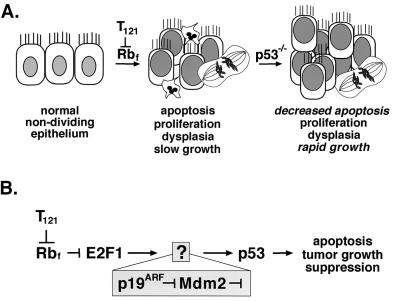
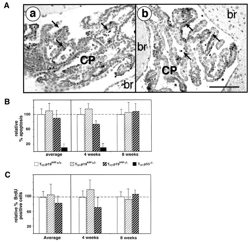
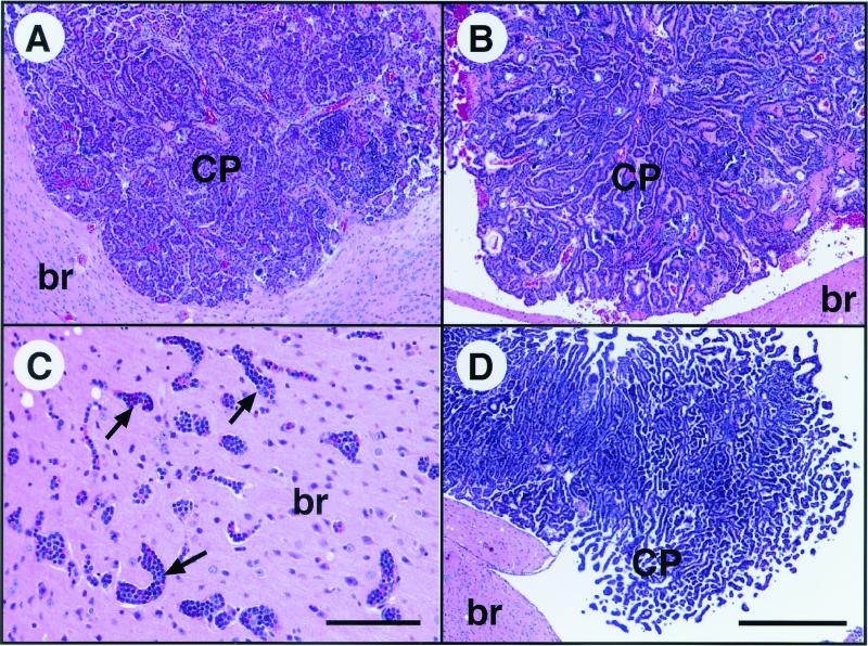
Recent studies have shown the p19(ARF) tumor suppressor to be involved in the response to oncogenic stress by regulating the activity of p53. This response is mediated by antagonizing the function of Mdm2, a negative regulator of p53, indicating a pathway for tumor suppression that involves numerous genes altered in human tumors. We previously described a transgenic mouse brain tumor model in which oncogenic stress, provided by cell-specific inactivation of the pRb pathway, triggers a p53-dependent apoptotic response. This response suppresses the growth of developing tumors and thus represents a bona fide in vivo tumor suppressor activity. We further showed that E2F1, a transcription factor known to induce p19(ARF) expression, was required for the response. Here, we use a genetic approach to test whether p19(ARF) functions to transduce the signal from E2F1 to p53 in this tumor suppression pathway. Contrary to the currently accepted hypothesis, we show that a deficiency in p19(ARF) has no impact on p53-mediated apoptosis or tumor suppression in this system. All measures of p53 function, including the level of apoptosis induced by pRb inactivation, the expression of p21 (a p53-responsive gene), and the rate of tumor growth, were comparable in mice with and without a functional p19(ARF) gene. Thus, although p19(ARF) is required in some cell types to transmit an oncogenic response signal to p53, it is dispensable for this function in an in vivo epithelial system. These results underscore the complexity of p53 tumor suppression and further indicate the existence of distinct cell-specific pathways that respond to similar stimuli.


Figures

FIG. 1.
FIG. 1.
p53 tumor suppression in brain epithelium. The diagram depicts previously elucidated steps in the development of choroid plexus tumors in TgT121 mice. (A) Cell-specific expression of the T121 transgene induces proliferation of normally nondividing epithelial cells by inactivating the pRb family proteins pRb, p107, and p130. p53-dependent apoptosis is then activated, as evidenced by the 85% reduction in apoptosis in TgT121;p53−/− mice. (B) Using a genetic approach, we previously showed that E2F1 acts upstream of p53 to induce apoptosis in response to T121. Previously, E2F1 was shown to activate p19ARF transcription, and p19ARF was shown to induce p53 activities by regulating Mdm2 in cultured cells (see the introduction). In the current report, we tested whether this pathway is operative to induce p53-dependent apoptosis and suppression of brain epithelial tumors in vivo.
FIG. 2.
FIG. 2.
p19ARF transcripts are induced in aberrantly proliferating CP epithelium. RNA in situ hybridization was performed with an antisense p19ARF RNA probe. (A) Hematoxylin and eosin (H & E)-stained section from the same brain as in panel C shows the morphology of CP in the lateral ventricle and is in an orientation similar to that of dark-field panels B to D. The borders between brain parenchyma (br) and the ventricles (V) are depicted with white dashed lines in panels B to D. No p19ARF expression above background is detected in the CP of nontransgenic mice (B). (Compare the signal in brain with that in CP.) p19ARF transcripts are induced in TgT121 CP (C). T121-induced p19ARF expression is p53 independent; the level of p19ARF transcripts is similar in TgT121;p53−/− CP (D) and TgT121 CP (C). (A) Bar, 200 μm (magnification is the same in all panels). Sense probe hybridization showed no signal above background (not shown).
FIG. 3.
FIG. 3.
T121-induced apoptosis and cell proliferation do not require p19ARF. T121 expression causes abnormal CP cell proliferation and apoptosis. About 85% of the apoptosis is p53-dependent (27) and requires E2F1 (18). The TUNEL assay was used to detect apoptosis (A). Apoptosis levels and morphology were indistinguishable between TgT121;p19ARF−/− (a) and TgT121;p19ARF+/+ (b) CP. Apoptotic nuclei are stained and appear black; representative apoptotic cells are indicated by arrows. The bar (b) is equal to 100 μm; both panels are of the same magnification. Quantitative analysis of apoptosis (B) and proliferation (C) in the CP of three mice each at 4 and 8 weeks of age was carried out. Levels are compared to that of TgT121;p19ARF+/+ tissue, which is considered to represent 100%. Average relative values for all mice of each age and genotype are shown in the left panels. For these data, error bars indicate the variation among mice. In the right panels, data for each mouse are presented, with error bars indicating the field-to-field variation in counts taken from 10 fields per brain. Different mice were used for apoptosis and proliferation assays so as to avoid any impact of BrdU incorporation on apoptosis levels. There is no significant difference in apoptotic indexes between TgT121;p19ARF+/+ and TgT121;p19ARF−/− mice (P = 0.601). Importantly, the index of TgT121;p19ARF−/− CP was significantly higher than that of TgT121;p53−/− CP (P < 0.05). The level of CP cell proliferation was determined by immunostaining of BrdU incorporated in vivo (C). The data show that p19ARF deficiency does not significantly alter cell proliferation (P = 0.288).
FIG. 4.
FIG. 4.
Tumor growth and morphology are unaffected by p19ARF deficiency. H & E-stained CP tumors from 20-week-old TgT121;p19ARF+/+ (A) and TgT121;p19ARF−/− (B) mice are similar in size and morphology. In contrast, tumors become life-threatening by 4 weeks of age in TgT121;p53−/− mice (D). Leukemia caused by the p19ARF−/− mutation is frequently observed invading the brains of TgT121;p19ARF−/− mice (C), a pathology that is present only in mice with compound genotypes and is thought to further reduce survival time (Table 1). Representative leukemia-filled vessels in the brain are indicated by arrows. The bar in panel C is equal to 100 μm. The bar in panel D is equal to 200 μm; panels A, B, and D are of the same magnification. br, brain tissue.
FIG. 5.
FIG. 5.
p53-mediated transactivation of p21 is unaffected by p19ARF deficiency. Brains were examined from nontransgenic (A and B), TgT121;p19ARF+/+ (C and D), TgT121;p19ARF−/− (E and F), and TgT121;p53−/− (G and H) mice. RNA in situ hybridization was performed on brain sections using an antisense p21 RNA probe and viewed by dark-field microscopy (B, D, F, and H). Adjacent sections were stained with H & E and viewed by bright-field microscopy to show the location and morphology of the CP (A, C, E, and G). No p21 expression is detected in normal CP (B). p21 expression is induced by expression of T121 (D) and remains unchanged in the absence of p19ARF (F). T121-induced p21 expression in CP requires functional p53 (H) (18). The bar in panel H is equal to 200 μm; all panels are of the same magnification. br, brain tissue.

References

    1. Ashcroft, M., and K. H. Vousden. 1999. Regulation of p53 stability. Oncogene 18:7637–7643. - PubMed
    1. Bates, S., A. C. Phillips, P. A. Clark, F. Stott, G. Peters, R. L. Ludwig, and K. H. Vousden. 1998. p14ARF links the tumour suppressors RB and p53. Nature 395:124–125. - PubMed
    1. de Stanchina, E., M. E. McCurrach, F. Zindy, S. Y. Shieh, G. Ferbeyre, A. V. Samuelson, C. Prives, M. F. Roussel, C. J. Sherr, and S. W. Lowe. 1998. E1A signaling to p53 involves the p19(ARF) tumor suppressor. Genes Dev. 12:2434–2442. - PMC - PubMed
    1. Eischen, C. M., J. D. Weber, M. F. Roussel, C. J. Sherr, and J. L. Cleveland. 1999. Disruption of the ARF-Mdm2–p53 tumor suppressor pathway in Myc-induced lymphomagenesis. Genes Dev. 13:2658–2669. - PMC - PubMed
    1. El-Deiry, W. S., T. Tokino, V. E. Velculescu, D. B. Levy, R. Parsons, J. M. Trent, D. Lin, W. E. Mercer, K. W. Kinzler, and B. Vogelstein. 1993. WAF1, a potential mediator of p53 tumor suppression. Cell 75:817–825. - PubMed

Publication types

MeSH terms

Substances