. 2001 Dec;12(12):3733-43.
doi: 10.1091/mbc.12.12.3733.

Interactions between syntaxins identify at least five SNARE complexes within the Golgi/prevacuolar system of the Arabidopsis cell

Affiliations
Free PMC article

Interactions between syntaxins identify at least five SNARE complexes within the Golgi/prevacuolar system of the Arabidopsis cell

A A Sanderfoot et al. Mol Biol Cell. 2001 Dec.
Free PMC article

Erratum in

  • Correction.
    [No authors listed] [No authors listed] Mol Biol Cell. 2021 Oct 1;32(20):cor1. doi: 10.1091/mbc.12.12.3733-corr. Mol Biol Cell. 2021. PMID: 34570654 Free PMC article. No abstract available.

Abstract

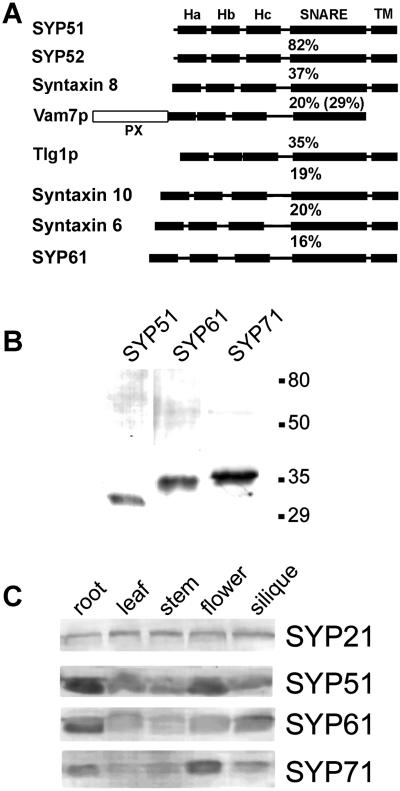
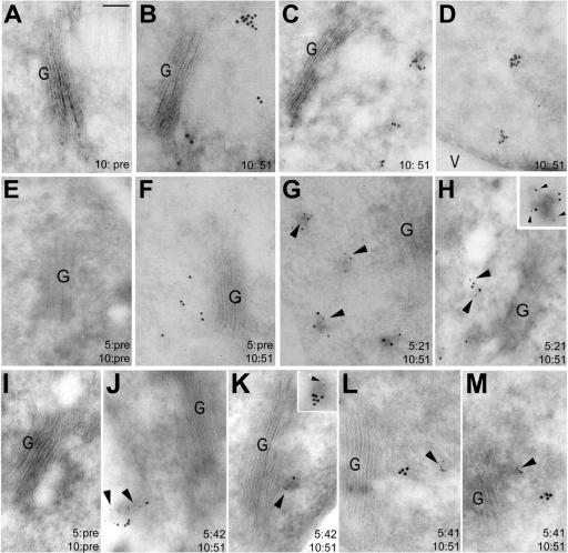
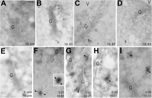
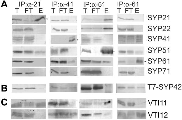
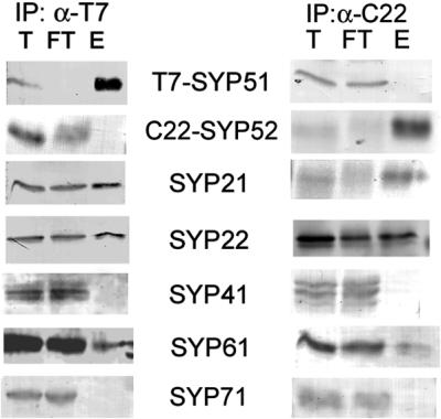
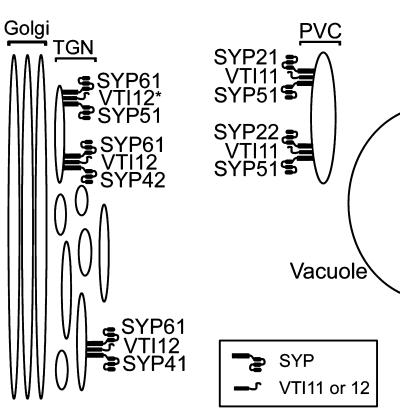
The syntaxin family of soluble N-ethyl maleimide sensitive factor adaptor protein receptors (SNAREs) is known to play an important role in the fusion of transport vesicles with specific organelles. Twenty-four syntaxins are encoded in the genome of the model plant Arabidopsis thaliana. These 24 genes are found in 10 gene families and have been reclassified as syntaxins of plants (SYPs). Some of these gene families have been previously characterized, with the SYP2-type syntaxins being found in the prevacuolar compartment (PVC) and the SYP4-type syntaxins on the trans-Golgi network (TGN). Here we report on two previously uncharacterized syntaxin groups. The SYP5 group is encoded by a two-member gene family, whereas SYP61 is a single gene. Both types of syntaxins are localized to multiple compartments of the endomembrane system, including the TGN and the PVC. These two groups of syntaxins form SNARE complexes with each other, and with other Arabidopsis SNAREs. On the TGN, SYP61 forms complexes with the SNARE VTI12 and either SYP41 or SYP42. SYP51 and SYP61 interact with each other and with VTI12, most likely also on the TGN. On the PVC, a SYP5-type syntaxin interacts specifically with a SYP2-type syntaxin, as well as the SNARE VTI11, forming a SNARE complex likely involved in TGN-to-PVC trafficking.


Figures

Figure 1
Figure 1
SYP5- and SYP6-type syntaxins of Arabidopsis. (A) Arabidopsis SYP51 (AF355755), SYP52 (AF355756), SYP61 (AF355754) human syntaxin 6 (O43752), syntaxin 8 (AF115323), syntaxin 10 (AF035531), yeast Tlg1p (NP_010756), and Vam7p (NP_011303) were aligned with the CUSTAL W algorithm. The aligned sequences were subjected to secondary structure predictions (NNPredict; Kneller et al., 1990). A comparison of the predicted secondary structures to the solved crystal structure of mammalian syntaxin 1 (Misura et al., 2000) produced the schematic diagrams of the predicted structure of the five proteins: Ha, Hb, and Hc are three N-terminal α-helices found in all syntaxins examined thus far, SNARE indicates the conserved coiled-coil domain found in all SNAREs, TM indicates the transmembrane helix found in the syntaxins, PX indicates the NADPH oxidase p40phox domain found at the N terminus of Vam7p. The percentage of amino acid identity of SYP51 to related syntaxins is shown above the schematic of each protein (percentage of identity of SYP51 and the SNARE-like domain of Vam7p shown in parentheses), whereas the percentage of identity of SYP61 to related syntaxins is shown below the schematics. Phylogenetic trees resulting from this alignment can be found at http://www.msu.edu/∼sanderfo/atsnare.htm. (B) Microsomal extracts of Arabidopsis root were separated by SDS-PAGE and transferred to nitrocellulose. Individual lanes were probed with affinity-purified rabbit antisera raised against Arabidopsis SYP51, SYP61, and SYP71. (C) Microsomal extracts were prepared from tissue extracted from each of the major organs of an Arabidopsis plant. Approximately equal amounts of total microsomal protein were separated by SDS-PAGE, transferred to nitrocellulose, and probed with antisera specific to the indicated Arabidopsis syntaxins. SYP21 is shown as a control because its expression is very consistent across different tissues.
Figure 2
Figure 2
SYP51 is found on the TGN and PVC. Cryosections of Arabidopsis root cells were immunolabeled with either SYP51 preimmune (A), affinity-purified SYP51 antibodies (B–D) followed by detection with 10 nm of colloidal gold. Double immunolabeling was performed with the use of cryosections from wild-type (E–H), or HA-SYP41 and T7-SYP42-expressing (I–M) roots. In all cases, SYP51 (or its preimmune) immunolabeling was detected with 10 nm of gold, whereas the second antibody was detected with 5 nm of gold (as indicated in the lower right on each panel). SYP51 preimmune (E and I) or affinity-purified antibodies (G and H, J–M, and insets) were detected with 10 nm of gold. A second labeling was then preformed with SYP21 preimmune (E and F) or affinity-purified antibodies (G and H, and inset), with a nonspecific control (I), T7-monoclonal antibodies (J and K), or HA-monoclonal antibodies (L and M), each detected with 5 nm of gold. All images were captured at the same magnification. G, Golgi; V, vacuole. Bar in A, 100 nm. Arrowheads are used to highlight the 5-nm gold particles.
Figure 3
Figure 3
SYP61 is found on the Golgi and TGN. Cryosections of Arabidopsis root cells were immunolabeled with either SYP61 preimmune (A) or affinity-purified SYP61 antibodies (B–D) followed by detection with 10 nm of colloidal gold. Double immunolabeling was performed with the use of cryosections from wild-type (E, F, and I) or from HA-SYP41- and T7-SYP42-expressing roots (G and H). SYP61 preimmune (E) or affinity-purified antibodies were detected with 10 nm of gold (F–H) or with 5 nm of gold (I). A second labeling was then performed with nonspecific (E), SYP21 affinity-purified (F), HA-monoclonal (G), or T7-monoclonal (H) antibodies followed by 5 nm of gold, or with SYP51 affinity-purified antibodies followed by 10 nm of gold (I). All images are shown at the same magnification. G, Golgi; V, vacuole. Bar in A, 100 nm. Arrowheads are used to highlight the 5-nm gold particles.
Figure 4
Figure 4
Interaction between Arabidopsis SYPs and VTI1-type SNAREs. Detergent extracts of Arabidopsis microsomes (see MATERIALS AND METHODS) from wild-type (A and C), or T7-SYP42-expressing (B) root cultures were immunoprecipitated with affinity-purified chicken anti-SYP21, affinity-purified rabbit anti-SYP41, anti-SYP51, or anti-SYP61. (A) Equal amounts of total (T) and flowthrough (FT), as well as approximately threefold more eluate (E) were separated by SDS-PAGE and then probed with rabbit antibodies specific for the indicated SYPs. Asterisks indicate the SYP used in a particular IP. (B) Extracts of plants expressing T7-SYP42 were used for IPs as in A to better differentiate between SYP41 and SYP42. (C) Extracts of wild-type plants were immunoprecipitated as in A except that 10-fold more eluate was loaded to better detect the small amounts of VTI11-type SNAREs coimmunoprecipitated in these experiments. Coimmunoprecipitation of SYP41 and VTI12 (and not VTI11) has been previously described in detail in Bassham et al. (2000).
Figure 5
Figure 5
Use of epitope tags to differentiate SYP51 and SYP52. Microsomes were prepared from Arabidopsis plants expressing T7-SYP51 and C22-SYP52. Detergent extracts of the microsomes were IPd with either immobilized T7-monoclonal (left) or CRUZ22-rabbit polyclonal antibodies (right). Equal amounts of the total (T) and flowthrough (FT), as well as approximately threefold more eluate (E) were separated by SDS-PAGE and then probed with rabbit antisera specific for the indicated proteins.
Figure 6
Figure 6
Schematic model of the Arabidopsis secretory system. A diagram of the late secretory pathway of Arabidopsis is shown. On two different domains of the TGN are found complexes containing SYP41 or 42, each in a complex containing VTI12 and SYP61. SYP51 is found on the SYP42-labeled domain of the TGN, as well as on the PVC. On the PVC, SYP51 is found in a complexes with VTI11 and either SYP21 or SYP22. SYP51 and SYP61 also form a complex, likely with VTI12 (although it is possible that this may be VTI11 instead, indicated by asterisk). The exact location of the SYP51/61 complex in unclear, but is drawn on the TGN because of the likelihood of containing VTI12 (see text).

References

    1. Antonin W, Holroyd C, Fasshauer D, Pabst S, Fischer Von Mollard G, Jahn R. A SNARE complex mediating fusion of late endosomes defines conserved properties of SNARE structure and function. EMBO J. 2000;19:6453–6464. - PMC - PubMed
    1. Bar-Peled M, Conceição AS, Frigerio L, Raikhel NV. Expression and regulation of aERD2, a gene encoding the KDEL receptor homolog in plants, and other genes encoding proteins involved in ER-Golgi vesicular trafficking. Plant Cell. 1995;7:667–676. - PMC - PubMed
    1. Bassham DC, Gal S, Conceição AS, Raikhel NV. An Arabidopsis syntaxin homologue isolated by functional complementation of a yeast pep12 mutant. Proc Natl Acad Sci USA. 1995;92:7262–7266. - PMC - PubMed
    1. Bassham DC, Sanderfoot AA, Kovaleva V, Zheng H, Raikhel NV. AtVPS45 complex formation at the trans-Golgi network. Mol Biol Cell. 2000;11:2251–2265. - PMC - PubMed
    1. Becherer KA, Reide SE, Emr SD, Jones EW. Novel syntaxin homologue, Pep12p, required for the sorting of lumenal hydrolases to the lysosome-like vacuole of yeast. Mol Biol Cell. 1996;7:579–594. - PMC - PubMed

Publication types

MeSH terms