. 2001 Dec;12(12):3839-51.
doi: 10.1091/mbc.12.12.3839.

APC2 Cullin protein and APC11 RING protein comprise the minimal ubiquitin ligase module of the anaphase-promoting complex

Affiliations
Free PMC article

APC2 Cullin protein and APC11 RING protein comprise the minimal ubiquitin ligase module of the anaphase-promoting complex

Z Tang et al. Mol Biol Cell. 2001 Dec.
Free PMC article

Abstract

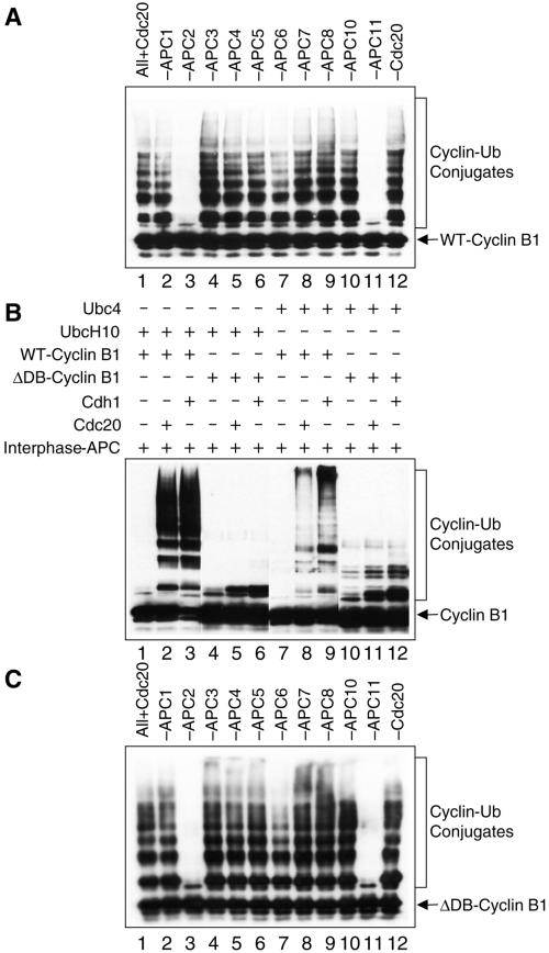
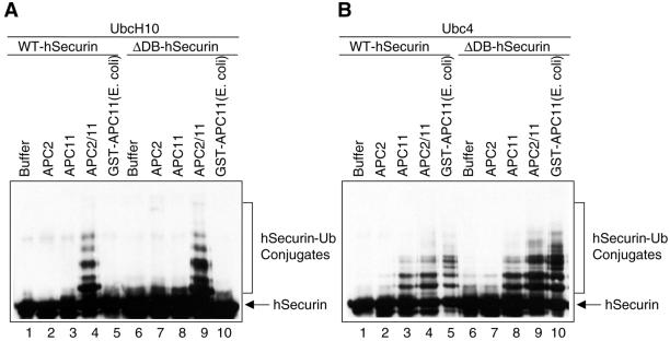
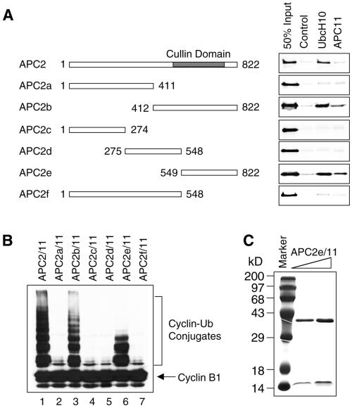
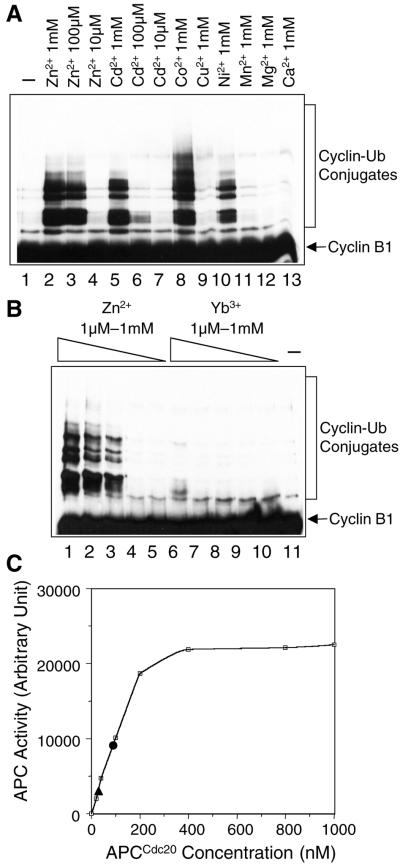
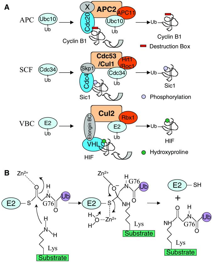
In mitosis, the anaphase-promoting complex (APC) regulates the onset of sister-chromatid separation and exit from mitosis by mediating the ubiquitination and degradation of the securin protein and mitotic cyclins. With the use of a baculoviral expression system, we have reconstituted the ubiquitin ligase activity of human APC. In combination with Ubc4 or UbcH10, a heterodimeric complex of APC2 and APC11 is sufficient to catalyze the ubiquitination of human securin and cyclin B1. However, the minimal APC2/11 ubiquitin ligase module does not possess substrate specificity, because it also ubiquitinates the destruction box deletion mutants of securin and cyclin B1. Both APC11 and UbcH10 bind to the C-terminal cullin homology domain of APC2, whereas Ubc4 interacts with APC11 directly. Zn(2+)-binding and mutagenesis experiments indicate that APC11 binds Zn(2+) at a 1:3 M ratio. Unlike the two Zn(2+) ions of the canonical RING-finger motif, the third Zn(2+) ion of APC11 is not essential for its ligase activity. Surprisingly, with Ubc4 as the E2 enzyme, Zn(2+) ions alone are sufficient to catalyze the ubiquitination of cyclin B1. Therefore, the Zn(2+) ions of the RING finger family of ubiquitin ligases may be directly involved in catalysis.


Figures

Figure 1
Figure 1
Reconstitution of the ubiquitin ligase activity of APC. (A) Hi5 insect cells were coinfected with baculoviruses encoding the indicated APC subunits. The expressed His6-tagged APC proteins were isolated from the insect cell lysate with the use of the Ni2+-NTA beads and assayed for ubiquitin ligase activity in the presence of UbcH10. The reaction mixture was separated on SDS-PAGE and blotted with α-Myc to detect the C-terminally Myc-tagged cyclin B1 protein. Omission of the APC2 (lane 3) or APC11 (lane 11) viruses from the coinfection resulted in the loss of ubiquitin ligase activity. (B) Ubiquitin ligase activity of the intact APC. Either UbcH10 (lanes 1–6) or Ubc4 (lanes 7–12) was used as the E2 enzyme. To determine the D-box dependency of APCCdc20 and APCCdh1, either the wild-type (WT) cyclin B1 (lanes 1–3 and 7–9) or the D-box deletion mutant (ΔDB) of cyclin B1 (lanes 4–6 and 10–12) was used as substrates. (C) Same as A except that ΔDB-cyclin B1 was used as the substrate instead of the wild-type cyclin B1 protein.
Figure 2
Figure 2
APC11 interacts with APC2 and APC10. (A) GST-APC11 baculovirus was coinfected with APC1/8 (a single baculovirus encoding both APC1 and APC8), APC2/7, APC3/6, APC4/5, or Cdh1 viruses in a pairwise manner into Sf9 cells. GST-APC11 and its interacting proteins were purified on glutathione beads, separated on SDS-PAGE, and stained with Coomassie Blue. GST was added to the lysates of APC1/8, APC2/7, APC3/6, APC4/5, and Cdh1 and used as controls. APC11 interacted strongly with APC2 (lane 3) and weakly with APC6 (lane 5). As determined by mass spectrometry, the band at 55 kDa belonged to α/β-tubulin, which presumably associated nonspecifically with GST-APC11. (B) His6-tagged APC10 baculovirus was coinfected with the GST-APC11 virus into Sf9 cells. His6-APC10 and its interacting proteins were purified on Ni2+-NTA beads, separated on SDS-PAGE, and stained with Coomassie Blue. As controls, Ni2+-beads were added to lysates infected with GST-APC11 alone. The α/β-tubulin proteins again copurified with APC10 and APC11. (C) To verify the interaction between APC10 and APC11, the same samples from B were blotted with α-GST, confirming the identity of the GST-APC11 band.
Figure 3
Figure 3
Heterodimeric complex of APC2 and APC11 possesses ubiquitin ligase activity. (A) Hi5 cells were either infected with His6-APC2 (lanes 2 and 7) and His6-APC11 (lanes 3 and 8) viruses individually, or coinfected with the His6-APC2 and His6-APC11 viruses (lanes 4 and 9). The APC2 and APC11 proteins were purified with Ni2+-NTA beads and assayed for ubiquitination activity with the use of UbcH10 as the E2 enzyme and wild-type human securin (lanes 1–5) or a D-box deletion mutant (ΔDB) of securin (lanes 6–10) as the substrates. Bacterial expressed GST-APC11 protein purified with glutathione-agarose beads was also tested for ligase activity (lanes 5 and 10). (B) Same as A except that Ubc4 was used as the E2 enzyme instead of UbcH10.
Figure 4
Figure 4
Ubc4 binds to the ring protein APC11 whereas UbcH10 binds to the cullin protein APC2. (A) Xenopus homolog of UbcH10, UbcX, was immunodepleted from mitotic Xenopus egg extracts with the use of a polyclonal α-UbcX antibody coupled to Affiprep protein A beads (compare lanes 1 and 2). The preimmune serum was used as the control. The kinetics of cyclin B1 degradation was assayed in the control-depleted extract (top), the UbcX-depleted extract (middle), and the UbcX-depleted extract with physiological amount of purified UbcX added back (bottom). (B) Binding assays among Ubc4, UbcH10, APC2, and APC11. Purified His6-tagged Ubc4 and UbcH10 proteins were immobilized on Ni2+-NTA beads and incubated with 35S-labeled APC2 or APC11 proteins. After washing, the 35S-labeled proteins bound to beads were analyzed by SDS-PAGE followed by autoradiography. (C) Sequence alignment of the RING-binding loops of human Ubc4, UbcH7, and UbcH10.
Figure 5
Figure 5
Cullin domain of APC2 is sufficient for binding to both APC11 and UbcH10. (A) Purified His6-tagged APC11 and UbcH10 proteins were immobilized on Ni2+-NTA beads and incubated with 35S-labeled APC2 or various APC2 truncation mutant proteins. After washing, the proteins retained on beads were analyzed by SDS-PAGE followed by autoradiography. (B) Complex of the cullin domain of APC2 and APC11 possesses ubiquitin ligase activity. A series of APC2 truncation mutants were expressed in insect cells together with APC11. APC proteins were purified with Ni2+-NTA beads and assayed for ubiquitination activity. (C) Purified APC2e/11 complex was analyzed by SDS-PAGE followed by Coomassie staining.
Figure 6
Figure 6
One APC11 protein molecule binds three Zn2+ ions. (A) Sequence alignment of APC11 and Rbx1 from various organisms (Hs, Homo sapiens; Dm, Drosophila melanogaster; Sc, Saccharomyces cerevisiae; and Sp, S. pombe). The residues that coordinate the two Zn2+ ions of the canonical RING finger motif are labeled as open and closed circles, respectively. The residues for coordinating the third Zn2+ ion are labeled as open triangles. (B) Autoubiquitination assay of the APC11 mutants. 35S-labeled APC11 mutants were translated in vitro in reticulocyte lysate and incubated with a mixture of ubiquitin, ATP, and E1 in the presence and absence of Ubc4. The reaction mixture was separated on SDS-PAGE followed by autoradiography. The Zn2+-coordinating residues are labeled as in A. The mutations that affect Zn2+-binding are indicated by +, whereas the mutations that do not affect zinc binding are marked by −.
Figure 7
Figure 7
Zn2+ ions alone stimulate the ubiquitination activity of Ubc4. (A) Various concentrations of Zn2+, Cd2+, and other divalent cations were added to a reaction mixture containing E1, ubiquitin, Ubc4, ATP, and cyclin B1. Ubiquitination of cyclin B1 was analyzed by immunoblotting with α-Myc. (B) Various concentrations of Zn2+ and Yb3+ were added to a reaction mixture containing E1, ubiquitin, Ubc4, ATP, and cyclin B1. Ubiquitination of cyclin B1 was analyzed by immunoblotting with α-Myc. (C) Comparison of the ubiquitin ligase activities of the intact APCCdc20, the reconstituted APC, and zinc ions alone. The ligase activity of APCCdc20 was plotted against the concentration used in the assay. The activities of the reconstituted APC at 5 μM and zinc ions alone at 100 μM were indicated by closed circle and triangle, respectively.
Figure 8
Figure 8
(A) APC belongs to the cullin-RING family of ubiquitin ligases. (B) Proposed role of Zn2+ in Ubc4-catalyzed ubiquitination reactions. See DISCUSSION for details.

References

    1. Aristarkhov A, Eytan E, Moghe A, Admon A, Hershko A, Ruderman JV. E2-C, a cyclin-selective ubiquitin carrier protein required for the destruction of mitotic cyclins. Proc Natl Acad Sci. 1996;93:4294–4299. - PMC - PubMed
    1. Burton JL, Solomon MJ. D box and KEN box motifs in budding yeast HSl1p are required APC-mediated degradation and direct binding to Cdc20p and Cdh1p. Genes Dev. 2001;15:2381–2395. - PMC - PubMed
    1. Deshaies RJ. SCF and Cullin/Ring H2-based ubiquitin ligases. Annu Rev Cell Dev Biol. 1999;15:435–467. - PubMed
    1. Fang S, Jensen JP, Ludwig RL, Vousden KH, Weissman AM. Mdm2 is a RING finger-dependent ubiquitin protein ligase for itself and p53. J Biol Chem. 2000;275:8945–8951. - PubMed
    1. Fang G, Yu H, Kirschner MW. Direct binding of CDC20 protein family members activates the anaphase-promoting complex in mitosis and G1. Mol Cell. 1998;2:163–171. - PubMed

Publication types

MeSH terms