. 2001 Dec;12(12):3852-63.
doi: 10.1091/mbc.12.12.3852.

TGFbeta and PTHrP control chondrocyte proliferation by activating cyclin D1 expression

Affiliations
Free PMC article

TGFbeta and PTHrP control chondrocyte proliferation by activating cyclin D1 expression

F Beier et al. Mol Biol Cell. 2001 Dec.
Free PMC article

Abstract

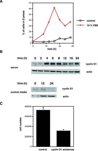
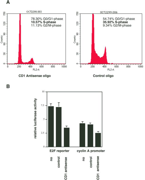
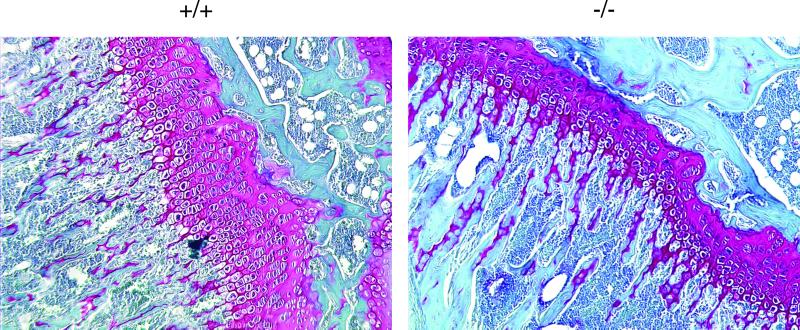
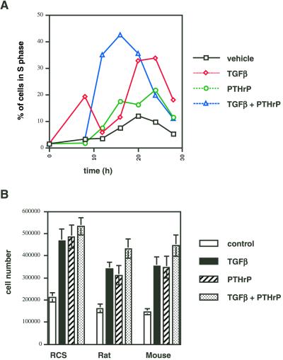
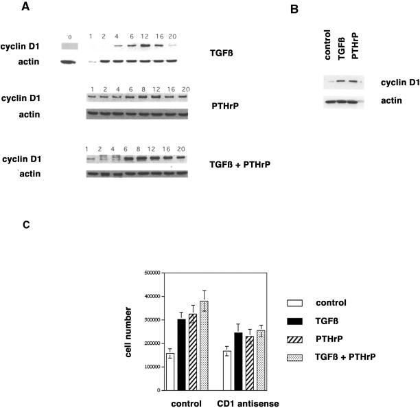
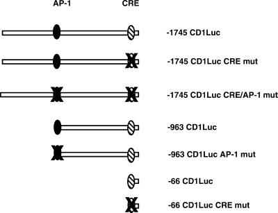
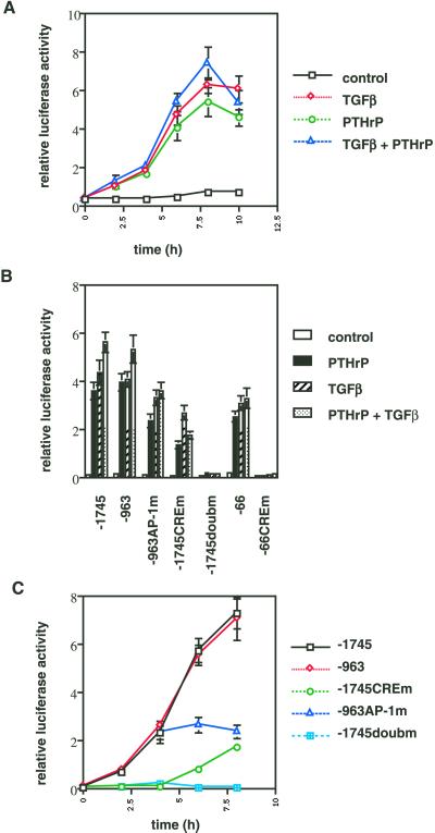
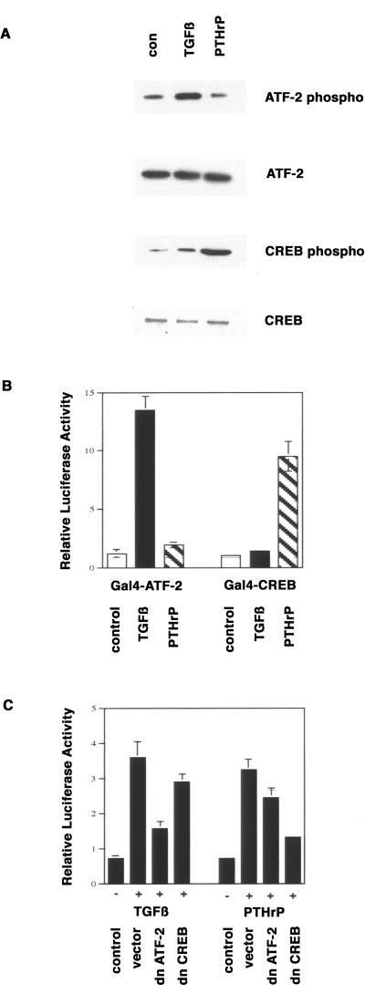
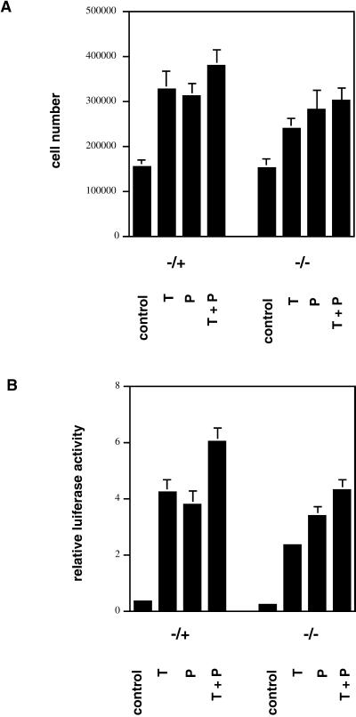
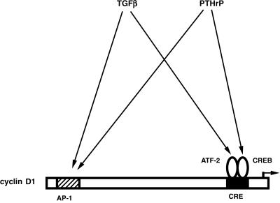
Exact coordination of growth plate chondrocyte proliferation is necessary for normal endochondral bone development and growth. Here we show that PTHrP and TGFbeta control chondrocyte cell cycle progression and proliferation by stimulating signaling pathways that activate transcription from the cyclin D1 promoter. The TGFbeta pathway activates the transcription factor ATF-2, whereas PTHrP uses the related transcription factor CREB, to stimulate cyclin D1 promoter activity via the CRE promoter element. Inhibition of cyclin D1 expression with antisense oligonucleotides causes a delay in progression of chondrocytes through the G1 phase of the cell cycle, reduced E2F activity, and decreased proliferation. Growth plates from cyclin D1-deficient mice display a smaller zone of proliferating chondrocytes, confirming the requirement for cyclin D1 in chondrocyte proliferation in vivo. These data identify the cyclin D1 gene as an essential component of chondrocyte proliferation as well as a fundamental target gene of TGFbeta and PTHrP during skeletal growth.


Figures

Figure 1
Figure 1
Cyclin D1 is required for serum-induced RCS proliferation. (A) RCS cells were serum-starved for 3 d, followed by stimulation with 10% FBS or control serum-free medium. Cells were labeled with BrdU in 4-h intervals. The percentage of cells incorporating BrdU was determined by FACS and is shown. (B) RCS cells were serum-starved for 3 d, followed by stimulation with 10% FBS or control medium for the indicated intervals, and harvested for the analyses of cyclin D1 protein expression by Western blot. Equal gel loading (5 × 10⁵ cells/lane) was documented by probing for actin protein expression. (C) RCS cells (150,000/well) were plated, serum-starved for 3 d, and restimulated with 10% FBS in the presence of control or cyclin D1 antisense oligonucleotides. Cells were counted after 48 h of stimulation. The average and SD from two independent experiments (performed in triplicate wells each) are shown.
Figure 2
Figure 2
Cyclin D1 is required for G1 progression and E2F-dependent gene expression in RCS cells. (A) Subconfluent RCS cells were incubated with cyclin D1 antisense or control oligo nucleotides for 24 h and harvested for FACS analysis. Cell cycle distribution was determined by propidium iodine staining of nuclear DNA. (B) Plasmids encoding the firefly luciferase gene under control of an E2F-responsive promoter or the cyclin A promoter were transfected into RCS cells, together with the plasmid pRlSV40. Cells were incubated without oligonucleotides (no), control oligonucleotides, or cyclin D1 antisense oligonucleotides for 48 h. Cells were harvested, and firefly luciferase activity was measured and standardized to Renilla luciferase activity to yield the relative luciferase activity.
Figure 3
Figure 3
Cyclin D1 is required for chondrocyte proliferation in vivo. Growth plates (stained red) from tibias of 6-week-old wild-type (+/+) or cyclin D1–deficient (−/−) mice were analyzed by histology with the use of hematoxylin/Fast Green/Saffronin O staining. The zone of proliferative chondrocytes (small, flattened chondrocytes, arranged in columns, on the right side of each growth plate) is reduced by 50% in −/− mice.
Figure 4
Figure 4
Induction of chondrocyte proliferation by TGFβ and PTHrP. (A) RCS cells were serum-starved for 3 d, stimulated with control medium, TGFβ (1 ng/ml), PTHrP (10−8 M), or both, and labeled with BrdU in 4-h intervals. The percentage of cells incorporating BrdU was determined by FACS and is shown. (B) RCS cells and primary rat and mouse chondrocytes were plated in 24-well plates (105 cells/well), stimulated with control medium, TGFβ (1 ng/ml), PTHrP (10−8 M), or both for 3 d, and then counted with the use of a hemacytometer.
Figure 5
Figure 5
Induction of cyclin D1 expression by TGFβ and PTHrP. (A) RCS cells were serum-starved for 3 d, stimulated with control medium, TGFβ (1 ng/ml), PTHrP (10−8 M), or both for the indicated intervals, and harvested for the analyses of cyclin D1 protein expression by Western blot. Equal gel loading (5 × 10⁵ cells/lane) was documented by probing for actin protein expression. (B) Primary rat chondrocytes were serum-starved for 1 d, stimulated with control medium, TGFβ (1 ng/ml), or PTHrP (10−8 M) for 8 h, and harvested for the analyses of cyclin D1 protein expression by Western blot. Equal gel loading (5 × 10⁵ cells/lane) was documented by probing for actin protein expression. (C) Primary rat chondrocytes were plated in 24-well plates (105 cells/well). The cells were incubated with control or cyclin D1 antisense oligonucleotides and stimulated with control medium, TGFβ (1 ng/ml), PTHrP (10−8 M), or both for 3 d, and then counted with the use of a hemacytometer.
Figure 6
Figure 6
Cyclin D1 promoter constructs. Overview of the human cyclin D1 promoter constructs used in this study. The described AP-1 and CRE elements are indicated.
Figure 7
Figure 7
Cyclin D1 promoter induction by PTHrP and TGFβ requires the AP-1 and CRE sites. (A) The plasmid −1745 CD1Luc was transfected into RCS cells, together with the plasmid pRlSV40. After transfection, cells were serum-starved for 3 d and stimulated with control medium, TGFβ (1 ng/ml), PTHrP (10−8 M), or both for 0, 2, 4, 6, 8, and 10 h. Cells were harvested at these time points, and firefly luciferase activity was measured and standardized to Renilla luciferase activity to yield the relative luciferase activity. (B) The cyclin D1 promoter constructs −1745 CD1Luc (−1745), −963 CD1Luc (−963), −963 CD1Luc AP-1mut (−963AP-1 m), −1745 CD1Luc CREmut (−1745CREm), −1745 CD1LucCRE/AP-1mut (−1745doubm), −66 CD1Luc (−66), and −66 CD1Luc CREmut (−66CREm) were transfected into RCS cells, together with pRlSV40. After transfection, cells were serum-starved for 3 d and stimulated with control medium, TGFβ (1 ng/ml), PTHrP (10−8 M), or both for 8 h. Cells were harvested, and firefly luciferase activity was measured and standardized to Renilla luciferase activity to yield the relative luciferase activity. (C) The cyclin D1 promoter constructs −1745 CD1Luc (−1745), −963 CD1Luc (−963), −963 CD1Luc AP-1mut (−963AP-1 m), −1745 CD1Luc CREmut (−1745CREm), and −1745 CD1LucCRE/AP-1mut (−1745doubm) were transfected into RCS cells, together with pRlSV40. After transfection, cells were serum-starved for 3 d and stimulated with control medium or a combination of TGFβ (1 ng/ml) and PTHrP (10−8 M) for 0, 2, 4, 6, or 8 h. Cells were harvested at the indicated time points, and firefly luciferase activity was measured and standardized to Renilla luciferase activity to yield the relative luciferase activity
Figure 8
Figure 8
Activation of the cyclin D1 CRE by TGFβ and PTHrP is mediated by ATF-2 and CREB. (A) RCS cells were serum-starved for 3 d and stimulated for 8 h with control medium, TGFβ (1 ng/ml), PTHrP (10−8 M), or both. Cells were harvested for the analyses of protein expression of ATF-2 and of phosphorylated forms of ATF-2 and CREB. (B) The plasmids pFA-ATF-2 and pFA-CREB (encoding Gal4-ATF-2 and Gal4-CREB fusion proteins, respectively) were transfected into RCS cells together with the Gal4 reporter plasmid pFRluc and pRlSV40. After transfection, cells were serum-starved for 3 d and stimulated with control medium, TGFβ (1 ng/ml), or PTHrP (10−8 M) for 8 h. Cells were harvested, firefly luciferase activity was measured and standardized to Renilla luciferase activity to yield the relative luciferase activity. (C) The plasmid −1745 CD1Luc was transfected into RCS cells together with pRlSV40 and empty expression vector or expression vectors for dominant-negative forms of ATF-2 and CREB. After transfection, cells were serum-starved for 3 d and stimulated with control medium, TGFβ (1 ng/ml), PTHrP (10−8 M), or both for 8 h. Cells were harvested, and firefly luciferase activity was measured and standardized to Renilla luciferase activity to yield the relative luciferase activity.
Figure 9
Figure 9
ATF-2 is necessary for the proliferative response of chondrocytes to TGFβ. (A) Chondrocytes from heterozygous (−/+) or homozygous (−/−) ATF-2–deficient mice were plated in 24-well plates (105 cells/well), stimulated with control medium, TGFβ (1 ng/ml; T), PTHrP (10−8 M; P), or both (T+P) for 3 d, and counted with the use of a hemacytometer. (B) The plasmid −1745 CD1Luc was transfected into primary chondrocytes from heterozygous (−/+) or homozygous (−/−) ATF-2–deficient mice, together with pRlSV40. After transfection, cells were serum-starved for 3 d and stimulated with control medium, TGFβ (1 ng/ml; T), PTHrP (10−8 M; P), or both (T+P) for 8 h. Cells were harvested, firefly luciferase activity was measured and standardized to Renilla luciferase activity to yield the relative luciferase activity.
Figure 10
Figure 10
Model of regulation of the cyclin D1 promoter by PTHrP and TGFβ. Both growth factors induce cyclin D1 promoter activity through the AP-1 and CRE elements. TGFβ effects require the transcription factor ATF-2, whereas the effects of PTHrP are mediated by the related transcriptional regulator CREB.

Similar articles

Cited by

References

    1. Ahn S, Olive M, Aggarwal S, Krylov D, Ginty DD, Vinson C. A dominant-negative inhibitor of CREB reveals that it is a general mediator of stimulus-dependent transcription of c-fos. Mol Cell Biol. 1998;18:967–977. - PMC - PubMed
    1. Albanese C, Johnson J, Watanabe G, Eklund N, Vu D, Arnold A, Pestell RG. Transforming p21ras mutants and c-Ets-2 activate the cyclin D1 promoter through distinguishable regions. J Biol Chem. 1995;270:23589–23597. - PubMed
    1. Amizuka N, Warshawsky H, Henderson JE, Goltzman D, Karaplis AC. Parathyroid hormone-related peptide-depleted mice show abnormal epiphyseal cartilage development and altered endochondral bone formation. J Cell Biol. 1994;126:1611–1623. - PMC - PubMed
    1. Ballock RT, Heydemann A, Wakefield LM, Flanders KC, Roberts AB, Sporn MB. TGF-beta 1 prevents hypertrophy of epiphyseal chondrocytes: regulation of gene expression for cartilage matrix proteins and metalloproteases. Dev Biol. 1993;158:414–429. - PubMed
    1. Battegay EJ, Raines EW, Seifert RA, Bowen-Pope DF, Ross R. TGF-beta induces bimodal proliferation of connective tissue cells via complex control of an autocrine PDGF loop. Cell. 1990;63:515–524. - PubMed

Publication types

MeSH terms

LinkOut - more resources