. 2001 Dec;12(12):3973-86.
doi: 10.1091/mbc.12.12.3973.

Dominant-lethal alpha-tubulin mutants defective in microtubule depolymerization in yeast

Affiliations
Free PMC article

Dominant-lethal alpha-tubulin mutants defective in microtubule depolymerization in yeast

K R Anders et al. Mol Biol Cell. 2001 Dec.
Free PMC article

Abstract

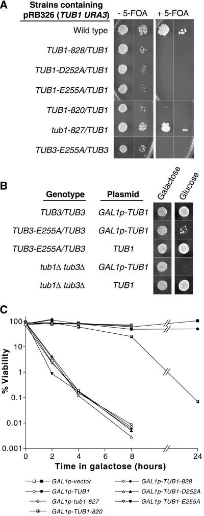
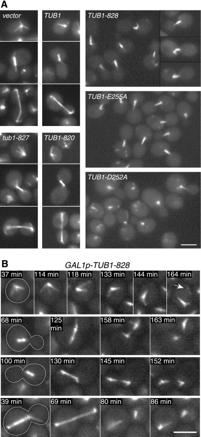
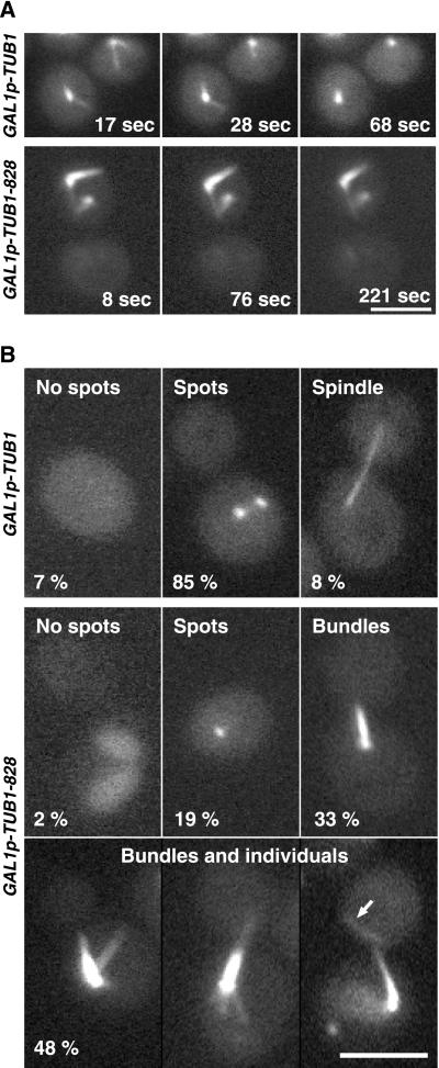
The dynamic instability of microtubules has long been understood to depend on the hydrolysis of GTP bound to beta-tubulin, an event stimulated by polymerization and necessary for depolymerization. Crystallographic studies of tubulin show that GTP is bound by beta-tubulin at the longitudinal dimer-dimer interface and contacts particular alpha-tubulin residues in the next dimer along the protofilament. This structural arrangement suggests that these contacts could account for assembly-stimulated GTP hydrolysis. As a test of this hypothesis, we examined, in yeast cells, the effect of mutating the alpha-tubulin residues predicted, on structural grounds, to be involved in GTPase activation. Mutation of these residues to alanine (i.e., D252A and E255A) created poisonous alpha-tubulins that caused lethality even as minor components of the alpha-tubulin pool. When the mutant alpha-tubulins were expressed from the galactose-inducible promoter of GAL1, cells rapidly acquired aberrant microtubule structures. Cytoplasmic microtubules were largely bundled, spindle assembly was inhibited, preexisting spindles failed to completely elongate, and occasional, stable microtubules were observed unattached to spindle pole bodies. Time-lapse microscopy showed that microtubule dynamics had ceased. Microtubules containing the mutant proteins did not depolymerize, even in the presence of nocodazole. These data support the view that alpha-tubulin is a GTPase-activating protein that acts, during microtubule polymerization, to stimulate GTP hydrolysis in beta-tubulin and thereby account for the dynamic instability of microtubules.


Figures

Figure 1
Figure 1
Location of α-tubulin mutations in this study. (A) Two α-β dimers longitudinally aligned in a protofilament show the location of yeast α-tubulin mutations relative to the interdimer interface. The structure shown is bovine tubulin (Nogales et al., 1998b) with amino acid side chains drawn that correspond to those identical residues mutated in the following S. cerevisiae alleles: TUB1-820 (E156A, E157A) and tub1-827 (R244A, D246A) shown in orange, and TUB1-828 (D252A, E255A) (Richards et al., 2000), TUB1-D252A, TUB1-E255A, and TUB3-E255A shown in red. Structure is oriented with the plus end up and the outside face of the microtubule to the right. Guanine nucleotides are shown in yellow. Structure drawn with Molscript (Kraulis, 1991). (B) Alignment of α-tubulin, β-tubulin, and FtsZ sequences in the region of longitudinal interaction. Residues shared by the tubulins and FtsZ are shaded. Residues mutated in the yeast tubulin alleles described in A are orange or red. The residue mutated in the GTPase-defective allele ftsZ2 (D212G) is green (Dai et al., 1994). Amino acid sequences (Swiss-Prot ID: TUBA1_YEAST, TUBA3_YEAST, TBA_PIG, TBB_YEAST, TBB_PIG, FTSZ_ECOLI) were aligned with Clustal W (Thompson et al., 1994). Because bovine tubulin sequence is unknown, porcine tubulin sequence is applied to the bovine crystal structure (Nogales et al., 1998b).
Figure 2
Figure 2
Dosage-dependent, dominant lethality of α-tubulin alleles containing D252A and/or E255A mutations. (A) Extra copies of TUB1 confer viability to dominant-lethal α-tubulin alleles. Strains containing pRB326 (TUB1, URA3) plasmid were grown overnight in liquid medium supplemented with uracil to allow the loss of the plasmid then spotted to plates containing 5-FOA to select for Ura(−) cells that had lost the plasmid. Approximately 104 and 102 cells were spotted to SC-lys,-leu,-ura without 5-FOA (SC-ura for TUB3-E255A/TUB3) and to SC supplemented with 5-FOA and grown for 3 d. Strains shown are DBY6600, DBY6628, DBY9557, DBY9559, DBY8247, DBY6627, and DBY9578. (B) TUB3-E255A/TUB3 strain is inviable when ectopic GAL1p-TUB1 expression is repressed by glucose. Strains containing pRB2785 (GAL1p-TUB1) or pRB326 (TUB1) plasmids were grown overnight in galactose-containing medium then ∼104 cells were spotted to SC-ura plates containing galactose or glucose and grown for 3 d. Strains shown are DBY9566, DBY9565, DBY9578, DBY9561, and DBY9562. (C) Viability after transient expression of plasmid-borne GAL1p-TUB1 alleles. Raffinose-containing cultures were treated with galactose to induce expression of GAL1p-TUB1 alleles then plated to glucose-containing plates to repress GAL1p-TUB1 allele expression. Viability is expressed in percentage of cells plated that resulted in colonies. Open symbols indicate that strain DBY9579 contains the designated plasmid. Black- and pattern-filled symbols indicate that strain DBY9580 (with a GFP::TUB1-LEU2 insertion), contains the designated plasmid.
Figure 3
Figure 3
Morphology of the microtubule cytoskeleton after expression of mutant GAL1p-TUB1 alleles. (A) GFP-labeled microtubules in cells exposed to galactose for 2 h then washed into glucose medium and incubated 1 h. Cells are strain DBY9580 containing one of the following plasmids, pTS210 (GAL1p-vector), pRB2785 (GAL1p-TUB1), pRB2956 (GAL1p-TUB1-820), pRB2958 (GAL1p-tub1-827), pRB2949 (GAL1p-TUB1-828), pRB2953 (GAL1p-TUB1-E255A), or pRB2951 (GAL1p-TUB1-D252A). [Nonmicrotubular spots of GFP fluorescence are occasionally seen with all plasmids except GAL1p-vector, similar to those described by Carminati and Stearns (1997).] (B) Time-lapse series showing the appearance of aberrant microtubule morphologies upon expression of GAL1p-TUB1-828. Galactose (2%) and glucose (0.2%) were added to a raffinose culture of strain DBY9580 + pRB2949 (GAL1p-TUB1-828) at time zero. Cells were mounted onto a microscope slide containing 2% galactose, 0.2% glucose medium and GFP fluorescence was visualized every 20 s. Arrow indicates loose microtubule that became unattached to the spindle pole body. Videos 3B_1.mov, 3B_2.mov, 3B_3.mov, and 3B_4.mov contain entire series from 30 to 164 min of galactose exposure. Bars, 3 μm.
Figure 4
Figure 4
Cell viability and microtubule structures after expression of GFP::TUB1-828 and GFP::TUB1. (A) Viability after transient expression of GAL1-driven TUB1 alleles. Raffinose cultures were treated with galactose then plated to glucose-containing plates. Viability is expressed in percentage of cells plated that resulted in colonies. Strain is DBY6597 containing pTS210 (GAL1p-vector), pRB2785 (GAL1p-TUB1), pRB2949 (GAL1p-TUB1-828), pRB2960 (GAL1p- GFP::TUB1), and pRB2963 (GAL1p- GFP::TUB1-828). (B) GFP-labeled microtubules after galactose-in-duction of GFP::TUB1 and GFP::TUB1-828 expression. Haploid spore clones from A (strains DBY9586-7) were grown in raffinose then treated with 2% galactose, 0.2% glucose for 3.5 h. Bar, 3 μm.
Figure 5
Figure 5
Expression of TUB1-828 during G1 α-factor arrest results in unattached microtubules and inhibited spindle assembly upon release from α-factor arrest. (A and B) GFP-labeled microtubules in strain DBY9580, containing either pTS210 (GAL1p-vector), pRB2785 (GAL1p-TUB1), or pRB2949 (GAL1p-TUB1-828, also shown with differential interference contrast), grown in raffinose and arrested with α-factor then subjected to galactose for 2 h. In B, cells were released from α-factor arrest by washing into glucose medium and incubated for 90 min. Arrows indicate free microtubules not attached to the presumed spindle pole body. Videos 5A_TUB1.mov (GAL1p-TUB1) and 5A_828.mov (GAL1p-TUB1-828) illustrate microtubule dynamics under these conditions and movement of the free microtubules in A, 5B_828.mov (GAL1p-TUB1-828) shows cells in B. Bar, 5 μm. (C) Distribution of microtubule structures and cell shapes in cultures 90 min after release from α-factor arrest shown in B. Drawings above each column indicate presence of bud and appearance of microtubules. Aberrant microtubule bundles and unattached microtubules indicated in the four columns on the right. (D) Anti-tubulin antibodies reveal unattached microtubules and aberrant microtubule bundles in cells expressing TUB1-828 in the absence of GFP::TUB1. Strain DBY9585, containing plasmid pRB2949 (GAL1p-TUB1-828), was arrested with α-factor in raffinose, subjected to galactose for 2 h (top row) then washed into galactose medium and grown for 3.5 h (bottom row). Control strains DBY9583 and DBY9584, containing plasmids pTS210 (GAL1p-vector) and pRB2785 (GAL1p-TUB1), appeared normal as in A and B (our unpublished data). Bar, 3 μm.
Figure 6
Figure 6
Expression of TUB1-828 after spindles have assembled disrupts spindle integrity and inhibits normal spindle elongation. Strain DBY9580, containing either pTS210 (GAL1p-vector), pRB2785 (GAL1p-TUB1), or pRB2949 (GAL1p-TUB1-828), was released from G1 α-factor arrest into medium containing hydroxyurea for 2.5 h. Galactose was added and the cells incubated for 2 h. Cells were then released from hydroxyurea-induced arrest into glucose medium at time zero. α-Factor was included to prevent a second cell division. (A) Cell cycle progression after release from hydroxyurea-induced arrest. Numbers of nuclei were determined by 4′,6′-diamidino-2-phenylindole staining and fluorescence microscopy. At least 100 cells were counted for each time point. (B) GFP-labeled microtubules 90–120 min after release from hydroxyurea. Arrows indicate thinning or disappearance of intranuclear microtubules that span the distance between spindle pole bodies; arrowheads indicate bundled cytoplasmic microtubules. Bar, 3 μm.
Figure 7
Figure 7
Time-lapse imaging showing the dynamics of GFP-labeled microtubules. Strain DBY9580, containing either pTS210 (GAL1p-vector), pRB2785 (GAL1p-TUB1), or pRB2949 (GAL1p-TUB1-828), was grown in raffinose medium then supplemented with 2% galactose, 0.2% glucose for 1.75 h and imaged within 1 h (GAL1p-TUB1-828) or within 2 h as described in MATERIALS AND METHODS. (A) Still frames from time-lapse series, supplied as videos 7A_MT1.mov, 7A_MT2.mov, 7A_MT3.mov, 7A_MT4-5.mov, 7A_MT6.mov, and 7A_MT7.mov. Black frames in the long-term series indicate removal of nonfocused images. Bar, 3 μm. (B) Graphs showing microtubule length over time for the individual microtubules (MT1-MT7) labeled in A. Open circles indicate microtubules in the focal plane, and filled circles indicate a microtubule partially out of the focal plane. A best-fit line was drawn for each growth and shrinkage phase, and its slope is indicated below each plot. Inset graph indicates the length of microtubule MT7 during 36 min of observation.
Figure 8
Figure 8
Effect of nocodazole in cells expressing TUB1-828. Strains DBY9589 (GAL1p-TUB1) and DBY9590 (GAL1p-TUB1-828) were exposed to galactose for 2.5 h then placed onto a glucose medium containing 15 μg/ml nocodazole. (A) Visualization of GFP-labeled microtubules after placement of cells onto nocodazole. Time-lapse series are available in videos 8A_TUB1.mov (GAL1p-TUB1) and 8A_828.mov (GAL1p-TUB1-828). (B) Microtubule structures after 20-min exposure to nocodazole. Percentage of culture is indicated for each morphological class. At least 100 cells were counted for each strain. Arrow indicates microtubule unattached to spindle pole body. Bars, 3 μm.
Figure 9
Figure 9
Visualization of the effect of TUB3-E255A on microtubules after GAL1-shutoff of ectopic TUB1 expression. (A) Experimental scheme. Strain DBY9576, containing the lethal allele TUB3-E255A, is viable in galactose medium because suppressing quantities of wild-type TUB1 are expressed from the GAL1 promoter. When placed in glucose medium, transcription of the GAL1p-TUB1 copy is repressed, and the cells become inviable. A copy of GFP::TUB1 allows visualization of the microtubules. Strain DBY9574, which contains TUB3, serves as a control. (B) Microtubules in TUB3 and TUB3-E255A strains after 8 h of glucose repression of GAL1-driven TUB1. Time-lapse series showing the dynamics of the representative microtubules MT1 and MT2 are contained in the videos 9B_MT1.mov and 9B_MT2.mov. Microtubule lengths are plotted over time. Open circles indicate microtubules in the focal plane, and filled circles indicate a microtubule partially out of the focal plane. For MT2 plot, dotted line connects in-focus data points. Arrows indicate anaphase spindles with poles brighter than along length, and arrowhead indicates unattached microtubule. Bar, 3 μm.

References

    1. Arnal I, Karsenti E, Hyman AA. Structural transitions at microtubule ends correlate with their dynamic properties in Xenopus egg extracts. J Cell Biol. 2000;149:767–774. - PMC - PubMed
    1. Byers B, Goetsch L. Duplication of spindle plaques and integration of the yeast cell cycle. Cold Spring Harb Symp Quant Biol. 1974;38:123–131. - PubMed
    1. Byers B, Goetsch L. Behavior of spindles and spindle plaques in the cell cycle and conjugation of Saccharomyces cerevisiae. J Bacteriol. 1975;124:511–523. - PMC - PubMed
    1. Caplow M, Ruhlen RL, Shanks J. The free energy for hydrolysis of a microtubule-bound nucleotide triphosphate is near zero: all of the free energy for hydrolysis is stored in the microtubule lattice. J Cell Biol. 1994;127:779–788. - PMC - PubMed
    1. Caplow M, Shanks J. Mechanism of the microtubule GTPase reaction. J Biol Chem. 1990;265:8935–8941. - PubMed

Publication types

MeSH terms