. 2001 Dec;12(12):4030-43.
doi: 10.1091/mbc.12.12.4030.

Traction forces mediated by alpha6beta4 integrin: implications for basement membrane organization and tumor invasion

Affiliations
Free PMC article

Traction forces mediated by alpha6beta4 integrin: implications for basement membrane organization and tumor invasion

I Rabinovitz et al. Mol Biol Cell. 2001 Dec.
Free PMC article

Abstract

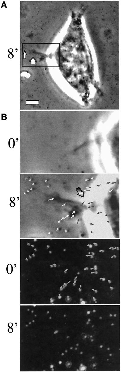
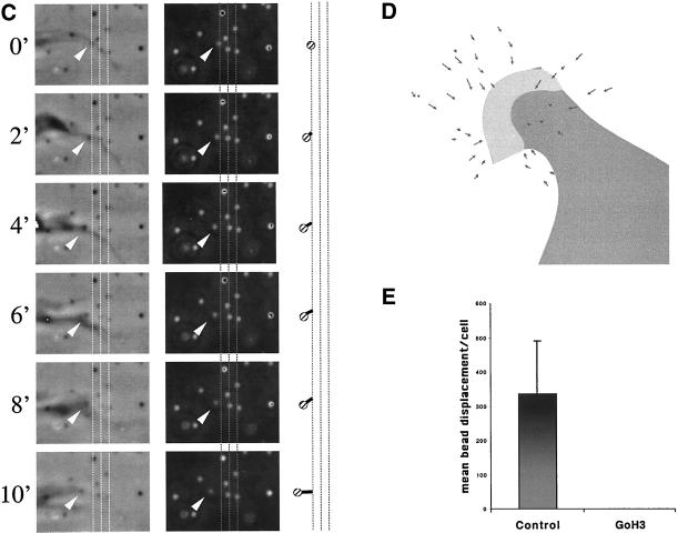
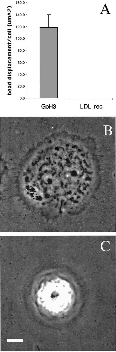
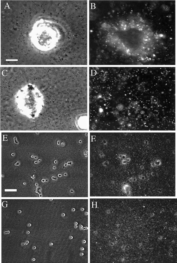
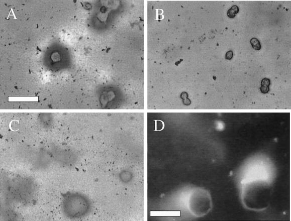
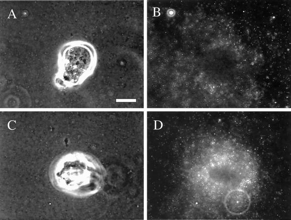
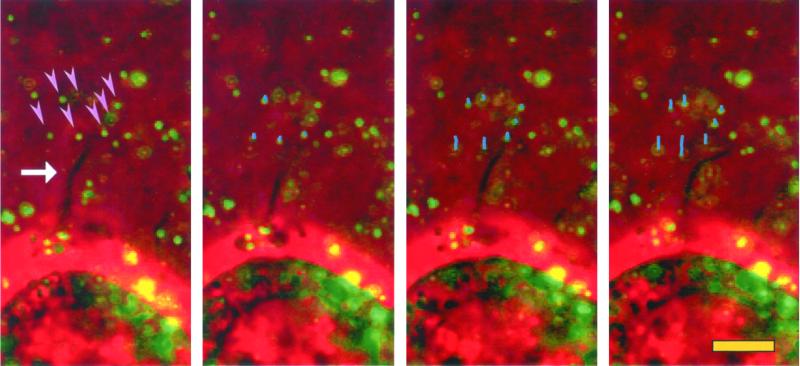
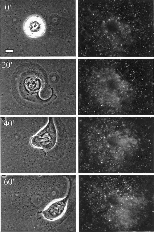
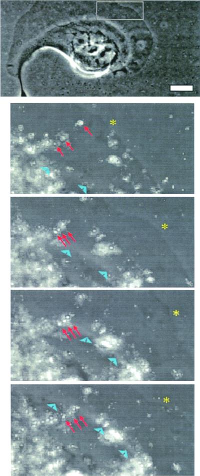
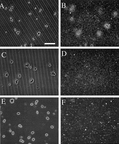
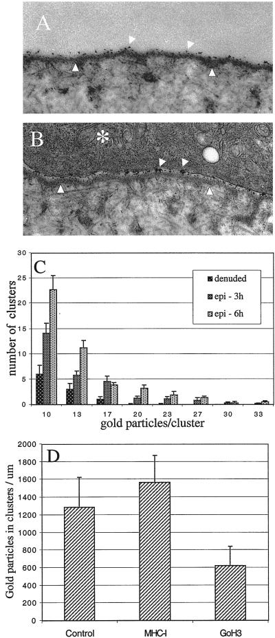
The integrin alpha6beta4, a laminin receptor that stabilizes epithelial cell adhesion to the basement membrane (BM) through its association with cytokeratins, can stimulate the formation and stabilization of actin-rich protrusions in carcinoma cells. An important, unresolved issue, however, is whether this integrin can transmit forces to the substrate generated by the acto-myosin system. Using a traction-force detection assay, we detected forces exerted through alpha6beta4 on either laminin-1 or on an anti-alpha6 antibody, demonstrating that this integrin can transmit forces without the need to engage other integrins. These alpha6beta4-dependent traction forces were organized into a compression machine localized to the base of lamellae. We hypothesized that the compression forces generated by alpha6beta4 result in the remodeling of BMs because this integrin plays a major role in the interaction of epithelial and carcinoma cells with such structures. Indeed, we observed that carcinoma cells are able to remodel a reconstituted BM through alpha6beta4-mediated compression forces by a process that involves the packing of BM material under the cells and the mechanical removal of BM from adjacent areas. The distinct signaling functions of alpha6beta4, which activate phosphoinositide 3-OH kinase and RhoA, also contribute to remodeling. Importantly, we demonstrate remodeling of a native BM by epithelial cells and the involvement of alpha6beta4 in this remodeling. Our findings have important implications for the mechanism of both BM organization and tumor invasion.


Figures

Figure 1
Figure 1
Integrin α6β4 mediates traction forces on laminin. Clone A colon carcinoma cells were plated on a polyacrylamide-flexible substrate containing fluorescent beads (2 μm diameter) and coated with laminin-1. (A and B) Cells were incubated for 1 h at 37°C and analyzed by time-lapse video microscopy by using phase contrast and fluorescent illumination. (A) Typical morphology of cell plated on a flexible laminin substrate. Notice that small lamellae (thick arrow) and filopodia (thin arrow) are formed. Bar, 10 μm. (B) Frame sequence of a protruding lamella magnified from the rectangular area in A. The upper two panels show a phase contrast image of the protruding lamella, whereas the lower ones show the corresponding fluorescent image of the underlying beads. Times 0′ an 8′ are shown. The entire frame sequence can be observed in the accompanying video (Figure 1b.mov). A vector map of bead displacement was built using both phase contrast and fluorescent images (middle two panels) by connecting the initial and final positions of each bead at the beginning and end of the frame sequence; the arrows indicate the direction of displacement and the thick-hatched arrow indicates the region were opposing forces are focused. (C) Frame sequence of discrete traction forces produced by filopodia. The columns of frames in the left and middle panels were photographed using phase contrast and fluorescence optics, respectively. The graphic on the right column represents the displacement of a small number of beads produced by the filopodia in the left columns. Grid lines are spatial references. Video Figure 1c.mov contains the corresponding frame sequence. (D and E) Cells were incubated 1 h at 37°C in the presence of GoH3 antibody or rat IgG control, and photographed using phase contrast and fluorescence illumination. The cells were treated with trypsin/EDTA to produce a relaxation of the substrate. The position of beads before and after trypsin/EDTA treatment was registered and a map of vectors representing the magnitude and direction of displacement was built for every cell. (D) Example of a vector map of such bead displacement. In E, the average bead displacement/cell (μm2) was calculated for cells in the presence or absence of the GoH3 antibody (see MATERIALS AND METHODS for calculation procedure).
Figure 1
Figure 1
Integrin α6β4 mediates traction forces on laminin. Clone A colon carcinoma cells were plated on a polyacrylamide-flexible substrate containing fluorescent beads (2 μm diameter) and coated with laminin-1. (A and B) Cells were incubated for 1 h at 37°C and analyzed by time-lapse video microscopy by using phase contrast and fluorescent illumination. (A) Typical morphology of cell plated on a flexible laminin substrate. Notice that small lamellae (thick arrow) and filopodia (thin arrow) are formed. Bar, 10 μm. (B) Frame sequence of a protruding lamella magnified from the rectangular area in A. The upper two panels show a phase contrast image of the protruding lamella, whereas the lower ones show the corresponding fluorescent image of the underlying beads. Times 0′ an 8′ are shown. The entire frame sequence can be observed in the accompanying video (Figure 1b.mov). A vector map of bead displacement was built using both phase contrast and fluorescent images (middle two panels) by connecting the initial and final positions of each bead at the beginning and end of the frame sequence; the arrows indicate the direction of displacement and the thick-hatched arrow indicates the region were opposing forces are focused. (C) Frame sequence of discrete traction forces produced by filopodia. The columns of frames in the left and middle panels were photographed using phase contrast and fluorescence optics, respectively. The graphic on the right column represents the displacement of a small number of beads produced by the filopodia in the left columns. Grid lines are spatial references. Video Figure 1c.mov contains the corresponding frame sequence. (D and E) Cells were incubated 1 h at 37°C in the presence of GoH3 antibody or rat IgG control, and photographed using phase contrast and fluorescence illumination. The cells were treated with trypsin/EDTA to produce a relaxation of the substrate. The position of beads before and after trypsin/EDTA treatment was registered and a map of vectors representing the magnitude and direction of displacement was built for every cell. (D) Example of a vector map of such bead displacement. In E, the average bead displacement/cell (μm2) was calculated for cells in the presence or absence of the GoH3 antibody (see MATERIALS AND METHODS for calculation procedure).
Figure 2
Figure 2
α6β4 integrin can relay forces directly onto the substrate. A polyacrylamide-flexible substrate containing fluorescent beads (2 μm diameter) was cross-linked with either anti-rat or anti-mouse antibodies. A431 cells were coated with the rat GoH3 mAb or the mouse anti-LDL receptor antibody, plated on the gel, and incubated for 1 h at 37°C. (A) Average bead displacement/cell (μm2) was analyzed by time-lapse video-microscopy by using phase contrast and fluorescent illumination as described in MATERIALS AND METHODS. The cells coated with the GoH3 antibody spread on the gel and produced numerous filopodia and lamellae (B), in contrast to the cell coated with the anti-LDL receptor antibody (C).
Figure 3
Figure 3
Mechanical remodeling of reconstituted BM is dependent on the α6β4 integrin. Clone A cells were plated on a film of Matrigel containing fluorescent beads and incubated in the presence of GoH3 (C and D and G and H) or control antibody (A and B and E and F) for 4 h at 37°C. The cells were photographed at high (A–D) and low (E–H) magnifications by using phase contrast (A and C) or fluorescence (B and D) microscopy. Notice the inhibition of bead concentration around the cells in the presence of the GoH3 antibody. Bars, 10 μm (A–D), 100 μm (E–H).
Figure 4
Figure 4
Remodeling of reconstituted BM involves condensation of material gathered from adjacent areas and is dependent on the integrin α6β4. (A and B) Clone A cells were plated on a film of Matrigel in the presence of either GoH3 (B) or control (A) antibody and incubated for 4 h. The cells were fixed and stained with Coomassie Blue. Notice the condensation of matrix material that forms around the cells and the clearing of material that occurs in adjacent areas. Bar, 75 μm. (C) At a short time of incubation (1 h), the cells have already produced a condensation ring around them. In this photograph, the cells were removed using EDTA to reveal the underlying matrix. (D) Fluorescence image of Clone A cells that were plated on a film of Matrigel containing FITC-conjugated laminin and incubated for 8 h. Notice that the fluorescent laminin condenses around the cells. Some intact Matrigel can be seen as a light veil at the top of this image. Bar, 25 μm.
Figure 5
Figure 5
Expression of the α6β4 integrin in breast carcinoma cells increases their ability to remodel reconstituted BM. MDA-MB-435 transfectants expressing the α6β4 integrin (C and D) or mock transfectants (A and B) were plated on Matrigel containing fluorescent beads and incubated at 37°C for 4 h. The cells were photographed using phase contrast (A and C) or fluorescence (B and D) microscopy. Note the intense concentration of beads surrounding the α6β4 transfectants in comparison with the mock transfectants. Bar, 10 μm.
Figure 6
Figure 6
Remodeling of reconstituted BM by filopodia. Clone A cells were plated on Matrigel containing fluorescent beads. The cells were analyzed by video microscopy with both phase contrast and fluorescence illumination. The frame sequence shown was taken at 2-min intervals. Only a few beads (arrowheads) from a small area surrounding a filopodium (arrow) are pulled toward the cell at the bottom. The lines in the second and subsequent frames represent the movement tracks of the beads indicated in the first frame. Beads distal to filopodia show little movement. In the video (Figure 6.mov), the arrows point to the area where filopodia activity is occurring.
Figure 7
Figure 7
Fan-shaped lamellae are powerful compressors of reconstituted BM. Time-lapse analysis of clone A cells stimulated with PMA (25 ng/ml) by using phase contrast (left) or fluorescent (right) illumination. Panels shown represent frames taken at 15-min intervals. Bar, 10 μm. In Figure 7.mov, the phase contrast and fluorescent images frames were captured 2-min intervals and then merged. Notice the strong compression that occurs under the lamella, relaxing partially after the base of the lamella passes over the compressed region. Also, note that although most of the direction of compression occurs perpendicular to the lamella, strong compression parallel to the lamella is evident at the end of the video.
Figure 8
Figure 8
Matrigel compression ends at the base of lamellae. Clone A cells were stimulated with PMA (25 ng/ml) and analyzed using time-lapse video microscopy. Both phase contrast and fluorescent images were taken at 2-min intervals and merged digitally. Top panel shows a phase contrast image of the cell and bottom panels show a magnified frame sequence of the region specified by the rectangle in the top panel. The frame sequence shows the compression of a group of beads (arrows) until the base of the lamellae (arrowheads) passes over them, a time at which the beads partially relax (bottom). This observation was commonly seen for other beads undergoing compression beneath the lamella. Asterisk denotes the leading edge. Bar, 10 μm.
Figure 9
Figure 9
Signaling pathways regulated by the α6β4 integrin are necessary for BM remodeling. Clone A cells were plated on Matrigel containing fluorescent beads and incubated for 4 h in the presence of either DMSO (A and B); Y27632, a Rho-kinase inhibitor (30 μM) (C and D); or LY294002, a PI3-K inhibitor (20 μm). The cells were photographed using either phase contrast (A, C, and E) and fluorescence (B, D, and F) microscopy. Bar, 100 μm. Notice that the inhibitors inhibit the remodeling process strongly.
Figure 10
Figure 10
α6β4 integrin functions in the remodeling of corneal BM. Deepithelialized corneas were labeled with gold colloid particles (5 nm) and recombined with fresh cornea epithelium as described in MATERIALS AND METHODS. The recombined corneas were incubated at 37°C for either 3 or 6 h, in the either presence or absence of the GoH3 mAb. (A) Electron micrograph of a recombined cornea showing areas of BM (arrowheads) either denuded (A) or covered with epithelium (B, asterisk). Notice that the distribution of the gold particles on the BM is homogeneous in A and aggregated in B (arrows). Bar, 0.5 μm. (C and D) Quantitative analysis of gold particle distribution. (C) Density of aggregates per unit length of BM (μm), was estimated at 3 and 6 h after recombining the epithelium with the denuded BM. Shown is the distribution of different size aggregates (D) Number of particles present in aggregates (>10 gold particles) per unit area of BM was quantified in recombined corneas that were incubated for 6 h in the presence or absence of the GoH3 mAb or anti-rabbit MHC-I antibody.

References

    1. Burton K, Park JH, Taylor DL. Keratocytes generate traction forces in two phases. Mol Biol Cell. 1999;10:3745–3769. - PMC - PubMed
    1. Colognato H, Winkelmann DA, Yurchenco PD. Laminin polymerization induces a receptor-cytoskeleton network. J Cell Biol. 1999;145:619–631. - PMC - PubMed
    1. Davis GE, Camarillo CW. Regulation of endothelial cell morphogenesis by integrins, mechanical forces, and matrix guidance pathways. Exp Cell Res. 1995;216:113–123. - PubMed
    1. Dembo M, Oliver T, Ishihara A, Jacobson K. Imaging the traction stresses exerted by locomoting cells with the elastic substratum method. Biophys J. 1996;70:2008–2022. - PMC - PubMed
    1. Dembo M, Wang YL. Stresses at the cell-to-substrate interface during locomotion of fibroblasts. Biophys J. 1999;76:2307–2316. - PMC - PubMed

Publication types

MeSH terms