. 2001 Dec;12(12):4114-28.
doi: 10.1091/mbc.12.12.4114.

HRD4/NPL4 is required for the proteasomal processing of ubiquitinated ER proteins

Affiliations
Free PMC article

HRD4/NPL4 is required for the proteasomal processing of ubiquitinated ER proteins

N W Bays et al. Mol Biol Cell. 2001 Dec.
Free PMC article

Abstract

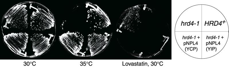
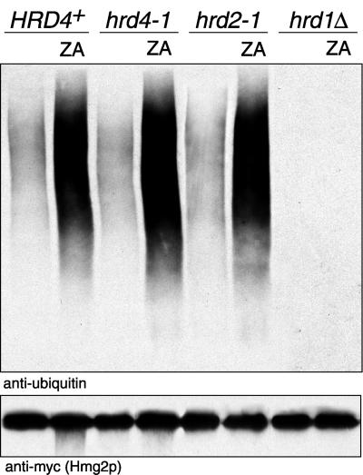
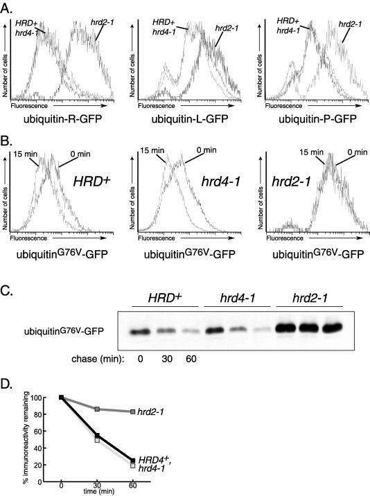
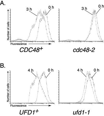
We isolated a temperature-sensitive mutant, hrd4-1, deficient in ER-associated degradation (ERAD). The HRD4 gene was identical to NPL4, a gene previously implicated in nuclear transport. Using a diverse set of substrates and direct ubiquitination assays, our analysis revealed that HRD4/NPL4 is required for a poorly characterized step in ERAD after ubiquitination of target proteins but before their recognition by the 26S proteasome. Our data indicate that this lack of proteasomal processing of ubiquitinated proteins constitutes the primary defect in hrd4/npl4 mutant cells and explains the diverse set of hrd4/npl4 phenotypes. We also found that each member of the Cdc48p-Ufd1p-Npl4p complex is individually required for ERAD.


Figures

Figure 1
Figure 1
hrd4–1 phenotypes and complementation by NPL4. The indicated yeast strains were incubated as designated by each labeled plate. (YCP) indicates the hrd4–1 strain was transformed with an ARS/CEN yeast genomic library plasmid, isolated for its ability to complement the hrd4–1 mutation and containing a genomic fragment bearing the NPL4 gene. (YIP) indicates a plasmid bearing only the NPL4 and URA3 genes was integrated at the ura3–52 locus in a hrd4–1 strain. “Lovastatin” indicates the addition of 200 μg/ml lovastatin to the indicated plate.
Figure 2
Figure 2
Hrd4p/Npl4p is required for degradation of ER proteins. (A) Both panels are stationary chase assays of protein degradation. The indicated yeast strains were grown into early stationary phase (12 h after strains reached an OD600 of 0.1). Equal numbers of cells were then lysed and subjected to SDS-PAGE, followed by immunoblotting for the myc-epitope tag. (B) Degradation of Hmg2-GFP was measured by cycloheximide chase, followed by flow cytometry. Cycloheximide was added to log-phase (OD600 < 0.2) cells 3 h before collection and analysis by flow cytometry. Each histogram represents 20,000 cells. (C) CPY* degradation in the indicated strains was assayed by cycloheximide chase. Equal numbers of log-phase (OD600 < 0.5) cells were collected at the indicated times post-cycloheximide addition, lysed and analyzed by SDS-PAGE, followed by immunoblotting for the HA-epitope tag present on the CPY*-HA protein. (D) Cycloheximide chase as in C, except that immunoblotting with the use of anti-Fur4p antibodies was performed to detect UP* (E) Steady-state cell fluorescence for the indicated strains expressing the UPR reporter construct, PKAR2-GFP, was analyzed by flow cytometry during log-phase growth. All experiments were performed at the permissive temperature of 30°C.
Figure 3
Figure 3
A block in nuclear transport has no effect on the degradation of an ER membrane protein. (A) The indicated strains expressing the constitutively-degraded 6MYC-Hmg2p were grown at the permissive temperature of 23°C. Log phase (OD600 = 0.1) cells were then shifted to the restrictive temperature of 37°C for 3.5 h before the addition of cycloheximide and subsequent chase at the indicated times. During this time, wild-type cells doubled at the normal rate, while the temperature-sensitive hrd4–1 and nup116Δ cells failed to complete even one additional doubling after temperature shift. Equal numbers of cells were collected at the indicated times following addition of cycloheximide and lysed. These lysates were then separated by SDS-PAGE, and immunoblotting for the myc epitope tag was performed. (B) Nup116 protein is absent in nup116Δ cells. Lysates from the indicated strains were subjected to SDS-PAGE and immunoblotting using anti-Nup116p antibodies.
Figure 4
Figure 4
Unsaturated fatty acids or OLE1 overexpression do not suppress the degradation defect in hrd4–1 cells. (A-C) Serial dilutions of the indicated strains were grown at the conditions labeling each panel. “+ UFA” indicates 0.5 mM each of oleic acid and palmitoleic acid were added to the labeled plates. “Lovastatin” indicates the addition of 200 μg/ml lovastatin.
Figure 5
Figure 5
Hmg2p is fully ubiquitinated in a hrd4–1 mutant strain. Ubiquitination of 1myc-Hmg2p was determined by immunoprecipitation of 1myc-Hmg2p from lysates obtained from log-phase cells with the use of anti-Hmg2p antisera. Immunoprecipitated material was separated by SDS-PAGE and followed by immunoblotting for ubiquitin and the myc epitope tag as indicated.
Figure 6
Figure 6
Degradation of Deg1-GFP is unaffected by the hrd4–1 mutation. (A) Cycloheximide chase of Deg1-GFP was performed by the addition of cycloheximide to log-phase cells at the indicated times before collection. Equal numbers of cells were lysed and analyzed by SDS-PAGE, followed by immunoblotting for GFP with the use of anti-GFP antibody. (B) Loss of Deg1-GFP immunoreactivity during cycloheximide chase. Densitometric analysis of data obtained in (A) was used to determine loss of GFP immunoreactivity in each indicated strain at 0, 30, and 60 min. (C) Steady-state fluorescence of the Deg1-GFP protein was determined for the indicated strains by flow cytometry. Fluorescence was identical for the following strains: HRD4+ Deg1-GFP, hrd4–1 Deg1-GFP, and a HRD4+ strain transformed with no GFP construct at all (cell autofluorescence). All experiments were performed at the permissive temperature of 30°C.
Figure 7
Figure 7
Degradation of several cytosolic proteins requires the 26S proteasome, but not Hrd4p. (A) Steady-state fluorescence of GFP fusions bearing different N-terminal amino acids was measured by flow cytometry. (B) Degradation of the UFD substrate, ubiquitinG76V-GFP, was examined by cycloheximide chase followed by flow cytometry. (C) Cycloheximide chase of ubiquitinG76V-GFP followed by immunoblotting with the use of anti-GFP antibody. (D) Loss of ubiquitinG76V-GFP immunoreactivity during cycloheximide chase was determined by densitometric analysis of data obtained in (C).
Figure 8
Figure 8
CDC48 and UFD1 are required for ERAD. Hmg2-GFP was transformed into strains with the indicated CDC48 (A) and UFD1 (B) alleles. Degradation of Hmg2-GFP was then measured by cycloheximide chase followed by flow cytometry. All experiments were performed at the permissive temperature of 30°C.

References

    1. Arias IM, Doyle D, Schimke RT. Studies on the synthesis and degradation of proteins of the endoplasmic reticulum of rat liver. J Biol Chem. 1969;244:3303–3315. - PubMed
    1. Bays NW, Gardner RG, Seelig LP, Joaezerio C, Hampton RY. Hrd1p/Der3p is a membrane-anchored ubiquitin ligase required for ER-associated degradation. Nat Cell Biol. 2001;3:24–29. - PubMed
    1. Bordallo J, Plemper RK, Finger A, Wolf DH. Der3p-Hrd1p is required for endoplasmic reticulum-associated degradation of misfolded lumenal and integral membrane proteins. Mol Biol Cell. 1998;9:209–222. - PMC - PubMed
    1. Brodsky JL, McCracken AA. ER protein quality control and proteasome-mediated protein degradation. Semin Cell Dev Biol. 1999;10:507–513. - PubMed
    1. Chen P, Johnson P, Sommer T, Jentsch S, Hochstrasser M. Multiple Ubiquitin-Conjugating Enzymes Participate in the In Vivo Degradation of the Yeast Mat-α-2 Repressor. Cell. 1993;74:357–369. - PubMed

Publication types

MeSH terms