. 2001 Dec 6;414(6864):660-665.
doi: 10.1038/414660a.

AID is required to initiate Nbs1/gamma-H2AX focus formation and mutations at sites of class switching

Affiliations

AID is required to initiate Nbs1/gamma-H2AX focus formation and mutations at sites of class switching

Simone Petersen et al. Nature. .

Abstract

Class switch recombination (CSR) is a region-specific DNA recombination reaction that replaces one immunoglobulin heavy-chain constant region (Ch) gene with another. This enables a single variable (V) region gene to be used in conjunction with different downstream Ch genes, each having a unique biological activity. The molecular mechanisms that mediate CSR have not been defined, but activation-induced cytidine deaminase (AID), a putative RNA-editing enzyme, is required for this reaction. Here we report that the Nijmegen breakage syndrome protein (Nbs1) and phosphorylated H2A histone family member X (gamma-H2AX, also known as gamma-H2afx), which facilitate DNA double-strand break (DSB) repair, form nuclear foci at the Ch region in the G1 phase of the cell cycle in cells undergoing CSR, and that switching is impaired in H2AX-/- mice. Localization of Nbs1 and gamma-H2AX to the Igh locus during CSR is dependent on AID. In addition, AID is required for induction of switch region (S mu)-specific DNA lesions that precede CSR. These results place AID function upstream of the DNA modifications that initiate CSR.


Figures
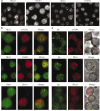
Figure 1
Figure 1
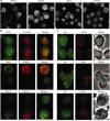
DNA repair foci in wild-type B lymphocytes after stimulation with LPS and IL-4 for 72 h. a, Distribution of Brca1, Rad51, Nbs1 and γ-H2AX in activated wild-type B cells. Confocal images were optically sectioned at 0.5-μm intervals and merged into a maximum projection. b, Double staining with Nbs1 (green) together with γ-H2AX (red), Rad51 (red) or Brca1 (red). The images were merged to determine co-localization (yellow). c, Co-localization of DNA repair foci with the Igh locus. B cells were stained with anti-γ-H2AX, anti-Nbs1, or anti-Brca1 antibodies (ICC (red)) followed by DNA FISH (green) detection of the Ch region. Cells were visualized by phase-contrast microscopy and the images were merged to determine co-localization (yellow). Fluorescence images in b and c represent a single optical section.
Figure 2
Figure 2
DNA repair foci associated with CSR form predominantly in the G1 phase of the cell cycle. a, Representative ICC-FISH images of G1 and S/G2/M sorted cells. Wild-type B cells stimulated for 48 h with LPS and IL-4 were labelled with Hoechst and live cells were electronically separated into G1 (purity >95%) and S/G2/M (purity >80%) cell cycle fractions. Sorted populations were analysed by ICC-FISH for co-localization (yellow) of γ-H2AX, Nbs1 and Brca1 with Ch. b, Percentage of sorted cells in a given optical section that contain Nbs1, γ-H2AX and Brca1 foci (whole bar), and percentage of sorted cells in which DNA repair foci co-localize with Ch (hatched section). Brca1 and γ-H2AX foci were found disproportionately in a greater fraction of S/G2/M cells relative to G1 (γ-H2AX, 31% compared with 15%; Brca1, 59% compared with 16%, respectively), whereas the percentage of G1 cells (11%) and S/G2/M (14%) cells with Nbs1 foci was similar. More than 1,000 cells were analysed.
Figure 3
Figure 3
Kinetics of Ch-associated foci accumulation in wild-type mice and impaired switching in H2AX–/– mice. a, Actin control, germline sterile Iγ1–Cγ1 transcripts, mature switch V(D)J–Cγ1 transcripts (assayed by PCR with reverse transcription (RT-PCR)), and Sμ–Sγ1 DNA rearrangements assayed by digestion-circularization PCR (DC-PCR) at indicated times of LPS and IL-4 culture. Cell surface expression of IgG1 was detected by flow cytometry. Percentages from total lymphocyte-gated populations are indicated. RT-PCR, DC-PCR and FACS analysis were performed as described. b, Analysis by ICC-FISH for co-localization of Nbs1 foci with Ch, quantified as in Fig. 2b. c, Cell surface expression of IgG1 in B cells from H2AX–/– mice and littermates assayed 72 h after stimulation with LPS and IL-4. An aliquot of the samples (shown below) was used to simultaneously measure cell cycle distribution at 72 h. Numbers indicate the percentage of sub-G1, G1 and S/G2/M cells, respectively. d, Iγ1–Cγ1 transcripts, mature switch V(D)J–Cγ1 transcripts, and Sμ–Sγ1 DNA rearrangements in B cells of H2AX–/– and H2AX+/+ mice assayed at 0 and 72 h after LPS and IL-4 culture. DNA and RNA samples taken at 72 h were diluted as indicated.
Figure 4
Figure 4
Co-localization of Nbs1 and γ-H2AX at the Igh locus is dependent on AID. a, AID–/– B cells were stimulated for 72 h with LPS and IL-4 and the intracellular localization of Brca1, Rad51, Nbs1 and γ-H2AX was determined by immunofluorescence as in Fig. 1a. b, Double staining with Nbs1 (green) together with either γ-H2AX (red), Brca1 (red) or Rad51 (red) in AID–/– cultures. The images were merged to determine co-localization (yellow). c, Co-localization of DNA repair foci with Igh locus in activated AID–/– B cells, analysed by ICC-FISH as in Fig. 1c.
Figure 5
Figure 5
AID-dependent Sμ mutation induced by LPS and IL-4. a, Proportion of Eμ, Cμ and Sμ sequences carrying different numbers of mutations before and after stimulation with LPS and IL-4 for 72 h. Segment sizes in the pie charts are proportional to the number of sequences carrying the number of mutations indicated in the periphery of the charts. The total number of independent sequences analysed is indicated in the centre of each chart. b, Proportion of Sμ sequences carrying different numbers of mutations 72 h after stimulation with CpG. c, Distribution of point mutations. The region sequenced is indicated with the first base corresponding to position 4,600 in the μ region germline sequence (GenBank accession number J00440). H, position of HindIII restriction enzyme sites. Lower-case letters above the line indicate mutations found among the sequenced clones. Each lower-case letter represents an independent mutational event. Hot spots containing mutations are underlined.

References

    1. Muramatsu M, et al. Class switch recombination and hypermutation require activation-induced cytidine deaminase (AID), a potential RNA editing enzyme. Cell. 2000;102:553–563. - PubMed
    1. Carney JP, et al. The hMre11/hRad50 protein complex and Nijmegen breakage syndrome: linkage of double-strand break repair to the cellular DNA damage response. Cell. 1998;93:477–486. - PubMed
    1. Rogakou EP, Boon C, Redon C, Bonner WM. Megabase chromatin domains involved in DNA double-strand breaks in vivo. J. Cell. Biol. 1999;146:905–916. - PMC - PubMed
    1. Downs JA, Lowndes NF, Jackson SP. A role for Saccharomyces cerevisiae histone H2A in DNA repair. Nature. 2000;408:1001–1004. - PubMed
    1. Revy P, et al. Activation-induced cytidine deaminase (AID) de®ciency causes the autosomal recessive form of the Hyper-IgM syndrome (HIGM2). Cell. 2000;102:565–575. - PubMed

Publication types

MeSH terms