. 2002 Feb 22;277(8):6287-95.
doi: 10.1074/jbc.M106649200. Epub 2001 Dec 11.

Transforming growth factor-beta induction of smooth muscle cell phenotpye requires transcriptional and post-transcriptional control of serum response factor

Affiliations

Transforming growth factor-beta induction of smooth muscle cell phenotpye requires transcriptional and post-transcriptional control of serum response factor

Karen K Hirschi et al. J Biol Chem. .

Abstract

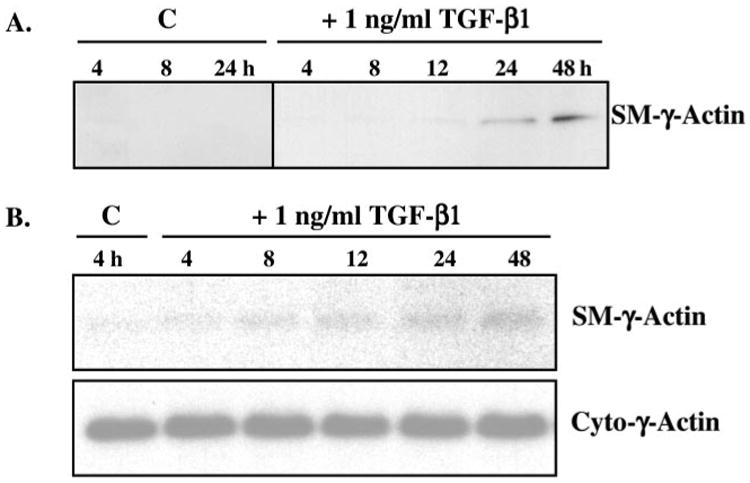
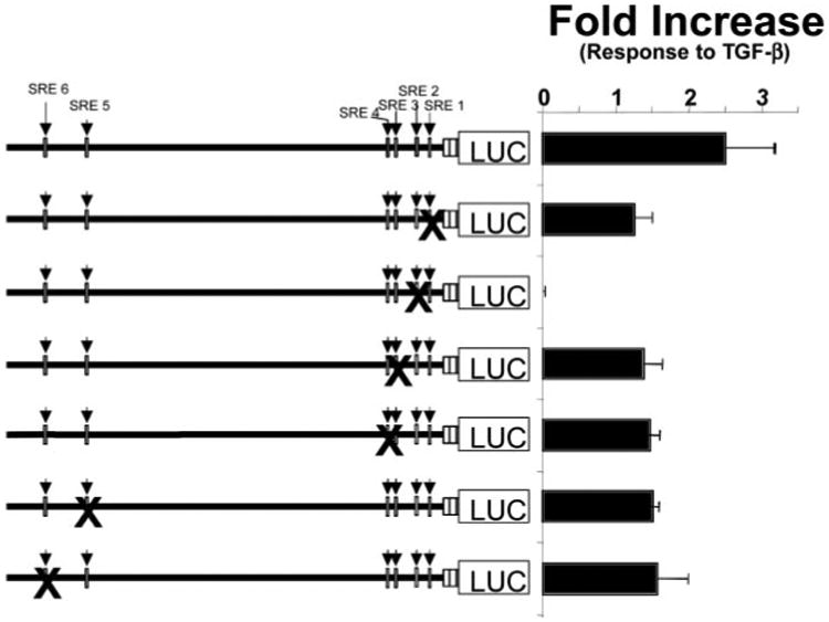
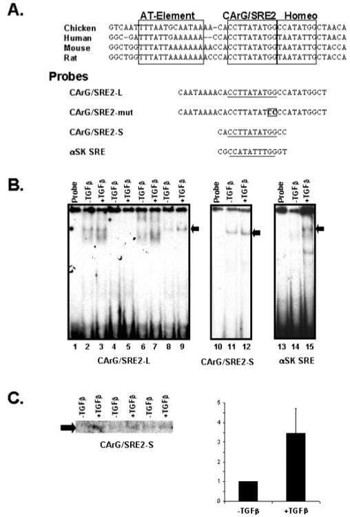
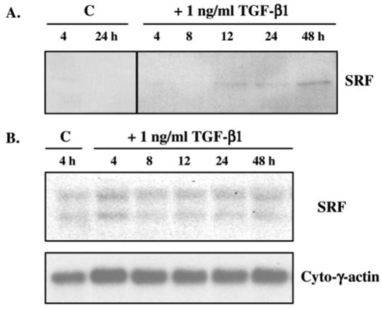
Transforming growth factor-beta induces a smooth muscle cell phenotype in undifferentiated mesenchymal cells. To elucidate the mechanism(s) of this phenotypic induction, we focused on the molecular regulation of smooth muscle-gamma-actin, whose expression is induced at late stages of smooth muscle differentiation and developmentally restricted to this lineage. Transforming growth factor-beta induced smooth muscle-gamma-actin protein, cytoskeletal localization, and mRNA expression in mesenchymal cells. Smooth muscle-gamma-actin promoter-luciferase reporter activity was enhanced by transforming growth factor-beta, and deletion analysis revealed that CArG box 2 in the promoter was necessary for this transcriptional activation. CArG motifs bind transcriptional activator serum response factor; gel shift analyses revealed increased binding of serum response factor-containing complexes to this site in response to transforming growth factor-beta, paralleled by increased serum response factor protein expression. Serum response factor expression was found to be up-regulated by transforming growth factor-beta via transcriptional activation of the gene and post-transcriptional regulation. Using mesenchymal cells stably transfected with wild type or dominant-negative serum response factor, we demonstrated that its expression is sufficient for induction of a smooth muscle phenotype in mesenchymal cells and is necessary for transforming growth factor-beta-mediated smooth muscle induction.


Figures

Fig. 1
Fig. 1. TGF-β induction of SM-γ-actin protein expression and cytoskeletal localization
10T1/2 mesenchymal cells were incubated with or without 1 ng/ml TGF-β1 for 24 h; bovine aortic endothelial cells and smooth muscle cells were incubated in control conditions for 24 h, as described under “Materials and Methods.” All cells were fixed in 4% paraformaldehyde and immunostained for SM-γ-actin (ICN antibody; 1:1000). 10T1/2 cells (C), as well as endothelial cells (A), did not express SM-γ-actin protein; however, TGF-β induced expression and cytoskeletal localization of SM-γ-actin in 10T1/2 cells (D) to a level similar to that of primary cultures of smooth muscle cells (B). Bar = 10 μm.
Fig. 2
Fig. 2. Time course of TGF-β induction of SM-γ-actin protein and mRNA expression
A, 10T1/2 mesenchymal cells were incubated with or without 1 ng/ml TGF-β1 for 0, 4, 8, 12, 24, and 48 h, after which total protein lysates were isolated and subjected to Western blot analyses (10 μg of total protein/lane). SM-γ-actin protein (∼43 kDa) expression was induced after 12–24 h of TGF-β treatment compared with control (C) conditions. B, 10T1/2 mesenchymal cells were treated with 1 ng/ml TGF-β1 (as in A), after which total RNA was isolated and subjected to Northern blot analyses (5 μg of total RNA/lane). SM-γ-actin mRNA (∼1.5 kb) expression was induced after ∼8 h. Blots were re-hybridized with a specific probe to cytoplasmic -γ-actin as a loading control. These data, taken together, suggest that SM-γ-actin gene expression is transcriptionally regulated in 10T1/2 cells by TGF-β.
Fig. 3
Fig. 3. TGF-β-induced transcriptional activation of the SM-γ-actin promoter
Chimeric deletion constructs, containing different lengths of SM-γ-actin promoter, ranging from full-length (2294 bp) to 65 bp, were transiently transfected (1 μg DNA/transfection) into 10T1/2 cells (70,000 cells/well) that were then incubated in the presence or absence of 1 ng/ml TGF-β1 for 48 h. Promoter activity was assessed in transfected cells by measurement of the firefly luciferase reporter. Each lysate was analyzed in triplicate, and luciferase activity was normalized to protein content. To determine the responsiveness of a promoter to TGF-β treatment, the luciferase activity generated in lysates of TGF-β-treated cells was directly compared with activity obtained with the same construct in untreated cells and is plotted as a fold increase response to TGF-β. The promoter constructs used in the experiments are diagrammed in A, with their response to TGF-β in 10T1/2 cells plotted (mean + S.D.) in B. There was a TGF-β-dependent response of 2–3-fold with promoter DNA containing the first 136 bp (SMGA −136). The TGF-β response was maintained at similar levels with the addition of DNA out to −2.3 kb flanking the gene.
Fig. 4
Fig. 4. CArG/SRE2 is required for TGF-β transcriptional regulation of SM-γ-actin promoter
Plasmids containing full-length SM-γ-actin promoter regions in which each of its six CArG/SRE motifs were individually mutated were transiently transfected into 10T1/2 cells. The TGF-β responsiveness of these mutated DNAs was evaluated by incubating cells in the presence or absence of 1 ng/ml TGF-β for 48 h and measuring the resultant luciferase activity in cell lysates. Mutations in CArG/SRE sequences 1, 3, 4, 5, and 6 did not abolish, but consistently reduced, TGF-β transcriptional activation by 40–60%. In contrast, mutations in the CArG/SRE2, which disrupt SRF binding to this cis-element, totally abolished TGF-β-induced transcriptional activation.
Fig. 5
Fig. 5. SRF binding activity at CArG/SRE2 is enhanced in response to TGF-β
Nuclear lysates were derived from 10T1/2 mesenchymal cells incubated with or without 1 ng/ml TGF-β for 48 h and utilized for gel shift analyses. A, an alignment of SM-γ-actin promoter sequences from chicken (14), human (18), mouse (49), and rat (75) containing the region surrounding the CARG/SRE2 motif is shown. The sequence from this highly conserved segment of the SM-γ-actin promoter contains an A-T-rich and homeodomain binding motifs in addition to the CArG/SRE2 element that function in SMGA transcription. Below the sequence alignment is the sequence of the oligonucleotide probes used for the gel shift experiments shown here. The CArG/ SRE2-L probe contains significant sequence 5′ and 3′ to the SRE element, whereas the CArG/SRE2-S probe contains the minimal SRF binding motif. The CArG/SRE2-mut probe contains a mutated SRE binding site and the α-SK SRE is a strong SRF binding site derived from the α-skeletal actin gene. B, multiple complexes were observed with 5 μg of nuclear lysate derived from treated and control cells using the CArG/SRE2-L probe (lanes 2 and 3). A 50× molar excess of unlabeled probe prevented the formation of binding complexes with the SM-γ-actin CArG/SRE2 probe (lanes 4 and 5), whereas a 50× excess of a probe that contained an SRF binding site derived from the α-skeletal actin gene efficiently inhibited the formation of one prominent complex (denoted with the arrow), indicating that this complex contained SRF as its principal binding component (lanes 6 and 7). A competitor oligonucleotide in which the CArG/SRE motif was mutated to prevent SRF binding did not inhibit the formation of the SRF complex with the native probe sequence; however the other complexes were efficiently prevented from forming (lanes 8 and 9). The arrow to the right of the autoradiograph denotes the position of the SRF containing complex. Using 5 μg of nuclear lysate the CArG/SRE2-S (lanes 11 and 12) and α-SK SRE (lanes 14 and 15) showed some varible nonspecific bands with a major band (denoted by the arrow) of SRF binding activity. C, quantitative assessment of SRF binding activity from 10T1/2 cells was performed by probing lysates from multiple, separate experiments with the CArG/ SRE2-S oligonucleotide probe and separating the resultant binding complexes on a single gel. SRF-binding complex formation with this probe using 3 μg of nuclear lysate from three separate experiments are shown. The radioactivity associated with the SRF complex band was then quantitated using a Bio-Rad PhosphorImager and the Molecular Analysis Software package (Bio-Rad). The amount of binding derived from treated cells was compared with that observed in control cells, which was designated as a value of 1. The relative binding activity was averaged and plotted ± the S.D.
Fig. 6
Fig. 6. TGF-β regulation of SRF expression
A, 10T1/2 cells were treated with 1 ng/ml TGF-β for up to 48 h, after which time total protein was isolated and subjected to Western blot analyses (10 μg of total protein/lane) using anti-SRF (ICN; 1:500). SRF protein (∼65 kDa) expression was induced after 8–12 h of TGF-β exposure compared with control (C) conditions. B, total RNA was isolated from similarly treated cells and probed, via Northern blot analysis, with a murine SRF cDNA. This probe recognized two mRNA species (∼4.5 and 2.5 kb) in the RNA populations derived from the 10T1/2 cells, which are the products of both differential polyadenylation and alternate splicing of the SRF gene transcript (42). Blots were re-hybridized with a specific probe to cytoplasmic γ-actin as a loading control. While there was a consistent 25–30% increase, in multiple experiments, in SRF mRNA in TGF-β-treated cells as compared with controls, which was detectable after 4 h, the mRNA levels did not exhibit a steady increase over the time of incubation as did the appearance of SRF protein.
Fig. 7
Fig. 7. SRF is necessary and sufficient for TGF-β induction of a SM phenotype
10T1/2 cells were transfected, via electroporation, with 5 μg of linearized HA-tagged expression plasmid containing no cDNA (vector control), 5 μg of linearized wtSRF cDNA, or 5 μg of linearized dnSRF cDNA. All cells were cotransfected with 0.5 μg of linearized pCI-neo plasmid, and stable transfectant clones were generated from each experimental group via selection in 1000 μg/ml G418-containing media. Total protein was isolated from each clone and screened via Western analyses (10 μg of total protein/lane) to assess the expression of SRF protein and the HA tag. A, transfectants expressing wtSRF (10T-wt SRF) and dnSRF (10T-dnSRF) exhibited SRF protein expression, and concomitant HA tag, via Western analyses; stable clones containing only the plasmid vectors (10T-v) did not exhibit SRF or HA expression. One representative clone of the 12 generated for each experimental group is shown. B, two or three clones from each group were cultured in the presence or absence of 1 ng/ml TGF-β1 for 24 h; protein was isolated from each and analyzed for expression of SM-γ-actin. Stable transfectants containing only vector (10T-v) exhibited a dose-dependent increase in SM-γ-actin protein expression in response to TGF-β. Transfectants expressing wtSRF (10T-wtSRF) exhibited elevated levels of SM-γ-actin in control conditions, which was further increased in response to TGF-β. Expression of dnSRF in mesenchymal cells prevented TGF-β induction of SM-γ-actin protein expression. Results generated from a representative clone for each experimental group are shown.

References

    1. Massaque J. Annu Rev Biochem. 1998;67:753–791. - PubMed
    1. Staehling-Hampton K, Hoffmann FM, Baylies MK, Rushton E, Bate M. Nature. 1994;372:783–786. - PubMed
    1. Shull MM, Ormsby I, Kier AB, Pawlowski S, Diebold RJ, Yin M, Allen R, Sidman C, Proetzel G, Calvin D, Annunziata N, Doetschman T. Nature. 1992;359:693–699. - PMC - PubMed
    1. Roberts A. Wound Repair Regen. 1995;3:408–418. - PubMed
    1. Dickson MC, Martin JS, Cousins FM, Kulkarni AB, Karlsson S, Akhurst RJ. Development (Camb) 1995;121:1845–1854. - PubMed

Publication types

LinkOut - more resources