. 2001 Dec 18;98(26):15050-5.
doi: 10.1073/pnas.261408198. Epub 2001 Dec 11.

A protein trap strategy to detect GFP-tagged proteins expressed from their endogenous loci in Drosophila

Affiliations

A protein trap strategy to detect GFP-tagged proteins expressed from their endogenous loci in Drosophila

X Morin et al. Proc Natl Acad Sci U S A. .

Abstract

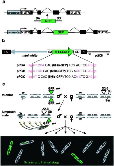
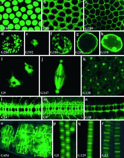
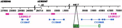
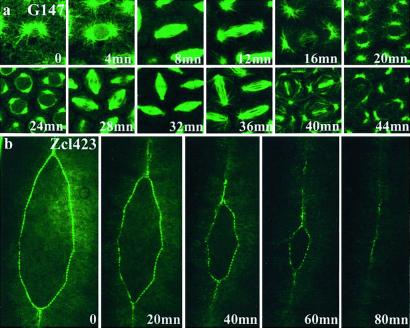
In Drosophila, enhancer trap strategies allow rapid access to expression patterns, molecular data, and mutations in trapped genes. However, they do not give any information at the protein level, e.g., about the protein subcellular localization. Using the green fluorescent protein (GFP) as a mobile artificial exon carried by a transposable P-element, we have developed a protein trap system. We screened for individual flies, in which GFP tags full-length endogenous proteins expressed from their endogenous locus, allowing us to observe their cellular and subcellular distribution. GFP fusions are targeted to virtually any compartment of the cell. In the case of insertions in previously known genes, we observe that the subcellular localization of the fusion protein corresponds to the described distribution of the endogenous protein. The artificial GFP exon does not disturb upstream and downstream splicing events. Many insertions correspond to genes not predicted by the Drosophila Genome Project. Our results show the feasibility of a protein trap in Drosophila. GFP reveals in real time the dynamics of protein's distribution in the whole, live organism and provides useful markers for a number of cellular structures and compartments.


Figures

Figure 1
Figure 1
The protein trap screen strategy. (a) Principle of the artificial exon: see text for details. (b) The PTTs. In addition to the 6His-GFP reporter flanked by splicing sequences, the P-element contains a miniwhite selection gene in the opposite orientation. In each of the three constructs GA, GB, and GC, the splice acceptor (ag | AT) and splice donor (AG | gt) consensus sequences are in a different reading frame relative to the 6His-GFP sequence. Although slightly different from the AG/GT acceptor splice consensus, AG/AT is the second most commonly found in Drosophila (31). (c) Crossing scheme used to generate GFP-positive flies. Flies are selected on the occurrence of a GFP signal. We used mutator lines with a “nonfluorescent” insertion on the third chromosome and no counter selection against the transposase or the starting chromosome. As a result, insertions on all three chromosomes can be recovered, including unstable insertions on the Delta2–3Sb chromosome or new insertions on the starting chromosome.
Figure 2
Figure 2
Subcellular distribution of trapped proteins. (ac) Examples of targeting of the trapped protein to the nucleus (a, line G280, His2Av), cytoplasm (b, line G89), and membrane (c, line G289). a and b are just before cellularization, and c is just after cellularization. (dh) GFP distribution in the giant nuclei of third-instar larval salivary glands of different “nuclear” lines. These cells contain polytene chromosome arms that retain the arrangement that they adopt in diploid interphase nuclei. Their nuclear architecture is easily visualized and consists of a chromosomal domain (d, line G280, His2Av:GFP), a large central domain occupied by the nucleolus (e, line G392), a meshwork-like extra-chromosomal nuclear domain (32) (f, line G180), delimited by the nuclear envelope (g, line G262, lamin:GFP and h, line G158, lamin C:GFP). Note the large nuclear dots in h. (i) In line G9, GFP is detected in the endoplasmic reticulum, surrounding a prophase nucleus in the syncitial blastoderm. “Holes” corresponding to the position of the two centrosomes within the endoplasmic reticulum can be seen. (jk) G147 produces a microtubule-associated fusion, seen here in a metaphase nucleus before cellularization (j) whereas the product of G138 is found in centrosomes only at a similar stage (k; the magnification is different between j and k). (ln) G9, G147, and G38 show a predominant GFP signal in axons in stage 16 embryos. (o) In G454, an insertion in Viking, a collagen IV type molecule, GFP labels the extracellular matrix. (pr) Insertions G5 (p, tropomyosin2), G129 (q), and G53 (r, kettin) reveal different subunits of the sarcomeric complex in adult thoracic indirect flight muscle fibers. (Magnifications: ac and k, ×500; dh, ×300; ij, ×1,000; ln, ×160; o, ×100; pr, ×1,000.)
Figure 3
Figure 3
Protein trap lines reveal genes not predicted in the genome annotation database. In line G108, the PTT is inserted at position 44617 of the genomic scaffold AE003567, downstream of predicted gene CG10649 and upstream of CG10668. blast searches of EST databases with CG10649 and CG10668 identify regions on the 5′ and 3′ ends of EST LD29922, respectively. Besides, the 5′-most part of LD29922 matches a third prediction, CG10647, further upstream, on the adjacent scaffold AE003566. Therefore, segments of all three predictions (CG10647, CG10649, and CG10668) are part of a single gene, which spans ≈120 kb. The insertion in line G108 reveals the expression of this gene: 3′ cDNA sequences fused to GFP match sequences of CG10668. Predicted genes are in blue, sequenced parts of the EST are in red, and the region found to be fused with GFP in the 3′ rapid amplification of cDNA ends experiment is in green.
Figure 4
Figure 4
Dynamics of GFP fusion distribution. (a) The distribution of the protein fusions produced in line G147 (microtubule-associated protein) was observed at different times during cell division in the syncitial embryo. (b) In line Zcl423, the GFP fusion is specifically expressed at the leading edge of epithelial cells during the zipper-like cell movements of dorsal closure. Anterior is up. (Magnifications: a, ×500; b, ×150.) Video versions of these and other time-lapse experiments can be viewed as Movies 1–4, which are published as supporting information on the PNAS web site, www.pnas.org.

References

    1. Chalfie M, Tu Y, Euskirchen G, Ward W W, Prasher D C. Science. 1994;263:802–805. - PubMed
    1. Cutler S R, Ehrhardt D W, Griffitts J S, Somerville C R. Proc Natl Acad Sci USA. 2000;97:3718–3723. - PMC - PubMed
    1. Misawa K, Nosaka T, Morita S, Kaneko A, Nakahata T, Asano S, Kitamura T. Proc Natl Acad Sci USA. 2000;97:3062–3066. - PMC - PubMed
    1. Ding D Q, Tomita Y, Yamamoto A, Chikashige Y, Haraguchi T, Hiraoka Y. Genes Cells. 2000;5:169–190. - PubMed
    1. Lindsey K, Wei W, Clarke M C, McArdle H F, Rooke L M, Topping J F. Transgenic Res. 1993;2:33–47. - PubMed

Publication types

MeSH terms

Substances