. 2001 Dec 18;98(26):14979-84.
doi: 10.1073/pnas.261463298. Epub 2001 Dec 11.

The gamma -secretase-cleaved C-terminal fragment of amyloid precursor protein mediates signaling to the nucleus

Affiliations

The gamma -secretase-cleaved C-terminal fragment of amyloid precursor protein mediates signaling to the nucleus

Y Gao et al. Proc Natl Acad Sci U S A. .

Abstract

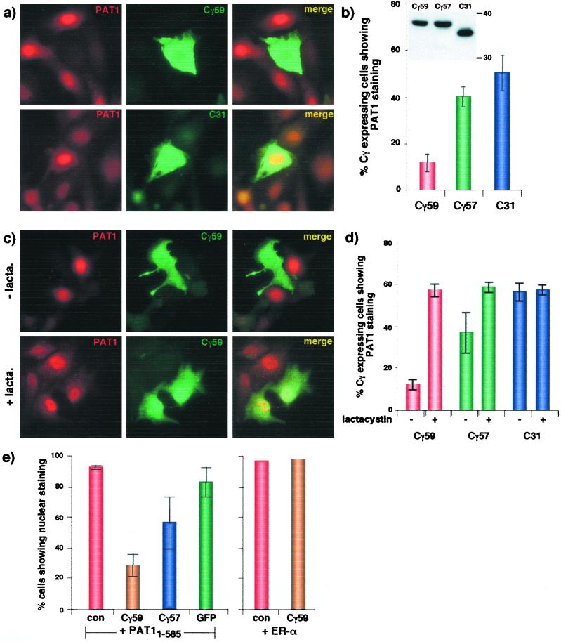
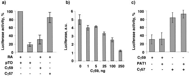
Sequential processing of the amyloid precursor protein (APP) by beta- and gamma-secretases generates the Abeta peptide, a major constituent of the senile plaques observed in Alzheimer's disease. The cleavage by gamma-secretase also results in the cytoplasmic release of a 59- or 57-residue-long C-terminal fragment (Cgamma). This processing resembles regulated intramembrane proteolysis of transmembrane proteins such as Notch, where the released cytoplasmic fragments enter the nucleus and modulate gene expression. Here, we examined whether the analogous Cgamma fragments of APP also exert effects in the nucleus. We find that ectopically expressed Cgamma is present both in the cytoplasm and in the nucleus. Interestingly, expression of Cgamma59 causes disappearance of PAT1, a protein that interacts with the APP cytoplasmic domain, from the nucleus and induces its proteosomal degradation. Treatment of cells with lactacystin prevents PAT1 degradation and retains its nuclear localization. By contrast, Cgamma57, a minor product of gamma-cleavage, is only marginally effective in PAT1 degradation. Furthermore, Cgamma59 but not Cgamma57 potently represses retinoic acid-responsive gene expression. Thus, our studies provide the evidence that, as predicted by the regulated intramembrane proteolysis mechanism, Cgamma seems to function in the nucleus.


Figures

Figure 1
Figure 1
PAT1 and its intracellular localization. (a) Schematic representation of putative motifs present in PAT1. See text for details. (b) Inducible expression of PAT1 and deletion mutants. MDCK cells stably transfected with PAT11–585, PAT11–270, or PAT11–411 were induced (+) or not (−) with tetracycline and analyzed by Western blotting with anti-PAT1 antibody (mAb37). The endogenous PAT1 shows as a doublet at 70 kDa. (c) MDCK cells were fixed in methanol and stained with anti-FLAG antibody (Left) and counterstained with 4′,6-diamidino-2-phenylindole (Right). (d) MDCK585 cells were fixed in 4% paraformaldehyde and treated without (Upper) or with (Lower) 0.5% Triton X-100 before staining with anti-FLAG antibody (×63 objective).
Figure 2
Figure 2
Cγ59 is selectively retained in the cytoplasm in MDCK411 cells. (a) Processing of APP. APP, initially cleaved by β-secretase close to the external surface of the membrane, can be cleaved by γ-secretase at two sites within the plane of the membrane. The major cleavage site (90–95%) produces Aβ-1–40 plus Cγ59, whereas the minor site yields minor Aβ-1–42 plus Cγ57. (b) Cells transiently transfected with GFP-Cγ59 were stained with antibody 369 that recognizes the cytoplasmic tail portion of APP. Note that Cγ59 is present in the nucleus as well as cytoplasm in MDCKTR and MDCK585 cells and mostly cytoplasmic in MDCK411 cells. (c) MDCK411 cells do not accumulate other nuclear proteins in the cytoplasm. MDCK585 (Upper) or MDCK411 cells (Lower) were transiently transfected with GFP-coilin or ER-α and stained with anti-FLAG antibody (Left and Center) or anti-ER-α antibody (Right). Left and Center show a view of the same field through rhodamine channel (Left) or observed for GFP signal (Center; ×63 objective).
Figure 3
Figure 3
Expression of Cγ59 causes disappearance of PAT1 from the nucleus. (a) MDCK585 cells transiently transfected with GFP-Cγ59 (Upper) or GFP-C31 (Lower) were labeled with anti-FLAG antibody followed by rhodamine-anti-mouse IgG (Left) or antibody 369 followed by FITC-anti-rabbit IgG (Center). (b) Quantification of data is shown on the right (means ± SD of at least three independent experiments). Inset shows that these three proteins were expressed at similar levels. (c) Lactacystin treatment prevents the disappearance of PAT1 in Cγ59-expressing MDCK585 cells. Details are as above. (d) Quantification of the data is shown on the right (means ± SD of at least three experiments). (e) MDCKTR cells were transiently cotransfected with PAT11–585 or ER-α together with other plasmids as indicated and labeled as above. con, Control (empty vector). Only those cells that showed signal for both proteins were quantified for nuclear vs. cytoplasmic localization of PAT1. Cγ59 did not affect the nuclear localization of ER-α.
Figure 4
Figure 4
Cγ59 potently represses RA-responsive transcription. (a) CV1 cells were cotransfected with a plasmid mixture of RARE-TK-Luc, RAR, and β-galactosidase (to normalize for transfection efficiency) together with the plasmids indicated below each bar. A 59-residue-long, γ-secretase-cleaved, C-terminal fragment of APP (Cγ59) but not the 57-residue-long fragment (Cγ57) represses transactivation. (b) Indicated amounts of GFP-Cγ59 plasmid DNA were cotransfected with the reporter plasmid DNA. The data show that Cγ59 represses transactivation in a dose-dependent manner. (c) Overexpression of PAT11–585 does not affect the repression by Cγ59. Cells were cotransfected with the reporter plasmid mix together with 250 ng each of Cγ and PAT11–585 or Cγ with pTO (parental plasmid vector). The normalized luciferase activity obtained with pTO plasmid is expressed as 100%. Bars = means ± SD of at least three experiments performed in duplicate except in b, which shows values from a single, representative experiment repeated twice.

References

    1. Price D L, Sisodia S S. Annu Rev Neurosci. 1998;21:479–505. - PubMed
    1. Selkoe D J. Trends Cell Biol. 1998;8:447–453. - PubMed
    1. De Strooper B, Annaert W. J Cell Sci. 2000;113:1857–1870. - PubMed
    1. Vassar R, Citron M. Neuron. 2000;27:419–422. - PubMed
    1. Haass C, De Strooper B. Science. 1999;286:916–919. - PubMed

Publication types

MeSH terms