. 2001 Dec 18;98(26):15294-9.
doi: 10.1073/pnas.261560998. Epub 2001 Dec 11.

Different responses of astrocytes and neurons to nitric oxide: the role of glycolytically generated ATP in astrocyte protection

Affiliations

Different responses of astrocytes and neurons to nitric oxide: the role of glycolytically generated ATP in astrocyte protection

A Almeida et al. Proc Natl Acad Sci U S A. .

Abstract

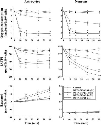
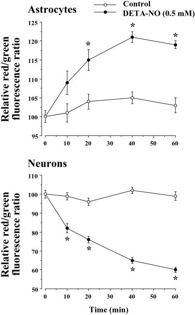
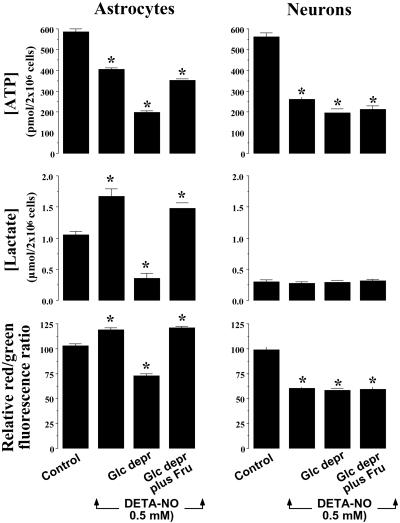
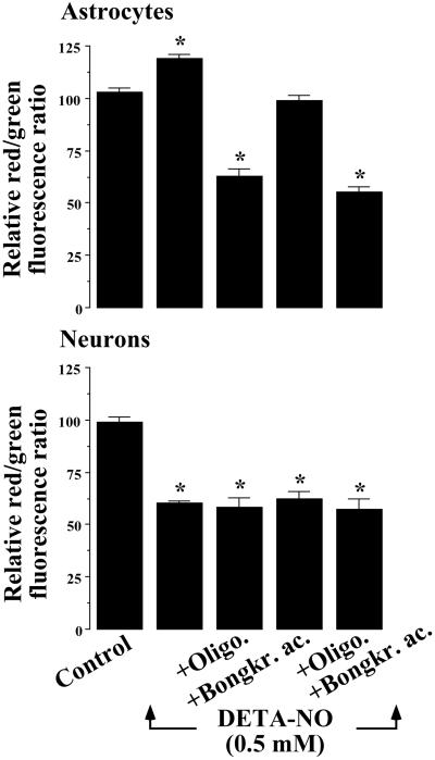
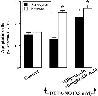
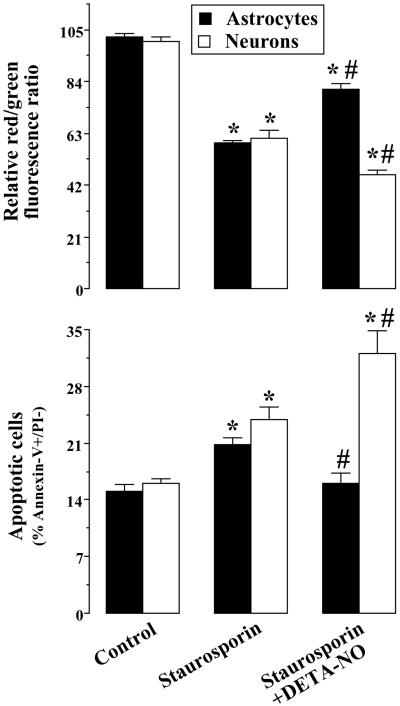
It was recently proposed that in Jurkat cells, after inhibition of respiration by NO, glycolytically generated ATP plays a critical role in preventing the collapse of mitochondrial membrane potential (Deltapsi(m)) and thus apoptotic cell death. We have investigated this observation further in primary cultures of rat cortical neurons and astrocytes-cell types that differ greatly in their glycolytic capacity. Continuous and significant ( approximately 85%) inhibition of respiration by NO (1.4 microM at 175 microM O(2)) generated by [(z)-1-[2-aminoethyl]-N-[2-ammonioethyl]amino]diazen-1-ium-1,2 diolate (DETA-NO) initially (10 min) depleted ATP concentrations by approximately 25% in both cell types and increased the rate of glycolysis in astrocytes but not in neurons. Activation of glycolysis in astrocytes, as judged by lactate production, prevented further ATP depletion, whereas in neurons, which do not invoke this mechanism, there was a progressive decrease in ATP concentrations over the next 60 min. During this time, there was a persistent mitochondrial hyperpolarization and absence of apoptotic cell death in astrocytes, whereas in the neurons there was a progressive fall in Deltapsi(m) and increased apoptosis. After glucose deprivation or treatment with inhibitors of the F(1)F(0)-ATPase and adenine nucleotide translocase, astrocytes responded to NO with a fall in Deltapsi(m) and apoptotic cell death similar to the response in neurons. Finally, although treatment of astrocytes with NO partially prevented staurosporin-induced collapse in Deltapsi(m) and cell death, NO and staurosporin synergized in decreasing Deltapsi(m) and inducing apoptosis in neurons. These results demonstrate that although inhibition of cellular respiration by NO leads to neurotoxicity, it may also result in initial neuroprotection, depending on the glycolytic capacity of the particular cell.


Figures

Figure 1
Figure 1
Inhibition of cellular respiration by NO stimulates glycolysis in astrocytes but not in neurons. Cell suspensions (2 × 10⁶ cells per ml) were incubated at 37°C in buffered Hanks' solution either in the absence (control) or presence of DETA-NO for the indicated times. Oxygen consumption experiments were performed at an initial O2 concentration of ≈200 μM. For ATP and lactate concentrations, aliquots of the cell suspensions were lysed in HClO4, neutralized with KHCO3, and used for metabolite determinations in the supernatants as described in Materials and Methods. *, P < 0.05 versus appropriate control values.
Figure 2
Figure 2
NO hyperpolarizes astrocytic mitochondria, but depolarizes neuronal mitochondria. Cell suspensions (2 × 10⁶ cells per ml) were incubated at 37°C in buffered Hanks' solution either in the absence (control) or presence of DETA-NO (0.5 mM) for the indicated times. In this buffer, 0.5 mM DETA-NO was found to generate a constant NO concentration of 1.4 μM. For Δψm measurements, cell suspensions were centrifuged, and the pellet was resuspended in buffered Hanks' solution containing JC-1 (3 μM) and incubated for a further 10-min at 37°C. Data acquisition was performed in a FACScalibur flow cytometer as detailed in Materials and Methods. Data are expressed as a percentage, with the red/green fluorescence ratio values of untreated cells (2.98 ± 0.10 for neurons, 3.11 ± 0.20 for astrocytes) considered as 100% and the red/green fluorescence ratio values of FCCP (5 μM)-treated cells (0.68 ± 0.04 for neurons, 0.84 ± 0.05 for astrocytes) considered as 0%. *, P < 0.05 versus appropriate control values.
Figure 3
Figure 3
NO-dependent glycolytic activation determines mitochondrial membrane potential. Cell suspensions (2 × 10⁶ cells per ml) were incubated at 37°C in buffered Hanks' solution either in the absence (control) or presence of DETA-NO (0.5 mM) for 60 min. For glucose-deprivation experiments (Glc depr), both the preincubation (45 min) and incubation (60 min) periods were carried out in glucose-free Hanks' solution. Under these conditions, the astrocytic content of glycogen was shown to be depleted. Where indicated, cells were incubated in the presence of 20 mM fructose throughout. ATP and lactate concentrations, as well as Δψm, were determined as described in the legends to Figs. 1 and 2. *, P < 0.05 versus appropriate control values.
Figure 4
Figure 4
F1F0-ATPase activity accounts for NO-mediated mitochondrial hyperpolarization. Cell suspensions (2 × 10⁶ cells per ml) were incubated at 37°C in buffered Hanks' solution either in the absence (control) or presence of DETA-NO (0.5 mM) for 60 min. Where indicated, the 60-min incubation period of the cells was carried out in the presence of oligomycin (8 μM), bongkrekic acid (10 μM), or both inhibitors together. Mitochondrial membrane potential measurements were made and expressed as described in the legend to Fig. 2. *, P < 0.05 versus appropriate control values.
Figure 5
Figure 5
F1F0-ATPase activity accounts for NO-mediated prevention from apoptotic cell death. Cell suspensions (2 × 10⁶ cells per ml) were incubated at 37°C in buffered Hanks' solution either in the absence (control) or presence of DETA-NO (0.5 mM) for 60 min. Where indicated, the 60-min incubation period of the cells was carried out in the presence of oligomycin (8 μM) plus bongkrekic acid (10 μM). Apoptotic cell death was determined after staining with FITC-conjugated annexin-V and PI. Data acquisition was carried out in a FACScalibur flow cytometer, as described in Materials and Methods. Annexin V+/PI cells were considered to be apoptotic. *, P < 0.05 versus appropriate control values.
Figure 6
Figure 6
NO partially protects astrocytes, but not neurons, from staurosporin-triggered Δψm collapse and apoptosis. Cell suspensions (2 × 10⁶ cells per ml) were incubated at 37°C in buffered Hanks' solution either in the absence (control) or presence of DETA-NO (0.5 mM) for 60 min. Where indicated, the 60-min incubation period of the cells were carried out in the presence of staurosporin (100 nM). Mitochondrial membrane potential measurements were carried out and expressed as described in the legend to Fig. 2. Apoptotic cell death was determined and expressed as described in the legend to Fig. 5. *, P < 0.05 versus appropriate control values. #, P < 0.05 versus appropriate staurosporin values.

References

    1. Moncada S, Palmer R M J, Higgs E A. Pharmacol Rev. 1991;43:109–142. - PubMed
    1. Melino G, Bernassola F, Catani M V, Rossi A, Corazzari M, Sabatini S, Vilbois F, Green D R. Cancer Res. 2000;60:2377–2383. - PubMed
    1. Sastry P S, Rao K S. J Neurochem. 2000;74:1–20. - PubMed
    1. Mitchell P. Nature (London) 1961;191:144–148. - PubMed
    1. Liu X, Kim C N, Yang J, Jemmerson R, Wang X. Cell. 1996;86:147–157. - PubMed

Publication types

LinkOut - more resources