. 2001 Dec 18;98(26):15239-44.
doi: 10.1073/pnas.261563198. Epub 2001 Dec 11.

Efficient assembly of an HIV-1/MLV Gag-chimeric virus in murine cells

Affiliations

Efficient assembly of an HIV-1/MLV Gag-chimeric virus in murine cells

B K Chen et al. Proc Natl Acad Sci U S A. .

Abstract

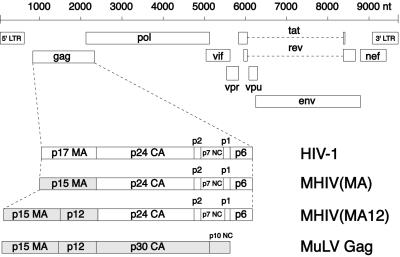
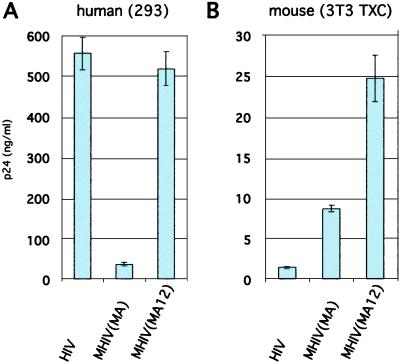
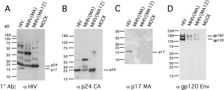
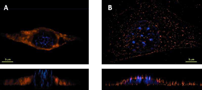
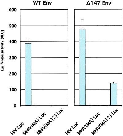
In human cells infected by HIV type 1 (HIV-1), the viral Gag protein directs the assembly of nascent viral particles at the plasma membrane. In murine cells, HIV-1 Gag fails to reach the plasma membrane and instead forms nonfunctional intracellular aggregates. The viral determinants of this species incompatibility are previously undefined. To address this problem, we replaced a region of HIV-1 Gag known to direct its localization, the matrix (MA) domain, with functionally homologous regions from Moloney murine leukemia virus (MLV), a murine retrovirus. An HIV-1 clone carrying such a chimeric Gag protein, designated murine HIV (MHIV), assembled more efficiently than nonchimeric HIV-1 and restored plasma membrane localization of Gag in murine cells. Increased efficiency of viral assembly in murine cells was observed from MHIV constructs carrying MLV MA in place of HIV-1 MA. Efficient processing of the HIV-1 capsid protein from the chimeric Gag polyprotein and subsequent infectivity of MHIV required the presence of MLV p12 in addition to MLV MA. These findings strongly suggest that the HIV-1 MA domain of HIV-1 Gag is responsible for the assembly defect in mouse cells. Although these MHIV do not recruit native HIV-1 Env efficiently, they are capable of single-round infection when produced by high-efficiency transfection of human 293 cells and provided with an HIV-1 Env lacking its cytoplasmic tail. With further adaptation, this chimeric MHIV approach may provide the basis for creating an infectious mouse model for HIV/AIDS.


Figures

Figure 1
Figure 1
Schematic illustration of the MHIV Gag chimeras. Genomic organization of HIV-1 (Upper). The HIV-1 Gag polyprotein is processed into p17 MA, p24 CA, p2, p7 NC, p1, and p6. Chimeric strategies replace the p17 MA with p15 MA of MLV Gag [MHIV(MA)] or both the p15 MA and p12 domains of MLV Gag [MHIV(MA12)]. The complete MLV Gag consists of p15 MA, p12, p30 CA, and p10 NC (Lower). Scale of viral genome sequence is shown in nucleotides (nt).
Figure 2
Figure 2
MHIV(MA12) releases greater levels of supernatant p24 than nonchimeric HIV-1 when transfected into CD4+, CXCR4+, cyclin T1-expressing murine 3T3 cells (3T3 TXC). Graphs are of p24 antigen harvested from supernatants from transfected human embryonic kidney cells 293 (A) and transfected 3T3 TXC cells (B). p24 ELISA was performed in triplicate as described in Materials and Methods. Note different y axis scales. Transfection efficiency and numbers of cells plated per transfection are significantly greater for human 293 cells, making quantitative comparisons between the two cell lines difficult. Results are representative of three independent experiments.
Figure 3
Figure 3
MHIV(MA12) is proteolytically processed in virus particles, but Env incorporation is inefficient. Western blot analysis of purified virus particles performed with human anti-HIV-1 patient serum (A), sheep anti-p24 (B), mouse anti-p17 (C), and sheep anti-gp120 HIV-1 Env (D). Virus construct-transfected or mock-transfected sample (MOCK) are indicated above each lane. Mock samples were transfected with equivalent amount of nonviral plasmid DNA.
Figure 4
Figure 4
Punctate membrane localization is restored in mouse cells by the MHIV(MA12) chimeric Gag protein. Shown are top and side views of three-dimensional image sets produced by deconvolution immunofluorescence microscopy of murine 3T3 TXC cells transiently transfected with nonchimeric HIV-1 (HIV Luc) (A) or Gag-chimeric HIV [MHIV(MA12) Luc] (B). (Upper) Top views of cells over the entire z stack of images. (Lower) Side view looking at the sum of several cross sections through the middle of the cell. Staining of Gag with polyclonal anti-p24 antibody was developed with Texas red-conjugated secondary antibody (shown in red), and DNA was stained with 4′,6-diamidino-2-phenylindole (shown in blue).
Figure 5
Figure 5
MHIV(MA12) Luc virus is able to undergo single-round infection when produced from human cells and provided with an HIV Env with a truncated cytoplasmic tail (Δ147 Env). Virus-containing supernatants were prepared from transfected 293 cells after cotransfection with an Env-deficient, luciferase-encoding provirus [HIV Luc, MHIV(MA) Luc, or MHIV(MA12) Luc] and an Env expression vector (WT Env or Δ147 Env). Infectivity of viruses was measured by luciferase assay of cell lysates prepared from infected CD4+ human osteosarcoma (HOS CD4) cells at 48 h after infection. Luciferase activity was measured in relative light units (RLU).

References

    1. Maddon P J, Dalgleish A G, McDougal J S, Clapham P R, Weiss R A, Axel R. Cell. 1986;47:333–348. - PubMed
    1. Feng Y, Broder C C, Kennedy P E, Berger E A. Science. 1996;272:872–877.
    1. Newstein M, Stanbridge E J, Casey G, Shank P R. J Virol. 1990;64:4565–4567. - PMC - PubMed
    1. Hart C E, Ou C Y, Galphin J C, Moore J, Bacheler L T, Wasmuth J J, Petteway S R, Jr, Schochetman G. Science. 1989;246:488–491. - PubMed
    1. Wei P, Garber M E, Fang S M, Fischer W H, Jones K A. Cell. 1998;92:451–462. - PubMed

Publication types

LinkOut - more resources