. 2001 Dec 17;20(24):6990-6.
doi: 10.1093/emboj/20.24.6990.

The structure of bovine IF(1), the regulatory subunit of mitochondrial F-ATPase

Affiliations

The structure of bovine IF(1), the regulatory subunit of mitochondrial F-ATPase

E Cabezón et al. EMBO J. .

Abstract

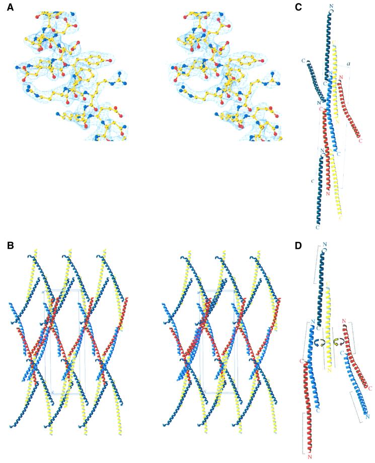
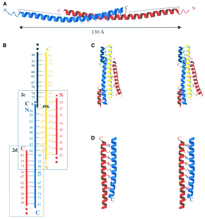
In mitochondria, the hydrolytic activity of ATP synthase is regulated by an inhibitor protein, IF(1). Its binding to ATP synthase depends on pH, and below neutrality, IF(1) is dimeric and forms a stable complex with the enzyme. At higher pH values, IF(1) forms tetramers and is inactive. In the 2.2 A structure of the bovine IF(1) described here, the four monomers in the asymmetric unit are arranged as a dimer of dimers. Monomers form dimers via an antiparallel alpha-helical coiled coil in the C-terminal region. Dimers are associated into oligomers and form long fibres in the crystal lattice, via coiled-coil interactions in the N-terminal and inhibitory regions (residues 14-47). Therefore, tetramer formation masks the inhibitory region, preventing IF(1) binding to ATP synthase.


Figures

None
Fig. 1. Crystal structure of bovine IF1-H49K. (A) Stereo view of the 2.2 Å resolution 2Fo – Fc electron density map calculated with CNS (contoured at 1.5 σ) from residues 27 to 41 of the protein. (B) Stereo view of the crystallographic packing. The four IF1 monomers in the asymmetric unit (A–D) are represented in red, sky blue, yellow and dark blue, respectively. The origin of the unit cell and the a, b and c axes are labelled ‘o’, ‘a’, ‘b’ and ‘c’, respectively. (C) View along the crystallographic b-axis. (D) Interactions between the two types of dimers. Dashed lines represent the minimal inhibitory sequence of IF1, which are masked in the tetramers. C- and N-termini are indicated with the letters C and N, respectively.
None
Fig. 2. Interhelical packing in IF1-H49K structure. (A) Ribbon diagram of the active dimer. Dashed lines represent the minimal inhibitory sequence. The disordered N-terminal residues 1–18 are shown as dotted lines. (B) Schematic representation of the interhelical packing. Residues involved in forming the coiled coils are represented with their sequence number. Helices are coloured as in Figure 1. Dotted lines represent the continuity of the helices. The position of lysine 49 in helix C (yellow) is shown in black, indicating the contacts with helices B (sky blue) and D (dark blue). Dashed boxes show the two areas of the protein represented in more detail in (C) and (D). (C) Stereo view of the interhelical packing in the N-terminus of the protein. (D) Stereo view of the interhelical packing in the C-terminus of the AB dimer.
None
Fig. 3. Covalent cross-linking of IF1 and IF1-H49K with dimethyl suberimidate. Experiments were carried out as described in Materials and methods. Samples treated with the cross-linking reagent were removed after 3 h of incubation and analysed by SDS–PAGE. Lanes 1–5 correspond to protein concentrations of 2, 1, 0.5, 0.2 and 0.1 mg/ml, respectively. IF1 and IF1-H49K are shown in (A) and (B), respectively. The positions of molecular weight markers are shown on the right in kDa. Arrows indicate the formation of higher oligomers.

References

    1. Abrahams J.P. and Leslie,A.G.W. (1996) Methods used in the structure determination of bovine mitochondrial F1-ATPase. Acta Crystallogr. D, 52, 30–42. - PubMed
    1. Brunger A.T. et al. (1998) Crystallography & NMR system: A new software suite for macromolecular structure determination. Acta Crystallogr. D, 54, 905–921. - PubMed
    1. Cabezón E., Butler,P.J.G., Runswick,M.J. and Walker,J.E. (2000a) Modulation of the oligomerization state of bovine F1-ATPase inhibitor protein, IF1, by pH. J. Biol. Chem., 275, 25460–25464. - PubMed
    1. Cabezón E., Arechaga,I., Butler,P.J.G. and Walker,J.E. (2000b) Dimerization of bovine F1-ATPase by binding the inhibitor protein, IF1. J. Biol. Chem., 275, 28353–28355. - PubMed
    1. CCP4 (1994) The CCP4 suite: programs for protein crystallography. Acta Crystallogr. D, 50, 760–763. - PubMed

Publication types