. 2001 Dec 17;20(24):6997-7007.
doi: 10.1093/emboj/20.24.6997.

Cell-to-cell movement and assembly of a plant closterovirus: roles for the capsid proteins and Hsp70 homolog

Affiliations

Cell-to-cell movement and assembly of a plant closterovirus: roles for the capsid proteins and Hsp70 homolog

D V Alzhanova et al. EMBO J. .

Abstract

Diverse animal and plant viruses are able to translocate their virions between neighboring cells via intercellular connections. In this work, we analyze the virion assembly and cell-to-cell movement of a plant closterovirus and reveal a strong correlation between these two processes. The filamentous virions of a closterovirus possess a long body formed by the major capsid protein (CP) and a short tail formed by the minor capsid protein (CPm). Genetic and biochemical analyses show that the functions of these virion components are distinct. A virion body is required primarily for genome protection, whereas a tail represents a specialized device for cell-to-cell movement. Furthermore, tail assembly is mediated by the viral Hsp70 homolog (Hsp70h) that becomes an integral part of the virion. Inactivation of the ATPase domain of Hsp70h results in assembly of tailless virions that are incapable of translocation. A dual role for the viral molecular chaperone Hsp70h in virion assembly and transport, combined with the previous finding of this protein in intercellular channels, allowed us to propose a model of closteroviral movement from cell to cell.


Figures

None
Fig. 1. (A) Genome map of the beet yellows virus (BYV). L-Pro, leader proteinase (the arrow below shows the site of L-Pro self-cleavage); MET, HEL and POL, the methyltransferase, RNA helicase and RNA polymerase domains of the BYV replicase, respectively; p6, a 6 kDa movement protein; p64, a 64 kDa movement protein; HSP70h, Hsp70 homolog; CPm and CP, the minor and major capsid proteins, respectively; p20 and p21, the 20 and 21 kDa proteins, respectively. (B) Electron micrograph of the BYV virion. The virion tail assembled by CPm was decorated with the CPm-specific antibodies and labeled with 10 nm gold particles (black dots). The virion body that is assembled by CP was not labeled. (C) Multiple alignment of the amino acid sequences of the CP and CPm of the BYV and grapevine leafroll-associated virus-2 (GLR) generated using the Macaw program (Schuler et al., 1991). The conserved amino acid motifs are boxed; the N-terminal motif unique to CPms (CPm box) is shown in gray. The four amino acid residues that are invariant among all capsid proteins of closteroviruses and conserved in most of the filamentous viruses are highlighted in bold and with asterisks. The numbers of amino acid residues in each protein are shown.
None
Fig. 2. Immunoblot analysis of the total protein extracts and virions derived from the transfected protoplasts. M, mock-transfected protoplasts; WT, transfection with the wild-type RNA transcripts derived from pBYV-4; R114D, transcripts harboring the corresponding mutation in the CP gene. Anti-CP serum was used to detect protein bands.
None
Fig. 3. Analysis of RNA encapsidation using RNase protection assays. The protoplast homogenates were incubated in the presence of endogenous RNases and RNase T1, and the protection of encapsidated RNA was assessed using RT–PCR and primer sets specific for the internal, 5′-terminal and 3′-terminal regions of the BYV genome. The products of RT–PCR were separated in a 1% agarose gel and visualized under UV following staining with ethidium bromide. SM, size markers; R114D and R128D, transfection with the RNA transcripts harboring corresponding mutations in the CP and CPm genes, respectively. Other designations are as in Figure 2.
None
Fig. 4. (A) Protein composition of the virions isolated from transfected protoplasts. R128D, transfection with RNA transcripts harboring the corresponding mutation in the CPm gene. The type of antiserum used for analysis is shown above the panels. Other designations are as in Figure 2. (B) Length distribution profile of the wild-type (WT) and tailless (CPm mutant R128D) virions.
None
Fig. 5. Infectivity of the virions tested in the second round of protoplast transfection. CP was detected in extracts of protoplasts using immunoblotting and anti-CP serum. (A) Time course of CP accumulation in protoplasts following transfection with wild-type virions isolated from protoplasts. The numbers correspond to days post-transfection. (B) Infectivity of the wild-type virions and tailless virions assembled by the wild-type CP in the presence of mutant CPm (R128D). Note that CP mutant R114D used as a negative control failed to produce any infectious virions.
None
Fig. 6. Diagrammatic summary of the phenotypes of CP and CPm mutants. Large unfilled boxes represent the conserved core parts of the CP and CPm, whereas lines represent variable N- and C-terminal regions. The CPm box is shown in gray; the letters S, R, G and D correspond to invariant serine, arginine, glycine and aspartic acid (Figure 1). Small vertical rectangles mark the positions of the mutants, with the upper rectangle for cell-to-cell movement phenotypes and the lower rectangles for the assembly phenotypes. The color code is shown at the bottom.
None
Fig. 7. Protein composition of the virions assembled in protoplasts by five movement-deficient CPm mutants. The names of mutants and type of antiserum used for analysis are shown above each panel.
None
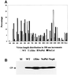
Fig. 8. Protein composition of the virions assembled by four BYV variants possessing mutant movement protein Hsp70h or lacking movement protein p6. ΔXho, deletion of most of the Hsp70h ORF; NoPh1 and NoCo1, mutations in the phosphate 1 and connect 1 motifs of Hsp70h, respectively; Nop6, inactivation of the p6 start codon. The names of mutants and type of antiserum used for analysis are shown above the panels.
None
Fig. 9. (A) Length distribution profile of the BYV virions formed by three Hsp70h mutants compared with that of the wild-type virions. (B) Infectivity of the virions tested in the second round of protoplast transfection. The designations of the Hsp70h and p6 mutations are the same as in Figure 8.
None
Fig. 10. Hypothetical model of BYV assembly and cell-to-cell movement. CW, cell wall separating adjacent plant cells.

References

    1. Agranovsky A.A., Boyko,V.P., Karasev,A.V., Koonin,E.V. and Dolja,V.V. (1991) Putative 65 kDa protein of beet yellows closterovirus is a homologue of HSP70 heat shock proteins. J. Mol. Biol., 217, 603–610. - PubMed
    1. Agranovsky A.A., Lesemann,D.E., Maiss,E., Hull,R. and Atabekov,J.G. (1995) ‘Rattlesnake’ structure of a filamentous plant RNA virus built of two capsid proteins. Proc. Natl Acad. Sci. USA, 92, 2470–2473. - PMC - PubMed
    1. Alzhanova D.V., Hagiwara,Y., Peremyslov,V.V. and Dolja,V.V. (2000) Genetic analysis of the cell-to-cell movement of beet yellows closterovirus. Virology, 267, 192–200. - PubMed
    1. Aranda M.A., Escaler,M., Wang,D. and Maule,A.J. (1996) Induction of HSP70 and polyubiquitin expression associated with plant virus replication. Proc. Natl Acad. Sci. USA, 93, 15289–15293. - PMC - PubMed
    1. Boorstein W.R., Ziegelhoffer,T. and Craig,E.A. (1994) Molecular evolution of the HSP70 multigene family. J. Mol. Evol., 38, 1–17. - PubMed

Publication types

Substances