. 2001 Dec 17;20(24):7096-107.
doi: 10.1093/emboj/20.24.7096.

Subunit interaction maps for the regulatory particle of the 26S proteasome and the COP9 signalosome

Affiliations

Subunit interaction maps for the regulatory particle of the 26S proteasome and the COP9 signalosome

H Fu et al. EMBO J. .

Abstract

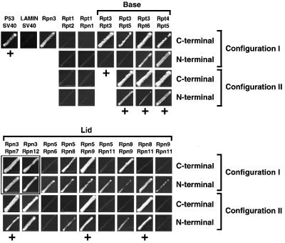
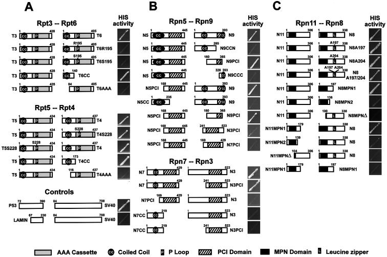
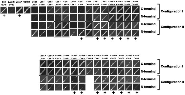
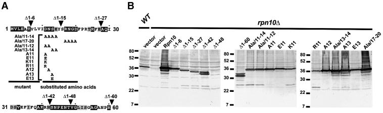
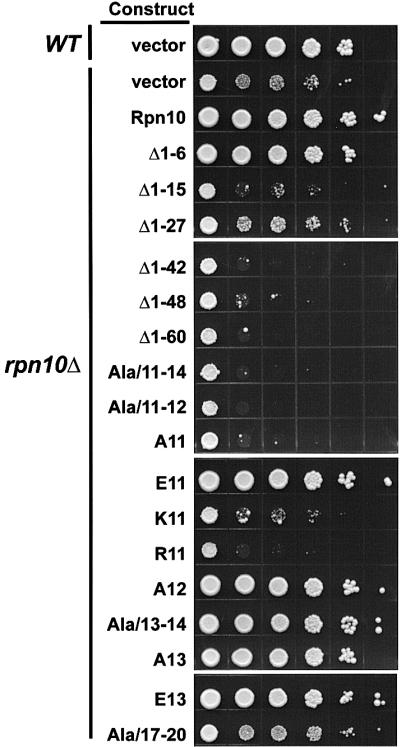
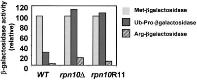
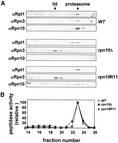
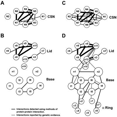
The 26S proteasome plays a major role in eukaryotic protein breakdown, especially for ubiquitin-tagged proteins. Substrate specificity is conferred by the regulatory particle (RP), which can dissociate into stable lid and base subcomplexes. To help define the molecular organization of the RP, we tested all possible paired interactions among subunits from Saccharomyces cerevisiae by yeast two-hybrid analysis. Within the base, a Rpt4/5/3/6 interaction cluster was evident. Within the lid, a structural cluster formed around Rpn5/11/9/8. Interactions were detected among synonymous subunits (Csn4/5/7/6) from the evolutionarily related COP9 signalosome (CSN) from Arabidopsis, implying a similar quaternary arrangement. No paired interactions were detected between lid, base or core particle subcomplexes, suggesting that stable contacts between them require prior assembly. Mutational analysis defined the ATPase, coiled-coil, PCI and MPN domains as important for RP assembly. A single residue in the vWA domain of Rpn10 is essential for amino acid analog resistance, for degrading a ubiquitin fusion degradation substrate and for stabilizing lid-base association. Comprehensive subunit interaction maps for the 26S proteasome and CSN support the ancestral relationship of these two complexes.


Figures

None
Fig. 1. RP subunit interactions of the yeast 26S proteasome as detected by Y2H. The 17 yeast RP subunits were tested for potential interaction in all possible paired BD–AD configurations (I and II, see text) by histidine auxotrophic growth. A subset of non-interacting pairs and all interacting pairs are shown with all tested configurations. Brackets: group interacting pairs within the base and lid. p53–SV40 (SV40 T-antigen) and LAMIN (lamin C)–SV40 represent known interacting and non-interacting protein partners. As shown, BD:Rpn3 can self-activate the HIS3 reporter. Interaction pairs with positive LacZ activity are indicated by a ‘+’ (see Table I). Boxes show interacting pairs involving BD:Rpn3, which were confirmed using different testing configurations and/or by LacZ assay.
None
Fig. 2. Detection of structural domains involved in various RP subunit interactions by Y2H using histidine auxotrophic growth. The organization of the various deletions and amino acid substitutions is indicated next to each protein. (A) Coiled-coil domains of Rpt4 (T4) and Rpt6 (T6) and the invariant K195 of Rpt6 are critical for Rpt3/6 or Rpt4/5 interaction. (B) PCI domains are essential for Rpn5/9 and Rpn3/7 interaction. (C) The sequences encompassing the MPN domains are involved in Rpn8/11 interaction.
None
Fig. 3. Interactions of Arabidopsis CSN subunits as detected by Y2H using histidine auxotrophic growth. Interacting pairs are shown with all tested configurations. Interactions with positive LacZ activity are indicated by a ‘+’ (see Table III). Boxes show interactions involving self-activated BD:Csn5A or BD:Csn5B, which were confirmed using different testing configurations and/or by LacZ assay.
None
Fig. 4. Yeast rpn10Δ expressing various N-terminal mutants of Rpn10. (A) A schematic of the N-terminal 60 amino acids of Rpn10 showing the various N-terminal deletion and substitution mutants. Black and gray boxes denote residues that are identical among Rpn10 proteins from yeast, human, Drosophila, Arabidopsis and Physcomitrella patens or identical in two or more species, respectively. Arrowheads indicate the beginning of each deletion mutant. The substitution mutants are shown below with the substitutions indicated under the corresponding wild-type residues. (B) Expression of various N-terminal mutations of Rpn10 in yeast. Crude extracts (15 µg) from wild-type (WT), rpn10Δ (vector) and rpn10Δ strains expressing various Rpn10 mutations were subjected to SDS–PAGE and immunoblotted with Arabidopsis Rpn10 antibodies.
None
Fig. 5. Growth sensitivity to amino acid analogs of yeast rpn10Δ strains expressing various Rpn10 N-terminal mutants. The Rpn10 mutants are shown in Figure 4. Wild-type (WT, vector), rpn10Δ (rpn10Δ, vector) and rpn10Δ strains expressing various N-terminal mutants were grown to the same cell density (A600 = 1.0), and spotted as 10-fold serial dilutions (left to right) onto SC media containing the amino acid analogs. Colony growth was observed after incubation at 30°C for 6 days.
None
Fig. 6. Steady-state levels of β-galactosidase derivatives in yeast rpn10Δ strains expressing wild-type Rpn10 (WT) or the Asp11 to arginine mutant rpn10R11. Steady-state levels of Met-βgal (light gray), Ub-Pro-βgal (dark gray) or Arg-βgal (striped) were quantified enzymatically. As shown here, elevated levels of β-galactosidase activity indicate that Ub-Pro-βgal is stabilized specifically in rpn10Δ and rpn10R11 mutants.
None
Fig. 7. Integrity of 26S proteasomes from rpn10Δ strains expressing Rpn10 variants. Partially purified 26S proteasomes were subjected to Mono Q FPLC using an NaCl gradient. (A) Fractions analyzed by SDS–PAGE and immunoblot analysis with antibodies against Rpn10 and Rpt1 from the RP base and Rpn3 from the RP lid. (B) Fractions tested for the CP by a peptidase assay using the fluorogenic peptide suc-LLVY-AMC (circles, WT; triangle, rpn10Δ, squares, rpn10R11). Whereas the lid, base and CP co-eluted when extracted from WT (A; top panel), the lid eluted at lower salt concentrations than the proteasome complex when extracted from a rpn10Δ strain or a rpn10R11 mutant (A; middle and lower panels).
None
Fig. 8. Subunit interaction maps of the CSN complex (A and C) and the 26S proteasome (B and D). Synonymous lid and CSN subunits (see Table II) are plotted at identical positions. (A and B) Subunit interactions detected here by Y2H. (C and D) Subunits interactions identified here in combination with those determined previously (Table IV). Black lines denote interactions detected directly by protein–protein interacting assays. Gray lines indicate interactions implied from various genetic studies (Table IV). Bold lines indicate conserved interactions between synonymous pairs in the RP lid and CSN.

References

    1. Aravind L. and Ponting,C.P. (1998) Homologues of 26S proteasome subunits are regulators of transcription and translation. Protein Sci., 7, 1250–1254. - PMC - PubMed
    1. Bailly E. and Reed,S.I. (1999) Functional characterization of rpn3 uncovers a distinct 19S proteasomal subunit requirement for ubiquitin-dependent proteolysis of cell cycle regulatory proteins in budding yeast. Mol. Cell. Biol., 19, 6872–6890. - PMC - PubMed
    1. Bech-Otschir D., Kraft,R., Huang,X., Henklein,P., Kapelari,B., Pollmann,C. and Dubiel,W. (2001) COP9 signalosome-specific phosphorylation targets p53 to degradation by the ubiquitin system. EMBO J., 20, 1630–1639. - PMC - PubMed
    1. Davy A. et al. (2001) A protein–protein interaction map of the Caenorhabditis elegans 26S proteasome. EMBO Rep., 2, 821–828. - PMC - PubMed
    1. Deng X.W. et al. (2000) Unified nomenclature for the COP9 signalosome and its subunits: an essential regulator of development. Trends Genet., 16, 202–203. - PubMed

Publication types

MeSH terms