Comparative Study
. 2001 Dec 17;20(24):7128-36.
doi: 10.1093/emboj/20.24.7128.

Glucocorticoid hormones inhibit food-induced phase-shifting of peripheral circadian oscillators

Affiliations
Comparative Study

Glucocorticoid hormones inhibit food-induced phase-shifting of peripheral circadian oscillators

N Le Minh et al. EMBO J. .

Abstract

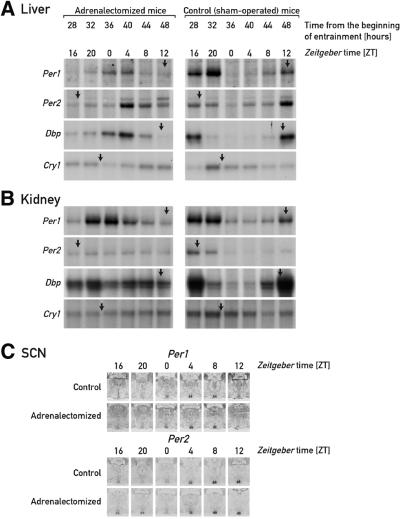
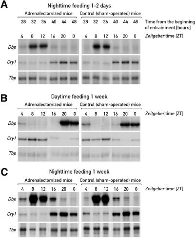
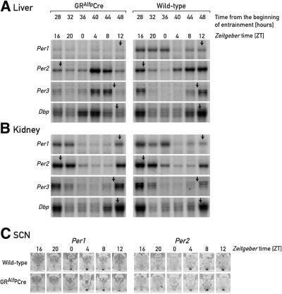
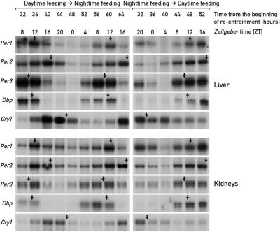
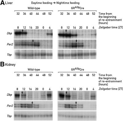
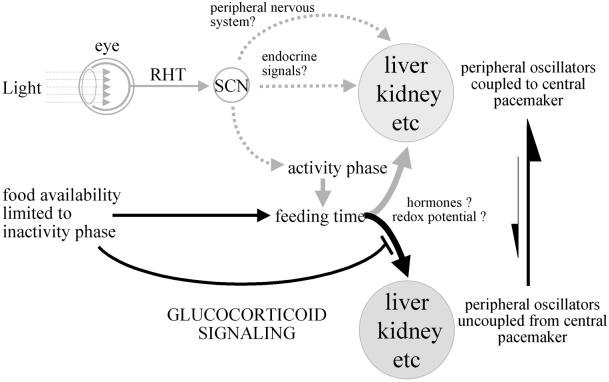
The circadian timing system in mammals is composed of a master pacemaker in the suprachiasmatic nucleus (SCN) of the hypothalamus and slave clocks in most peripheral cell types. The phase of peripheral clocks can be completely uncoupled from the SCN pacemaker by restricted feeding. Thus, feeding time, while not affecting the phase of the SCN pacemaker, is a dominant Zeitgeber for peripheral circadian oscillators. Here we show that the phase resetting in peripheral clocks of nocturnal mice is slow when feeding time is changed from night to day and rapid when switched back from day to night. Unexpectedly, the inertia in daytime feeding-induced phase resetting of circadian gene expression in liver and kidney is not an intrinsic property of peripheral oscillators, but is caused by glucocorticoid signaling. Thus, glucocorticoid hormones inhibit the uncoupling of peripheral and central circadian oscillators by altered feeding time.


Figures

None
Fig. 1. Accumulation of circadian transcripts in adrenalectomized and sham-operated mice at the onset of daytime feeding. Adrenalectomized mice and sham-operated mice, kept under a 12 h light–dark regimen (lights on ZT0), were switched from ad libitum to daytime feeding. On day D, food was removed at ZT12 and provided exclusively during the light phase of the following days. During the second day (D+1) of restricted feeding, animals were killed at 4 h intervals and the livers, kidneys and brains were collected for monitoring the accumulation of mRNAs specified by the three clock genes mPer1, mPer2 and mCry1, and the clock output gene Dbp. Relative transcript levels in liver (A) and kidney (B) were determined by RNase protection assays using radiolabeled antisense RNA probes. Tbp (Tatabox-binding protein) mRNAs served as a control for a transcript whose accumulation does not oscillate during the day (not shown). The approximate times at which peak levels of mRNA accumulation are observed in animals fed ad libitum or during the night are ZT12 (mPer1), ZT17 (mPer2), ZT11 (Dbp) and ZT22 (mCry1) (see Damiola et al., 2000 and Figure 2C). These times are indicated by arrows on top of the respective panels. The accumulation of mPer1 and mPer2 mRNA in the SCN was assessed by in situ hybridization to coronal brain sections, using 35S-labeled antisense RNA probes (C). Only the hypothalamus regions containing the SCN (small pairwise structures at the base of the hypothalamus) are depicted. At ZT20, the signal obtained for mPer2 mRNA is somewhat higher in the adrenalectomized animal compared with sham-operated animals, but this difference does not significantly change the phase of mPer2 mRNA accumulation.
None
Fig. 2. Hepatic accumulation of mCry1 and Dbp transcripts in night-time- and daytime-fed adrenalectomized and sham-operated mice. Adrenalectomized mice and sham-operated mice fed ad libitum were kept under a 12 h light–dark regimen (lights on ZT0) before they were switched to night-time feeding [(A) and (C)] or daytime feeding (B). One day (A) or 1 week [(B) and (C)] after restricted feeding had been initiated, animals were killed at 4 h intervals and the livers were collected for monitoring the accumulation of Dbp and mCry1 transcripts. Tbp mRNA was included as a transcript whose accumulation is neither circadian- nor feeding time-dependent.
None
Fig. 3. Diurnal corticosterone levels in animals fed ad libitum or during the day. Daytime-dependent corticosterone serum levels were determined in mice fed ad libitum (A), or in mice fed exclusively during the day for 7 days (B) or 1–2 days (C). For each time point, the blood was collected from five [(B) and (C)] to seven (A) individuals. As indicated in the text, due to the episodic nature of glucocorticoid secretion, the individual variability is small during nadir times but large during zenith times. In the steady-state profiles shown in (A) and (B), the values obtained for ZT16 and ZT20 are repeated to visualize the bimodal distributions.
None
Fig. 4. Accumulation of circadian transcripts in GRAlfpCre and wild-type mice at the onset of daytime feeding. GRAlfpCre and wild-type mice were switched to daytime feeding as explained in the legend to Figure 1. The hepatocytes of GRAlfpCre mice harbor a glucocorticoid receptor null allele and thus cannot respond to corticosterone signaling. The relative mRNA levels were determined by RNase protection assays for liver (A) and kidney (B), and by in situ hybridization for the SCN (C). For comparison, the approximate peak times of mRNA accumulation in animals fed ad libitum or during the night are indicated by arrows (see Figure 1).
None
Fig. 5. Accumulation of circadian transcripts in mice switched from daytime to night-time feeding or vice versa. Mice kept under a 12 h LD regimen were entrained to daytime feeding or night-time feeding for 8 days, and then switched to night-time or daytime feeding, respectively. At the times after feeding time reversal (re-entrainment) indicated, liver and kidney RNAs were subjected to RNase protection assays with the probes indicated on the left-hand side of the panels. Tbp mRNA was included as a transcript whose accumulation is neither circadian- nor feeding time-dependent (not shown). For comparison, the approximate peak times of mRNA accumulation in animals fed ad libitum or during the night are indicated by arrows.
None
Fig. 6. Accumulation of Dbp and mPer2 mRNA in GRAlfpCre and wild-type mice switched from daytime to night-time feeding. Mice kept under a 12 h LD regimen were entrained to daytime feeding during 8 days, and then switched to night-time feeding. At the times after the feeding time reversal (re-entrainment) indicated, liver (A) and kidney (B) RNAs were subjected to RNase protection assays with Dbp, mPer2 and Tbp probes. Tbp mRNA was included as a transcript whose accumulation is neither circadian- nor feeding time-dependent. For comparison, the approximate peak times of mRNA accumulation in animals fed ad libitum or during the night are indicated by arrows. Note that glucocorticoid signaling [compare left and right autoradiographs in (A)] has little if any effect on the rapid phase adjustments in mRNA accumulation cycles observed in mice switched from daytime to night-time feeding.
None
Fig. 7. The phase entrainment of circadian oscillators in peripheral organs. The scheme displays a hypothetical model of the synchronization of peripheral oscillators by the central SCN pacemaker and feeding time. The SCN is entrained by the photoperiod via synaptic connections with the retina (retino–hypothalamic tract, RHT). In turn, the SCN master clock entrains circadian gene expression in peripheral tissues (e.g. liver, kidney) through direct neuronal and humoral pathways, or indirectly by determining the activity phase and thus feeding time. When food, the dominant Zeitgeber for (at least some) peripheral oscillators, is only available during the resting phase (light phase in nocturnal rodents), the phases of these clocks are inverted. This process is slowed down by glucocorticoid signaling, probably through the abundant secretion of corticosterones during the dark phase in animals switched to daytime feeding. In contrast to the slow-phase adaptation of peripheral gene expression accompanying the switch from night-time to daytime feeding, during which peripheral oscillators become uncoupled from the central pacemaker, the switch back from daytime to night-time feeding causes an almost instantaneous phase inversion. The molecular mechanisms by which food resets the phase of peripheral oscillators are not known, but they may involve changes in the redox potential [NAD(P)H/NAD(P)+ ratio] or in hormones whose secretion is provoked or suppressed by food metabolites.

References

    1. Akashi M. and Nishida,E. (2000) Involvement of the MAP kinase cascade in resetting of the mammalian circadian clock. Genes Dev., 14, 645–649. - PMC - PubMed
    1. Albrecht U., Sun,Z.S., Eichele,G. and Lee,C.C. (1997) A differential response of two putative mammalian circadian regulators, mper1 and mper2, to light. Cell, 91, 1055–1064. - PubMed
    1. Balsalobre A., Damiola,F. and Schibler,U. (1998) A serum shock induces circadian gene expression in mammalian tissue culture cells. Cell, 93, 929–937. - PubMed
    1. Balsalobre A., Brown,S.A., Marcacci,L., Tronche,F., Kellendonk,C., Reichardt,H.M., Schutz,G. and Schibler,U. (2000a) Resetting of circadian time in peripheral tissues by glucocorticoid signaling. Science, 289, 2344–2347. - PubMed
    1. Balsalobre A., Marcacci,L. and Schibler,U. (2000b) Multiple signaling pathways elicit circadian gene expression in cultured Rat-1 fibroblasts. Curr. Biol., 10, 1291–1294. - PubMed

Publication types