. 2001 Dec 17;20(24):7137-48.
doi: 10.1093/emboj/20.24.7137.

The Saccharomyces cerevisiae Set1 complex includes an Ash2 homologue and methylates histone 3 lysine 4

Affiliations

The Saccharomyces cerevisiae Set1 complex includes an Ash2 homologue and methylates histone 3 lysine 4

A Roguev et al. EMBO J. .

Abstract

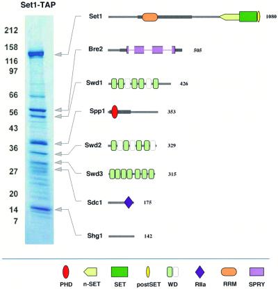
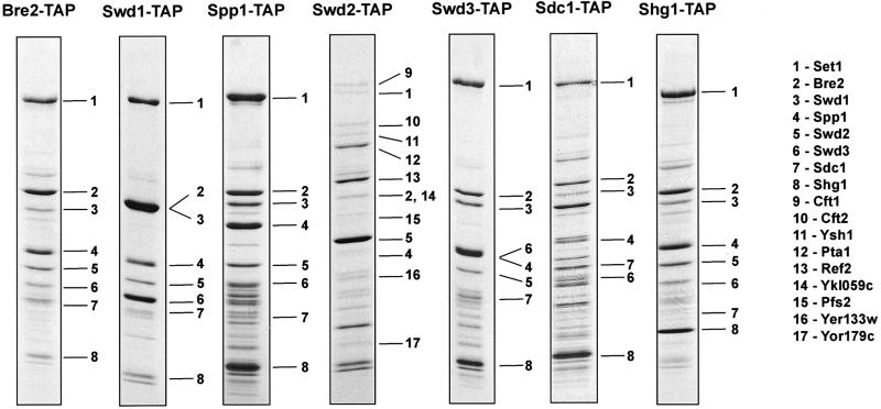
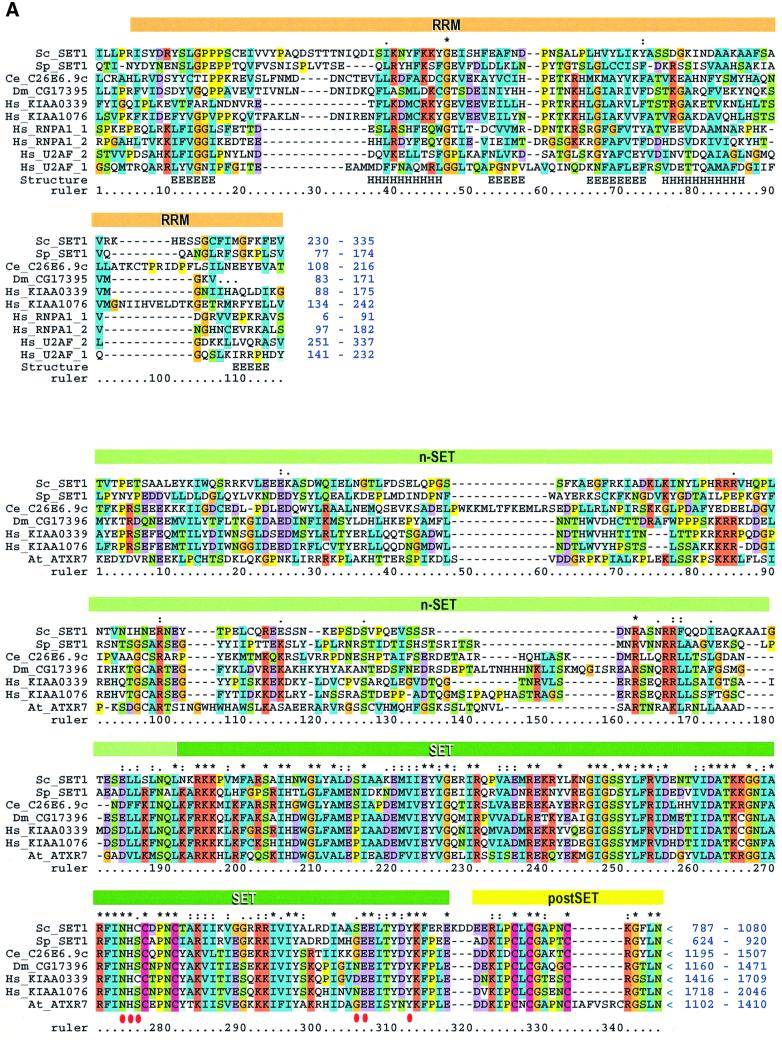
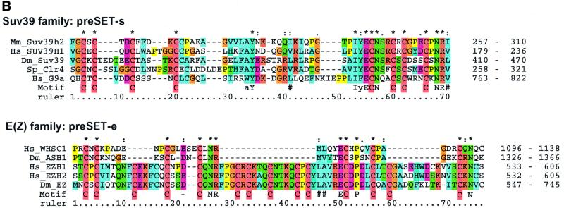
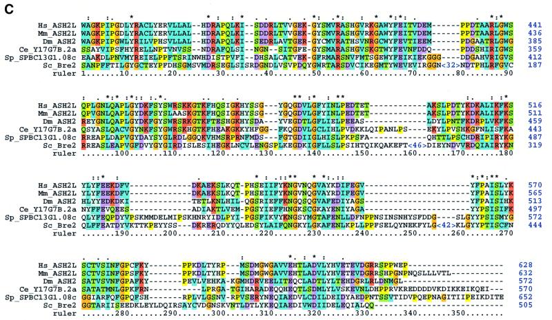
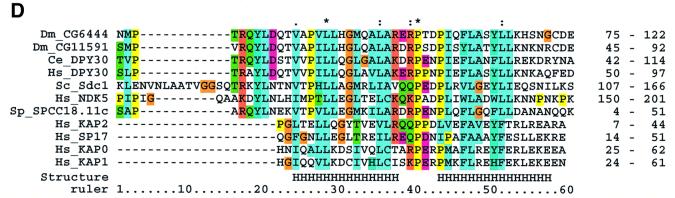
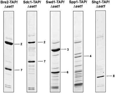
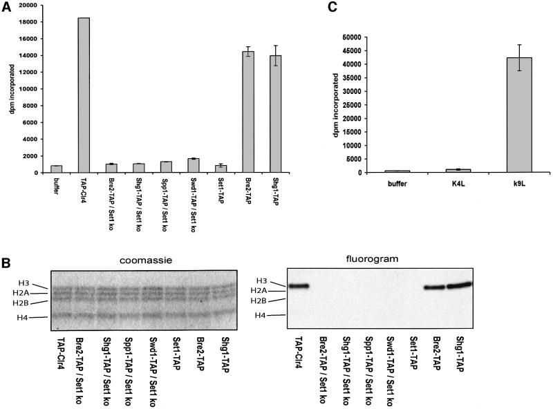
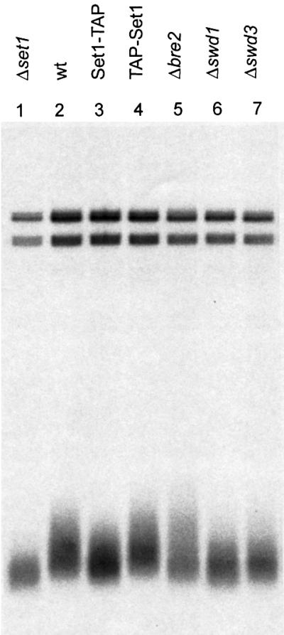
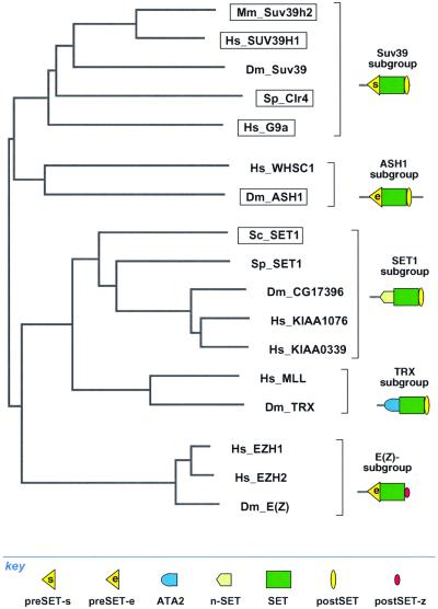
The SET domain proteins, SUV39 and G9a have recently been shown to be histone methyltransferases specific for lysines 9 and 27 (G9a only) of histone 3 (H3). The SET domains of the Saccharomyces cerevisiae Set1 and Drosophila trithorax proteins are closely related to each other but distinct from SUV39 and G9a. We characterized the complex associated with Set1 and Set1C and found that it is comprised of eight members, one of which, Bre2, is homologous to the trithorax-group (trxG) protein, Ash2. Set1C requires Set1 for complex integrity and mutation of Set1 and Set1C components shortens telomeres. One Set1C member, Swd2/Cpf10 is also present in cleavage polyadenylation factor (CPF). Set1C methylates lysine 4 of H3, thus adding a new specificity and a new subclass of SET domain proteins known to methyltransferases. Since methylation of H3 lysine 4 is widespread in eukaryotes, we screened the databases and found other Set1 homologues. We propose that eukaryotic Set1Cs are H3 lysine 4 methyltransferases and are related to trxG action through association with Ash2 homologues.


Figures

None
Fig. 1. The composition of the Set1 complex. The affinity-purified Set1-TAP complex was separated on 7–25% SDS–PAGE and visualized by staining with Coomassie Blue. Molecular weight markers indicated on the left in kilodaltons. All bands present in this gel were identified and those subsequently determined to be specific to Set1C by repetition and further affinity purification exercises are depicted to the right. Each protein is depicted with identifiable protein domains and motifs as indicated in the key below. The thickened grey lines in Set1 and Spp1 indicate regions of further conservation to S.pombe proteins, Set1 and SPBC13G1.08c, respectively and the thickened grey line in Bre2 indicates the extent of further homology to Ash2 either side of the SPRY domain (see Figure 3C). The length of each polypeptide (aa) is noted on the right. The domains indicated are: PHD finger (Pfam:PF00628); n-SET (N-terminal SET associated domain in SET1 family; see Figure 3A); SET domain (Pfam:PF00856); postSET, C-terminal SET-associated peptide (SMART:00508); WD domain (Pfam:PF00400); RIIa, protein kinase A regulatory subunit dimerization domain (Pfam:PF02197); RRM, RNA recognition motif (Pfam:PF00076) and SPRY, domain in SPlA kinase and the ryanodine receptor (Pfam:PF00622). The SPRY domain in Bre2 is interrupted by three insertions (see Figure 3C). WD40 domains showing significant alignment scores are shown in light green, inferred alignments in white (data not shown).
None
Fig. 2. Sequential affinity purification of Set1C. Each of the other seven members of Set1C was TAP-tagged and the purified complexes visualized by Coomassie Blue staining as indicated above each panel. The identity of each protein was established by mass spectroscopy and is indicated by numbers (see key to the right). Many unlabelled bands were also identified and were found to be highly abundant proteins (see Material and methods). The presence of the tag, after TEV cleavage, increases the size of the tagged protein by ∼10 kDa.
None
Fig. 3. Sequence analyses of the Set1C. (A) Multiple sequence alignment of Set1 family members showing the RRM region (upper) and n-SET/SET/postSET region (lower). The alignment is colour coded in order to highlight the conserved features according to Gibson et al. (1994). The amino acid co-ordinates in each sequence are indicated after each of the two sequence blocks. The RRM domain (also called RNP) is highlighted with an orange bar. Four RRMs from human U2AF and RNPA are included for comparison. Assignment of secondary structure elements (H, helix; E, strand) is based on the known RRM structure (Burd and Dreyfuss, 1994). Below, the n-SET region is denoted by a light green bar, the SET domain by a dark green bar and the postSET motif by a yellow bar. The conserved residues characteristic for the methyltransferase catalytic core of the SET domain (Rea et al., 2000) are indicated with red dots. The position of stop codons is indicated by ‘<’. (B) Multiple alignments of selected preSET regions are shown. At the top, the preSET regions found in SUV39 and G9a families, termed preSET-s, is shown. At the bottom, the preSET region found in E(Z) and ASH1 families, preSET-e, is shown. (C) Multiple alignment of the SPRY region of Bre2 with the Ash2 family. The region of homology shown extends further N- and C-terminally than the defined SPRY domain, which, in this alignment, starts at residue 61 and ends at 283. Three insertions in Bre2 of 32, 46 and 42 residues are indicated. (D) Multiple alignment of a region in Sdc1 with Dpy30 and other sequences, including four human protein kinase A factors for reference. This region contains a motif that is related to the dimerization domain (here called RIIa) of protein kinase A regulatory subunits. The position of two α-helices in the RIIa structure (pdb:1r2a) is shown. The database sources of all proteins used in this figure and elsewhere in this paper are given in Table II.
None
Fig. 4. Dissection of interactions within Set1C. The structure of Set1C was examined in set1 strains carrying TAP-tagged Set1C members, as indicated above each panel, by affinity purification. All protein identities, including many unlabelled unspecific bands were established by mass spectroscopy. Numbers are the same as in Figure 2 and are 2, Bre2; 3, Swd1; 4, Spp1; 6, Swd3; 7, Sdc1; 8, Shg1.
None
Fig. 5. Set1C specifically methylates lysine 4 of H3. Histone methyltransferase activity was assayed by incubation of an H3 tail peptide (A) or free histones (B), with affinity purified extracts prepared from yeast strains carrying the TAP tag fused to Clr4 or Set1C components as indicated above each lane. The extracts were prepared from either wild-type or set1 strains as indicated. The TAP-Clr4 extract was prepared from the wild-type strain carrying a TAP-Clr4 CEN plasmid. (A) Extracts were incubated with an H3 N-terminal peptide in the presence of S-adenosyl-L-[methyl-3H]methionine and incorporated radioactivity determined by filter binding. Buffer, incubation of all reagents without any added extract. (B) Extracts were incubated with free histones in the presence of S-adenosyl-L-[methyl-3H]methionine, followed by gel electrophoresis and Coomassie Blue staining (left) and fluorography (right). (C) Set1C, isolated from a wild-type strain including Bre2-TAP, was incubated with H3 N-terminal peptides carrying either a lysine 4 to leucine (K4L) or lysine 9 to leucine (K9L) mutation.
None
Fig. 6. Set1C activity is required for maintenance of telomere lengths. Telomeres were visualized by a Southern blotting strategy using a telomere specific probe. Genomic DNAs were isolated from the following strains: set1 (lane 1); wild type (lane 2); Set1-TAP (lane 3); TAP-Set1 (lane 4); bre2 (lane 5); swd1 (lane 6); swd3 (lane 7).
None
Fig. 7. Classification of SET domains by two different criterion yields the same groupings. At the left, a non-rooted tree of the SET domains, based on multiple sequence alignment of the SET domain, without inclusion of preSET and postSET regions, is shown. Four major groups are evident [SUV39, ASH1, SET1/TRX, E(Z)]. To the right, the distribution of flanking sequence elements is depicted. Known methyltransferases, present in three of the four major branches, are boxed.

References

    1. Aasland R., Gibson,T.J. and Stewart,A.F. (1995) The PHD finger: implications for chromatin-mediated transcriptional regulation. Trends Biochem. Sci., 20, 56–59. - PubMed
    1. Adamson A.L. and Shearn,A. (1996) Molecular genetic analysis of Drosophila ash2, a member of the trithorax group required for imaginal disc pattern formation. Genetics, 144, 621–633. - PMC - PubMed
    1. Altschul S.F., Madden,T.L., Schäffer,A.A., Zhang,J., Zhang,Z., Miller,W. and Lipman,D.J. (1997) Gapped BLAST and PSI-BLAST: a new generation of protein database search programs. Nucleic Acids Res., 25, 3389–3402. - PMC - PubMed
    1. Bannister A.J., Zegerman,P., Partridge,J.F., Miska,E.A., Thomas,J.O., Allshire,R.C. and Kouzarides,T. (2001) Selective recognition of methylated lysine 9 on histone H3 by the HP1 chromo domain. Nature, 410, 120–124. - PubMed
    1. Bateman A., Birney,E., Durbin,R., Eddy,S.R., Howe,K.L. and Sonnhammer,E.L.L. (2000) The Pfam protein families database. Nucleic Acids Res., 28, 263–266. - PMC - PubMed

MeSH terms