. 2001 Dec 17;20(24):7168-73.
doi: 10.1093/emboj/20.24.7168.

Repression of inflammatory responses in the absence of DNA binding by the glucocorticoid receptor

Affiliations

Repression of inflammatory responses in the absence of DNA binding by the glucocorticoid receptor

H M Reichardt et al. EMBO J. .

Abstract

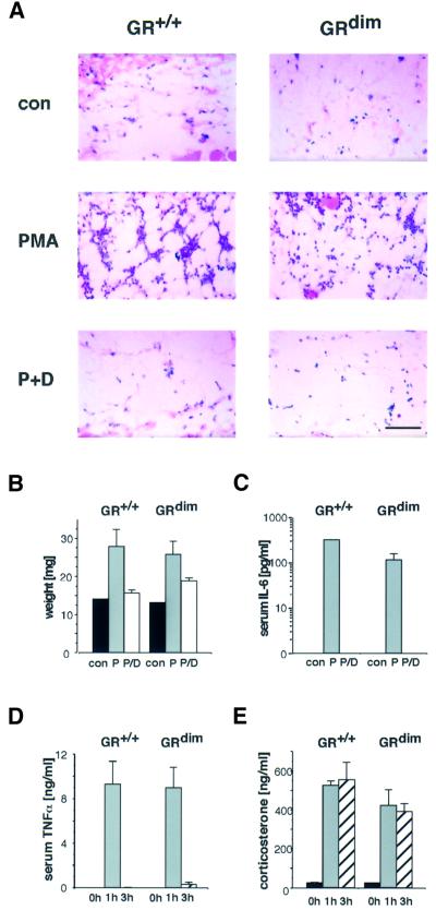
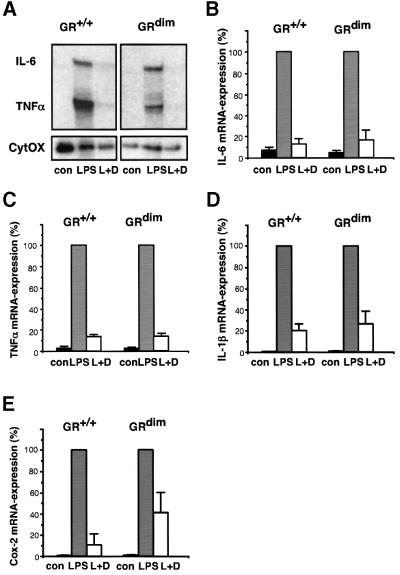
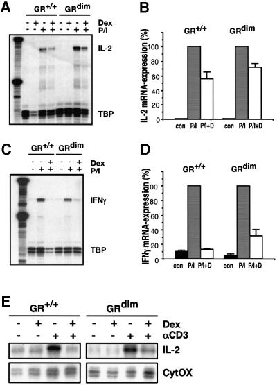
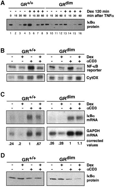
The glucocorticoid receptor (GR) acts both as a transcription factor itself on genes carrying GR response elements (GREs) and as a modulator of other transcription factors. Using mice with a mutation in the GR, which cannot activate GRE promoters, we examine whether the important anti-inflammatory and immune suppressive functions of glucocorticoids (GCs) can be established in this in vivo animal model. We find that most actions are indeed exerted in the absence of the DNA-binding ability of the GR: inhibition of the inflammatory response of locally irritated skin and of the systemic response to lipopolysaccharides. GCs repress the expression and release of numerous cytokines both in vivo and in isolated primary macrophages, thymocytes and CD4(+) splenocytes. A transgenic reporter gene controlled by NF-kappa B exclusively is also repressed, suggesting that protein- protein interaction with other transcription factors such as NF-kappa B forms the basis of the anti-inflammatory activity of GR. The only defect of immune suppression detected so far concerns the induced apoptosis of thymocytes and T lymphocytes.


Figures

None
Fig. 1. GCs potently suppress local and systemic inflammatory responses in GRdim mice. (A) Hematoxylin–eosin-stained sections of subdermal fat tissue derived from the back skin of wild-type (GR+/+) and GRdim mice treated with either vehicle acetone (con), 10 nmol PMA (P) or PMA plus 50 µg dexamethasone (P+D) for 6 h. Scale bar: 100 µm. (B) Vehicle acetone (con), 1 nmol PMA (P) or PMA plus 5 µg dexamethasone (P/D) were applied ectopically to the ears of the mice and the swelling measured after 6 h. (C) IL-6 levels in serum of mice treated as described in (A) were determined by ELISA. (D and E) Wild-type (GR+/+) or GRdim mice were injected with 100 µg of LPS per mouse and killed at the time points indicated. TNF-α serum levels were measured by ELISA (D) and corticosterone serum levels by RIA (E).
None
Fig. 2. Repression of cytokine genes by GC in peritoneal macrophages does not require the DNA-binding function of GR. (A) TNF-α and IL-6 mRNA levels were determined by RNase protection assay in peritoneal macrophages of either wild-type (GR+/+) or GRdim mice cultured under the following conditions: mock-treated (con), treated with 100 ng/ml LPS for 2 h (LPS) and LPS reatment in the presence of 10–6 M dexamethasone (L+D). Dexamethasone was added 1 h prior to LPS. mRNA levels for CytOx (cytochrome oxidase) were used for normalization. (B and C) Quantitative evaluation of the data in (A). The levels after induction by LPS were taken as 100%. mRNA levels for IL-1β (D) and cyclooxygenase-2 (Cox-2) (E) were determined by quantitative PCR using HPRT for normalization.
None
Fig. 3. Repression of cytokine genes by GCs in T lymphocytes does not require the DNA-binding function of GR. Primary thymocytes of either wild-type (GR+/+) or GRdim genotype were cultured in the absence (con) or presence of 10–6 M dexamethasone and 10 µg/ml PMA/0.5 µg/ml ionomycin (P/I) for 6 h. (A and C) IL-2 and IFN-γ mRNA levels were determined by RNase protection analysis and normalized to TATA box-binding protein (TBP). (B and D) Quantitative evaluation of the data shown in (A) and (C). The level after induction by P/I was taken as 100%. (E) IL-2 analysis of primary CD4+ splenocytes by RNase protection using CytOX RNA for normalization. Cells were cultured on αCD3-coated culture dishes in the absence or presence of 10–6 M dexamethasone for 4 h.
None
Fig. 4. Induction of IκBα expression is not required for repression of NF-κB activity in MEFs and CD4+ splenocytes. (A) GR+/+ and GRdim MEFs were treated with 10–7 M dexamethasone for 2 h followed by treatment with 50 ng/ml TNF-α. IκBα protein content was measured by immunoblotting. (B) CD4+ splenocytes from NF-κB transgenic reporter mice expressing either wild-type (GR+/+) or DNA binding-defective GR (GRdim) were cultured on αCD3-coated culture dishes in the absence or presence of 10–6 M dexamethasone for 4 h. Expression of the reporter gene (B) was determined by RNase protection analysis and normalized to CytOX. Relative IκBα mRNA levels (C) were determined by northern blot analysis and IκBα protein (D) was detected by western blot analysis.

References

    1. Auphan N., DiDonato,J.A., Rosette,C., Helmberg,A. and Karin,M. (1995) Immunosuppression by glucocorticoids: inhibition of NF-κB activity through induction of IκB synthesis. Science, 270, 286–290. - PubMed
    1. Baeuerle P.A. and Baltimore,D. (1996) NF-κB: ten years after. Cell, 87, 13–20. - PubMed
    1. Barnes P.J. (1998) Anti-inflammatory actions of glucocorticoids: molecular mechanisms. Clin. Sci., 94, 557–572. - PubMed
    1. Beato M., Herrlich,P. and Schütz,G. (1995) Steroid hormone receptors: many actors in search of a plot. Cell, 83, 851–857. - PubMed
    1. Brostjan C., Anrather,J., Csizmadia,V., Natarajan,G. and Winkler,H. (1997) Glucocorticoids inhibit E-selectin expression by targeting NF-κB and not ATF/c-Jun. J. Immunol., 158, 3836–3844. - PubMed

Publication types