. 2001 Dec 17;20(24):7184-96.
doi: 10.1093/emboj/20.24.7184.

Activation of AML1-mediated transcription by MOZ and inhibition by the MOZ-CBP fusion protein

Affiliations

Activation of AML1-mediated transcription by MOZ and inhibition by the MOZ-CBP fusion protein

I Kitabayashi et al. EMBO J. .

Abstract

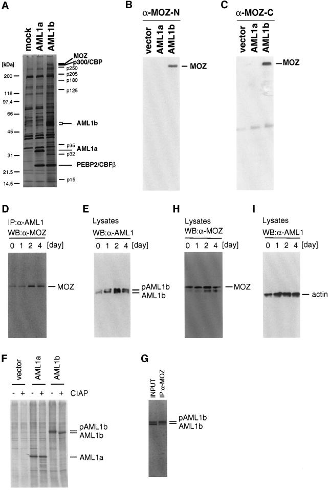
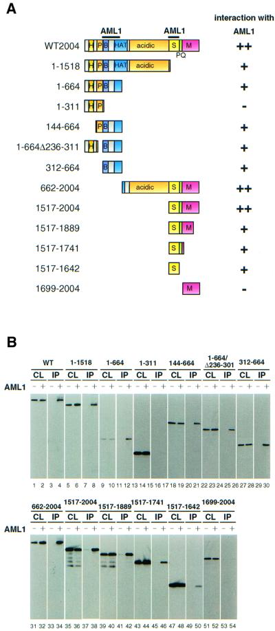
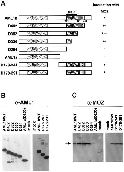
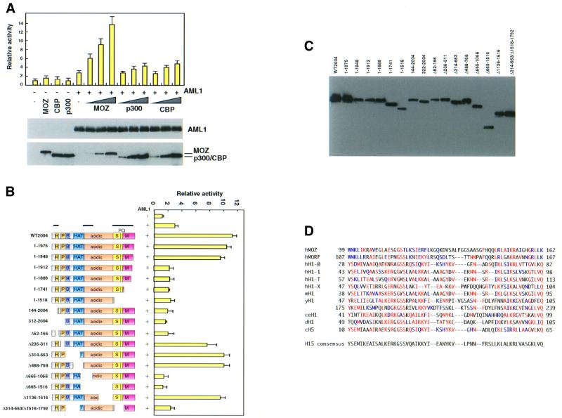
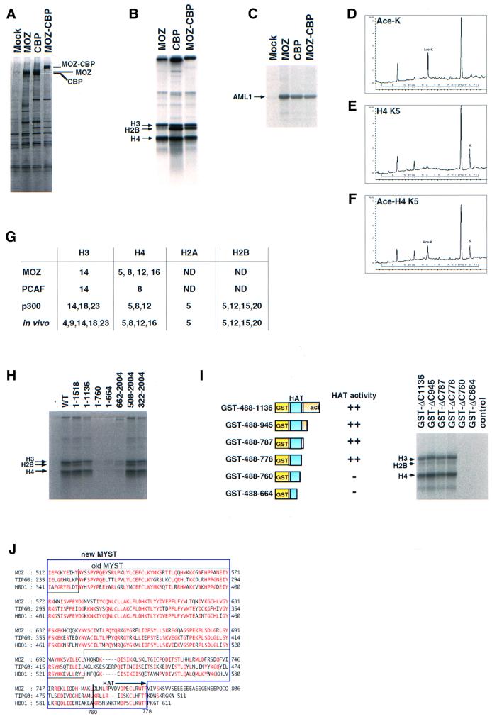
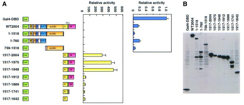
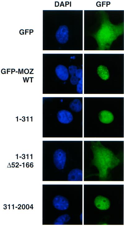
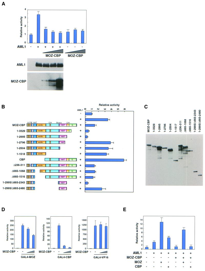
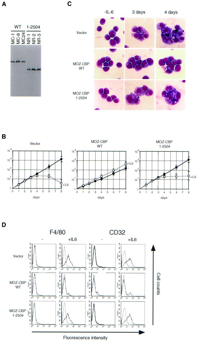
The AML1-CBF beta transcription factor complex is the most frequent target of specific chromosome translocations in human leukemia. The MOZ gene, which encodes a histone acetyltransferase (HAT), is also involved in some leukemia-associated translocations. We report here that MOZ is part of the AML1 complex and strongly stimulates AML1-mediated transcription. The stimulation of AML1-mediated transcription is independent of the inherent HAT activity of MOZ. Rather, a potent transactivation domain within MOZ appears to be essential for stimulation of AML1-mediated transcription. MOZ, as well as CBP and MOZ-CBP, can acetylate AML1 in vitro. The amount of AML1-MOZ complex increases during the differentiation of M1 myeloid cells into monocytes/macrophages, suggesting that the AML1-MOZ complex might play a role in cell differentiation. On the other hand, the MOZ-CBP fusion protein, which is created by the t(8;16) translocation associated with acute monocytic leukemia, inhibits AML1-mediated transcription and differentiation of M1 cells. These results suggest that MOZ-CBP might induce leukemia by antagonizing the function of the AML1 complex.


Figures

None
Fig. 1. MOZ forms a complex with AML1b. (A) FLAG-tagged AML1a and AML1b proteins were partially purified from lysates of infectants and vector control cells (mock) using anti-FLAG antibody (M2). (B and C) The fractions in (A) were analyzed by immunoblotting using polyclonal antibodies against the N- (B) and C-terminal region of MOZ (C). (D, E, H and I) AML1–MOZ complex during the differentiation of M1 cells. M1 cells were exposed to IL-6 for the indicated number of days. The cell lysates were prepared and immunoprecipitated using anti-AML1 antibody. The immunoprecipitates (D) or the cell lysates (E, H and I) were analyzed by immunoblotting with anti-MOZ-N (D and E), anti-AML1 antibodies (F) and anti-actin antibodies (I). (F) The M1 infectants that expressed Flag-tagged AML1a or AML1b were exposed to IL-6 for 4 days and labeled with [35S]methionine. The cell lysates were immunoprecipitated with anti-FLAG antibody. The immunoprecipitates were then incubated with (+) or without (–) calf intestine alkaline phosphatase (CIAP) and analyzed by 10% SDS–PAGE followed by autoradiography. (G) The M1 infectants that expressed Flag-tagged AML1b were exposed to IL-6 for 2 days. The cell lysates were immunoprecipitated with anti-MOZ antibody. The cell lysates (INPUT) and the immunoprecipitates (IP:α-MOZ) were analyzed by immunoblotting using horseradish peroxidase-conjugated anti-FLAG antibody.
None
Fig. 2. AML1 interacts with two regions of MOZ. (A) Schematic representation of the structure of MOZ deletion mutants used for determining the AML1-interacting domains. The H15 domain (H), the PHD-type zinc finger domain (P), the basic domain (B), the histone acetyltransferase (HAT) domain, the serine-rich region (S), the proline- and glutamine-rich region (PQ), the methionine-rich region (M) and the regions that bind to AML1 (bars) are indicated. (B) Regions of MOZ required for interaction with AML1. Bosc23 cells were transfected with deletion mutants of MOZ together with (+) or without (–) pLNCX-FLAG-AML1b. The cell lysates were immunoprecipitated with anti-FLAG antibody. The immunoprecipitates (IP) and the cell lysates (CL) were separated by SDS–PAGE and subjected to immunoblot analysis using the anti-HA antibody.
None
Fig. 3. MOZ interacts with the transactivation domain of AML1. (A) Schematic representation of the structure of AML1 deletion mutants. The runt homology domain (Runt), transcription activation domain (AD) and transcription inhibition domain (ID) are indicated. (B and C) Regions of AML1 required for interaction with MOZ. Bosc23 cells were transfected with FLAG-tagged AML1 mutants. The lysates of the transfectants were immunoprecipitated with the anti-FLAG antibody M2. The immunoprecipitates were analyzed by immunoblotting using anti-AML1 (B) and anti-MOZ (C) antibodies.
None
Fig. 4. MOZ strongly activates AML1-mediated transcription. (A) Activation of AML1 by MOZ. P19 cells were transfected with 50 ng of MPO-luc, 200 ng of pLNCX-AML1b and either 30 (lanes 6, 9 and 12), 100 (lanes 7, 10 and 13) or 350 ng (lanes 2–4, 8, 11 and 14) of pLNCX-HA-MOZ (lanes 2 and 6–8), pLNCX-HA-p300 (lanes 3 and 9–11) or pLNCX-HA-CBP (lanes 4 and 12–14) and 25 ng of pRL-tk in 24-well plates. Luciferase activity was assayed as described in Materials and methods. The lysates of the cells transfected as described above were also analyzed by immunoblotting with anti-AML1 and anti-HA antibodies (for MOZ, p300 and CBP). Essentially similar results were obtained using the CCP1-luc reporter instead of MPO-luc. Note that the expression of intrinsic AML1 could not be detected in P19. (B) Regions of MOZ required for activation of AML1. Saos-2 cells were transfected with 50 ng of MPO-luc, 200 ng of pLNCX-AML1b, 350 ng of pLNCX-HA-MOZ mutants and 25 ng of pRL-tk. The left panel shows a schematic representation of the structure of the deletion mutants. Bars above the panel indicate the region essential for stimulation of AML1-mediated transactivation. Essentially similar results were obtained using 3T3 cells. H, H15; P, PHD; B, basic; HAT, histone acetyltransferase; S, serine-rich; PQ, proline and glutamine; M, methionine-rich. (C) Expression of MOZ mutants. The Saos2 cells were transfected with HA-tagged MOZ mutants and the cell lysates were analyzed by immunoblotting with anti-HA antibody. (D) Comparison of amino acid sequences for the H15 domain of human MOZ (hMOZ), human MORF (hMORF) and linker histone H1 and H5 species. Amino acids that are identical and homologous to the H15 consensus are indicated in red and blue, respectively.
None
Fig. 5. MOZ acetylates histones H3 and H4. (A) Purification of MOZ, CBP and MOZ–CBP. Bosc23 cells were transfected with 10 µg of either the pLNCX vector (–), pLNCX-FLAG-MOZ (MOZ), pLNCX-FLAG-CBP (CBP) or pLNCX-FLAG-MOZ–CBP (MOZ–CBP) in a 10 cm dish. Proteins were immunoprecipitated using anti-FLAG antibody (M2)-conjugated beads and eluted by incubation with 0.2 mg/ml FLAG peptide. The protein constituents of the eluates were separated by 5–20% SDS–PAGE and stained with silver. (B and C) Acetylation of histones and AML1 by MOZ, CBP and MOZ–CBP. The affinity-purified MOZ, CBP and MOZ–CBP proteins were assayed for acetylation activity with histones H2B, H3 and H4 (B) or FLAG-AML1 (C). (D) HPLC charts of standard acetylated lysine. (E and F) Untreated and MOZ-acetylated histones H3 and H4 were subjected to N-terminal microsequencing. HPLC charts of fifth cycles for untreated (D) and MOZ-acetylated (E) histones H4 are shown. (G) Comparison of lysine residues acetylated in vivo, or in vitro by MOZ, PCAF or p300. The results of PCAF- and p300-acetylating residues were adopted from Schiltz et al. (1999). (H) HAT activity of MOZ mutants. Bosc23 cells were transfected with deletion mutants of MOZ as described above. The cell lysates were immunoprecipitated with anti-FLAG antibody and eluted by incubation with 0.2 mg/ml FLAG peptide. The eluates were analyzed for HAT activity. (I) HAT activity of GST–MOZ. GST–MOZ fusion proteins were produced in Escherichia coli, purified by using glutathione–Sepharose beads and analyzed for HAT activity. (J) Comparison of amino acid sequences for the HAT domains of MOZ, TIP60 and HBO1. Amino acids that are identical in more than two proteins are indicated in red.
None
Fig. 6. Transactivation domain of MOZ. (A) Transactivation by GAL4-MOZ. Saos-2 cells were transfected with 250 ng of Gal4-luc (pFR-luc), 50 ng of GAL4-MOZ mutants and 25 ng of pRL-tk. The luciferase assay was performed as described in Materials and methods. Results are given as relative activity, based on the control activity for GAL4-DBD (arbitrarily set at 1). (B) Expression of GAL4-MOZ mutants. The Saos2 cells were transfected with GAL4-MOZ mutants and the cell lysates were analyzed by immunoblotting with anti-GAL4 antibody.
None
Fig. 7. The H15 domain is important for nuclear localization of MOZ. COS7 cells were transfected with the wild-type and deletion mutants of GFP–MOZ fusion constructs. Left and right panels show DAPI staining and GFP fluorescence, respectively.
None
Fig. 8. MOZ–CBP inhibits AML1-mediated transcription. (A) Effects of MOZ–CBP on AML1 activity. Saos-2 cells were transfected with 50 ng of MPO-luc, 200 ng of pLNCX-AML1b (lanes 2–5), and either 30 (lanes 3 and 6), 100 (lanes 4 and 7) or 350 ng (lanes 5 and 8) of pLNCX-HA-MOZ–CBP and 25 ng of pRL. Luciferase assays were performed as described in Materials and methods. The lysates of the cells transfected as above were also analyzed by immunoblotting with anti-AML1 and anti-HA antibodies (for MOZ–CBP). (B) Effects of deletion mutants of MOZ–CBP on AML1 activity. Saos-2 cells were transfected with 50 ng of MPO-luc, 200 ng of pLNCX-AML1b, 350 ng of pLNCX-HA-MOZ–CBP mutants and 25 ng of pRL-tk. Luciferase assays were performed. (C) Expression of deletion mutants of MOZ–CBP. The Saos-2 cells were transfected with HA-tagged MOZ–CBP mutants and the cell lysates were analyzed by immunoblotting with anti-HA antibody. (D) Effects of MOZ–CBP on transactivation by using GAL4 fusions. Saos-2 cells were transfected with 250 ng of Gal4-luc (pFR-luc), 50 ng of either GAL4-MOZ (lanes 2–4), GAL4-CBP (lanes 6–8) or GAL4-VP16 (lanes 10–12), 50 (lanes 3, 7 and 11) or 200 ng (lanes 4, 8 and 12) of MOZ–CBP and 25 ng of pRL. Luciferase assays were performed. (E) Effects of MOZ–CBP on co-activation by MOZ and CBP. Saos-2 cells were transfected with 50 ng of MPO-luc, 200 ng of pLNCX-AML1b (lanes 2–7), 100 ng of pLNCX-HA-MOZ–CBP (lanes 5–7), 250 ng of either pLNCX-HA-MOZ (lanes 3 and 6) or pLNCX-HA-CBP (lanes 4 and 7) and 25 ng of pRL. A luciferase assay was performed.
None
Fig. 9. MOZ–CBP inhibits the differentiation of M1 cells. (A) Expression of wild-type and a deletion mutant (1–2504) of MOZ–CBP. M1 cells were transfected with linearized LNCX, LNCX-HA-MOZ–CBP or LNCX-HA-MOZ–CBP(1–2504) and selected with 0.8 mg/ml G418. Selected colonies were cloned. Cell lysates of M1 clones were separated by 5% SDS–PAGE and analyzed by western blotting using anti-HA antibody (3F10). (B) Growth curve of the M1 infectants in response to IL-6. Each of the three M1 clones in (A) was cultured in the presence or absence of 5 ng/ml of IL-6. Results represent the average values of the relative numbers of viable cells for these clones. (C) Morphology of the cells. The M1 clones, which expressed wild-type MOZ–CBP (MC-1) or deletion mutant (NR-1), were exposed to IL-6 for 3 or 4 days and stained with May–Gruenwald and Giemsa solutions. Similar results were obtained with other clones. (D) The expression of surface antigens. The M1 infectants were exposed to IL-6 for 3 days and the expression of F4/80 and CD32 was analyzed by the indirect immunofluorescent method. Representative results for MC-1 (wild-type) and NR-1 (1–2504) are shown.

References

    1. Borrow J., et al. (1996) The translocation t(8;16)(p11;p13) of acute myeloid leukaemia fuses a putative acetyltransferase to the CREB-binding protein. Nature Genet., 14, 33–41. - PubMed
    1. Carapeti M., Aguiar,R.C.T., Goldman,J.M. and Cross,N.C.P. (1998) A novel fusion between MOZ and the nuclear receptor coactivator TIF2 in acute myeloid leukemia. Blood, 91, 3127–3133. - PubMed
    1. Chaffanet M., Gressin,L., Preudhomme,C., Soenen-Cornu,V., Birnbaum,D. and Pebusque,M.-J. (2000) MOZ is fused to p300 in an acute monocytic leukemia with t(8;22). Genes Chromosomes Cancer, 28, 138–144. - PubMed
    1. Champagne N., Pelletier,N. and Yang,X.J. (2001) The monocytic leukemia zinc finger protein MOZ is a histone acetyltransferase. Oncogene, 20, 404–409. - PubMed
    1. Chen H., Lin,R.J., Schiltz,R.L., Chakravarti,D., Nash,A., Nagy,L., Privalsky,M.L., Nakatani,Y. and Evans,R.M. (1997) Nuclear receptor coactivator ACTR is a novel histone acetyltransferase and forms a multimeric activation complex with P/CAF and CBP/p300. Cell, 90, 569–580. - PubMed

Publication types

MeSH terms